Show PDB file:   
         Plain Text   HTML   (compressed file size)
QuickSearch:   
by PDB,NDB,UniProt,PROSITE Code or Search Term(s)  
(-)Asym./Biol. Unit
(-)Asym./Biol. Unit - sites
collapse expand < >
Image Asym./Biol. Unit
Asym./Biol. Unit  (Jmol Viewer)
Image Asym./Biol. Unit - sites
Asym./Biol. Unit - sites  (Jmol Viewer)

(-) Description

Title :  SELF-ASSEMBLY OF SPIDER SILK PROTEINS IS CONTROLLED BY A PH-SENSITIVE RELAY
 
Authors :  G. Askarieh, H. Hedhammar, K. Nordling, A. Rising, J. Johansson, S. D. K
Date :  10 Feb 10  (Deposition) - 12 May 10  (Release) - 26 May 10  (Revision)
Method :  X-RAY DIFFRACTION
Resolution :  2.30
Chains :  Asym./Biol. Unit :  A,B
Keywords :  Dragline Spider Silk, Self-Assembly, Ph-Dependence, Nt E79Q Mutant, Structural Protein (Keyword Search: [Gene Ontology, PubMed, Web (Google))
 
Reference :  G. Askarieh, M. Hedhammar, K. Nordling, A. Saenz, C. Casals, A. Rising, J. Johansson, S. D. Knight
Self-Assembly Of Spider Silk Proteins Is Controlled By A Ph-Sensitive Relay.
Nature V. 465 236 2010
PubMed-ID: 20463740  |  Reference-DOI: 10.1038/NATURE08962

(-) Compounds

Molecule 1 - MAJOR AMPULLATE SPIDROIN 1
    ChainsA, B
    EngineeredYES
    Expression SystemESCHERICHIA COLI
    Expression System Taxid562
    FragmentN-TERMINAL DOMAIN
    GeneMASP1
    MutationYES
    Organism ScientificEUPROSTHENOPS AUSTRALIS
    Organism Taxid332052

 Structural Features

(-) Chains, Units

  12
Asymmetric/Biological Unit AB

Summary Information (see also Sequences/Alignments below)
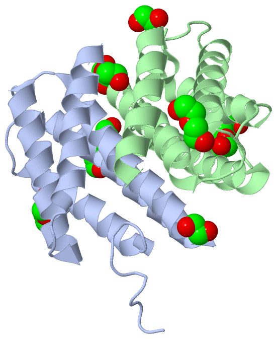
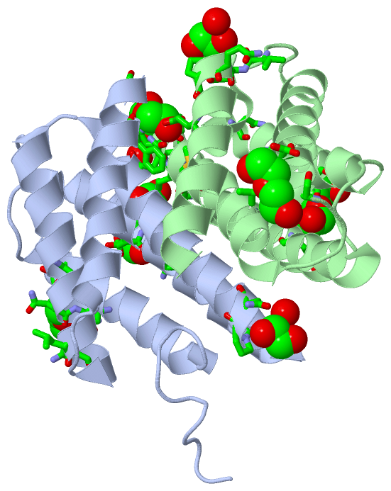
(-) Ligands, Modified Residues, Ions  (3, 8)

Asymmetric/Biological Unit (3, 8)
No.NameCountTypeFull Name
1EDO4Ligand/Ion1,2-ETHANEDIOL
2PEG2Ligand/IonDI(HYDROXYETHYL)ETHER
3PGE2Ligand/IonTRIETHYLENE GLYCOL

(-) Sites  (8, 8)

Asymmetric Unit (8, 8)
No.NameEvidenceResiduesDescription
1AC1SOFTWARESER A:62 , PRO A:63 , ASN A:64 , HOH A:143BINDING SITE FOR RESIDUE EDO A 1397
2AC2SOFTWAREGLN A:79 , PHE A:127 , GLY A:131 , MET B:71 , GLU B:119BINDING SITE FOR RESIDUE EDO A 138
3AC3SOFTWARESER A:22 , GLN A:25 , GLY A:26 , ALA A:100 , ASN A:103 , ALA A:104 , GLN A:107 , ASN B:12 , GLY B:14BINDING SITE FOR RESIDUE PEG A 139
4AC4SOFTWAREHOH A:142 , SER B:62 , PRO B:63 , ASN B:64BINDING SITE FOR RESIDUE EDO B 1397
5AC5SOFTWAREVAL B:112 , ASN B:113 , GLN B:114 , PRO B:115 , HOH B:167BINDING SITE FOR RESIDUE EDO B 138
6AC6SOFTWARESER B:89 , LEU B:90 , SER B:91BINDING SITE FOR RESIDUE PEG B 139
7AC7SOFTWAREASP A:39 , SER A:42 , THR A:43 , GLN A:46 , HOH A:155 , ARG B:60 , LYS B:65BINDING SITE FOR RESIDUE PGE B 140
8AC8SOFTWARELEU A:106 , GLN A:107 , ASN B:21 , GLN B:46 , VAL B:49 , GLN B:53BINDING SITE FOR RESIDUE PGE B 141

(-) SS Bonds  (0, 0)

(no "SS Bond" information available for 3LR8)

(-) Cis Peptide Bonds  (0, 0)

(no "Cis Peptide Bond" information available for 3LR8)

 Sequence-Structure Mapping

(-) SAPs(SNPs)/Variants  (0, 0)

(no "SAP(SNP)/Variant" information available for 3LR8)

(-) PROSITE Motifs  (0, 0)

(no "PROSITE Motif" information available for 3LR8)

(-) Exons   (0, 0)

(no "Exon" information available for 3LR8)

(-) Sequences/Alignments

Asymmetric/Biological Unit
   Reformat: Number of residues per line =  ('0' or empty: single-line sequence representation)
  Number of residues per labelling interval =   
  UniProt sequence: complete  aligned part    
   Show mapping: SCOP domains CATH domains Pfam domains Secondary structure (by author)
SAPs(SNPs) PROSITE motifs Exons
(details for a mapped element are shown in a popup box when the mouse pointer rests over it)
Chain A from PDB  Type:PROTEIN  Length:131
 aligned with Q05H60_9ARAC | Q05H60 from UniProtKB/TrEMBL  Length:394

    Alignment length:133
                                    27        37        47        57        67        77        87        97       107       117       127       137       147   
         Q05H60_9ARAC    18 GSSSLASHTTPWTNPGLAENFMNSFMQGLSSMPGFTASQLDDMSTIAQSMVQSIQSLAAQGRTSPNKLQALNMAFASSMAEIAASEEGGGSLSTKTSSIASAMSNAFLQTTGVVNQPFINEITQLVSMFAQAG 150
               SCOP domains ------------------------------------------------------------------------------------------------------------------------------------- SCOP domains
               CATH domains ------------------------------------------------------------------------------------------------------------------------------------- CATH domains
               Pfam domains ------------------------------------------------------------------------------------------------------------------------------------- Pfam domains
         Sec.struct. author ...--........hhhhhhhhhhhhhhhhh.....hhhhhhhhhhhhhhhhhhhhhhhhhh..hhhhhhhhhhhhhhhhhhhhhhh....hhhhhhhhhhhhhhhhhhhhh...hhhhhhhhhhhhhhhhhhh Sec.struct. author
                 SAPs(SNPs) ------------------------------------------------------------------------------------------------------------------------------------- SAPs(SNPs)
                    PROSITE ------------------------------------------------------------------------------------------------------------------------------------- PROSITE
                 Transcript ------------------------------------------------------------------------------------------------------------------------------------- Transcript
                 3lr8 A   1 GSG--NSHTTPWTNPGLAENFMNSFMQGLSSMPGFTASQLDDMSTIAQSMVQSIQSLAAQGRTSPNKLQALNMAFASSMAQIAASEEGGGSLSTKTSSIASAMSNAFLQTTGVVNQPFINEITQLVSMFAQAG 131
                              |  |   8        18        28        38        48        58        68        78        88        98       108       118       128   
                              3  4                                                                                                                               

Chain B from PDB  Type:PROTEIN  Length:125
 aligned with Q05H60_9ARAC | Q05H60 from UniProtKB/TrEMBL  Length:394

    Alignment length:125
                                    35        45        55        65        75        85        95       105       115       125       135       145     
         Q05H60_9ARAC    26 TTPWTNPGLAENFMNSFMQGLSSMPGFTASQLDDMSTIAQSMVQSIQSLAAQGRTSPNKLQALNMAFASSMAEIAASEEGGGSLSTKTSSIASAMSNAFLQTTGVVNQPFINEITQLVSMFAQAG 150
               SCOP domains ----------------------------------------------------------------------------------------------------------------------------- SCOP domains
               CATH domains ----------------------------------------------------------------------------------------------------------------------------- CATH domains
               Pfam domains ----------------------------------------------------------------------------------------------------------------------------- Pfam domains
         Sec.struct. author .hhhhhhhhhhhhhhhhhhhhh.....hhhhhhhhhhhhhhhhhhhhhhhhh...hhhhhhhhhhhhhhhhhhhhhhh....hhhhhhhhhhhhhhhhhhhh....hhhhhhhhhhhhhhhhhhh Sec.struct. author
                 SAPs(SNPs) ----------------------------------------------------------------------------------------------------------------------------- SAPs(SNPs)
                    PROSITE ----------------------------------------------------------------------------------------------------------------------------- PROSITE
                 Transcript ----------------------------------------------------------------------------------------------------------------------------- Transcript
                 3lr8 B   7 TTPWTNPGLAENFMNSFMQGLSSMPGFTASQLDDMSTIAQSMVQSIQSLAAQGRTSPNKLQALNMAFASSMAQIAASEEGGGSLSTKTSSIASAMSNAFLQTTGVVNQPFINEITQLVSMFAQAG 131
                                    16        26        36        46        56        66        76        86        96       106       116       126     

   Legend:   → Mismatch (orange background)
  - → Gap (green background, '-', border residues have a numbering label)
    → Modified Residue (blue background, lower-case, 'x' indicates undefined single-letter code, labelled with number + name)
  x → Chemical Group (purple background, 'x', labelled with number + name, e.g. ACE or NH2)
  extra numbering lines below/above indicate numbering irregularities and modified residue names etc., number ends below/above '|'

 Classification and Annotation

(-) SCOP Domains  (0, 0)

(no "SCOP Domain" information available for 3LR8)

(-) CATH Domains  (0, 0)

(no "CATH Domain" information available for 3LR8)

(-) Pfam Domains  (0, 0)

(no "Pfam Domain" information available for 3LR8)

(-) Gene Ontology  (0, 0)

Asymmetric/Biological Unit(hide GO term definitions)
    (no "Gene Ontology" information available for 3LR8)

 Visualization

(-) Interactive Views

Asymmetric/Biological Unit
  Complete Structure
    Jena3D(integrated viewing of ligand, site, SAP, PROSITE, SCOP information)
    WebMol | AstexViewer[tm]@PDBe
(Java Applets, require no local installation except for Java; loading may be slow)
    STRAP
(Java WebStart application, automatic local installation, requires Java; full application with system access!)
    RasMol
(require local installation)
    Molscript (VRML)
(requires installation of a VRML viewer; select preferred view via VRML and generate a mono or stereo PDF format file)
 
  Ligands, Modified Residues, Ions
    EDO  [ RasMol | Jena3D ]  +environment [ RasMol | Jena3D ]
    PEG  [ RasMol | Jena3D ]  +environment [ RasMol | Jena3D ]
    PGE  [ RasMol | Jena3D ]  +environment [ RasMol | Jena3D ]
 
  Sites
    AC1  [ RasMol ]  +environment [ RasMol ]
    AC2  [ RasMol ]  +environment [ RasMol ]
    AC3  [ RasMol ]  +environment [ RasMol ]
    AC4  [ RasMol ]  +environment [ RasMol ]
    AC5  [ RasMol ]  +environment [ RasMol ]
    AC6  [ RasMol ]  +environment [ RasMol ]
    AC7  [ RasMol ]  +environment [ RasMol ]
    AC8  [ RasMol ]  +environment [ RasMol ]
 
  Cis Peptide Bonds
(no "Cis Peptide Bonds" information available for 3lr8)
 

(-) Still Images

Jmol
  protein: cartoon or spacefill or dots and stick; nucleic acid: cartoon and stick; ligands: spacefill; active site: stick
Molscript
  protein, nucleic acid: cartoon; ligands: spacefill; active site: ball and stick

 Databases and Analysis Tools

(-) Databases

Access by PDB/NDB ID
  3lr8
    Family and Domain InformationProDom | SYSTERS
    General Structural InformationGlycoscienceDB | MMDB | NDB | OCA | PDB | PDBe | PDBj | PDBsum | PDBWiki | PQS | PROTEOPEDIA
    Orientation in MembranesOPM
    Protein SurfaceSURFACE
    Secondary StructureDSSP (structure derived) | HSSP (homology derived)
    Structural GenomicsGeneCensus
    Structural NeighboursCE | VAST
    Structure ClassificationCATH | Dali | SCOP
    Validation and Original DataBMRB Data View | BMRB Restraints Grid | EDS | PROCHECK | RECOORD | WHAT_CHECK
 
Access by UniProt ID/Accession number
  Q05H60_9ARAC | Q05H60
    Comparative Protein Structure ModelsModBase
    Genomic InformationEnsembl
    Protein-protein InteractionDIP
    Sequence, Family and Domain InformationInterPro | Pfam | SMART | UniProtKB/TrEMBL
 
Access by Enzyme Classificator   (EC Number)
  (no 'Enzyme Classificator' available)
    General Enzyme InformationBRENDA | EC-PDB | Enzyme | IntEnz
    PathwayKEGG | MetaCyc
 
Access by Disease Identifier   (MIM ID)
  (no 'MIM ID' available)
    Disease InformationOMIM
 
Access by GenAge ID
  (no 'GenAge ID' available)
    Age Related InformationGenAge

(-) Analysis Tools

Access by PDB/NDB ID
    Domain InformationXDom
    Interatomic Contacts of Structural UnitsCSU
    Ligand-protein ContactsLPC
    Protein CavitiescastP
    Sequence and Secondary StructurePDBCartoon
    Structure AlignmentSTRAP(Java WebStart application, automatic local installation, requires Java; full application with system access!)
    Structure and Sequence BrowserSTING
 
Access by UniProt ID/Accession number
  Q05H60_9ARAC | Q05H60
    Protein Disorder PredictionDisEMBL | FoldIndex | GLOBPLOT (for more information see DisProt)

 Related Entries

(-) Entries Sharing at Least One Protein Chain (UniProt ID)

UniProtKB/TrEMBL
        Q05H60_9ARAC | Q05H602lpi 2lpj 2lth 3lr2 3lr6 3lrd 4fbs

(-) Related Entries Specified in the PDB File

3lr2 NT WILD-TYPE
3lr6
3lrd