Show PDB file:   
         Plain Text   HTML   (compressed file size)
QuickSearch:   
by PDB,NDB,UniProt,PROSITE Code or Search Term(s)  
(-)Asymmetric Unit
(-)Asym. Unit - sites
(-)Biological Unit 1
(-)Biol. Unit 1 - sites
collapse expand < >
Image Asymmetric Unit
Asymmetric Unit  (Jmol Viewer)
Image Asym. Unit - sites
Asym. Unit - sites  (Jmol Viewer)
Image Biological Unit 1
Biological Unit 1  (Jmol Viewer)
Image Biol. Unit 1 - sites
Biol. Unit 1 - sites  (Jmol Viewer)

(-) Description

Title :  CRYSTAL STRUCTURE OF PUTATIVE SUGAR ISOMERASE. (YP_001305105.1) FROM PARABACTEROIDES DISTASONIS ATCC 8503 AT 1.44 A RESOLUTION
 
Authors :  Joint Center For Structural Genomics (Jcsg)
Date :  01 Feb 10  (Deposition) - 23 Feb 10  (Release) - 13 Jul 11  (Revision)
Method :  X-RAY DIFFRACTION
Resolution :  1.44
Chains :  Asym. Unit :  A
Biol. Unit 1:  A  (2x)
Keywords :  Putative Sugar Isomerase. , Structural Genomics, Joint Center For Structural Genomics, Jcsg, Protein Structure Initiative, Psi-2, Isomerase (Keyword Search: [Gene Ontology, PubMed, Web (Google))
 
Reference :  Joint Center For Structural Genomics (Jcsg)
Crystal Structure Of Putative Sugar Isomerase. (Yp_001305105. 1) From Parabacteroides Distasonis Atcc 8503 At 1. 44 A Resolution
To Be Published
PubMed: search

(-) Compounds

Molecule 1 - PUTATIVE SUGAR ISOMERASE
    ChainsA
    EngineeredYES
    Expression SystemESCHERICHIA COLI
    Expression System PlasmidSPEEDET
    Expression System StrainHK100
    Expression System Taxid562
    Expression System Vector TypePLASMID
    GeneBDI_3797
    Organism ScientificPARABACTEROIDES DISTASONIS
    Organism Taxid435591
    StrainATCC 8503 / DSM 20701 / NCTC 11152

 Structural Features

(-) Chains, Units

  1
Asymmetric Unit A
Biological Unit 1 (2x)A

Summary Information (see also Sequences/Alignments below)

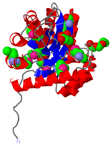
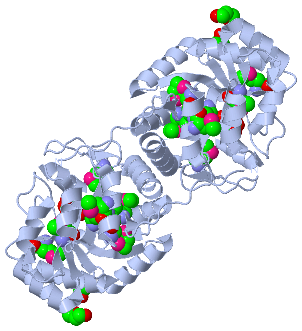
(-) Ligands, Modified Residues, Ions  (4, 11)

Asymmetric Unit (4, 11)
No.NameCountTypeFull Name
1CIT1Ligand/IonCITRIC ACID
2MSE8Mod. Amino AcidSELENOMETHIONINE
3PEG1Ligand/IonDI(HYDROXYETHYL)ETHER
4PGE1Ligand/IonTRIETHYLENE GLYCOL
Biological Unit 1 (4, 22)
No.NameCountTypeFull Name
1CIT2Ligand/IonCITRIC ACID
2MSE16Mod. Amino AcidSELENOMETHIONINE
3PEG2Ligand/IonDI(HYDROXYETHYL)ETHER
4PGE2Ligand/IonTRIETHYLENE GLYCOL

(-) Sites  (3, 3)

Asymmetric Unit (3, 3)
No.NameEvidenceResiduesDescription
1AC1SOFTWAREHOH A:6 , TYR A:51 , LYS A:76 , PRO A:108 , TYR A:110 , HIS A:158 , HIS A:160 , HIS A:192 , LYS A:217 , GLU A:257 , GLU A:259 , HOH A:285 , HOH A:370BINDING SITE FOR RESIDUE CIT A 1
2AC2SOFTWARELYS A:125 , GLU A:150 , THR A:220 , ASP A:221 , HOH A:297 , HOH A:526 , HOH A:575BINDING SITE FOR RESIDUE PEG A 2
3AC3SOFTWAREASP A:77 , ASN A:83 , HOH A:589 , HOH A:610 , HOH A:645BINDING SITE FOR RESIDUE PGE A 3

(-) SS Bonds  (0, 0)

(no "SS Bond" information available for 3LMZ)

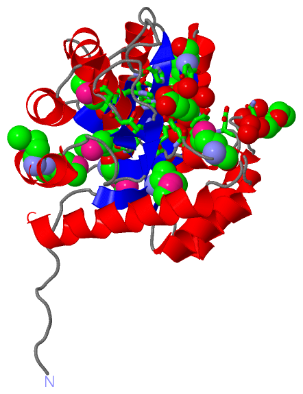
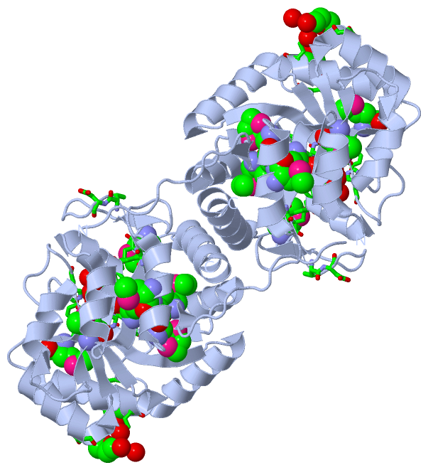
(-) Cis Peptide Bonds  (1, 1)

Asymmetric Unit
No.Residues
1Gly A:107 -Pro A:108

 Sequence-Structure Mapping

(-) SAPs(SNPs)/Variants  (0, 0)

(no "SAP(SNP)/Variant" information available for 3LMZ)

(-) PROSITE Motifs  (0, 0)

(no "PROSITE Motif" information available for 3LMZ)

(-) Exons   (0, 0)

(no "Exon" information available for 3LMZ)

(-) Sequences/Alignments

Asymmetric Unit
   Reformat: Number of residues per line =  ('0' or empty: single-line sequence representation)
  Number of residues per labelling interval =   
  UniProt sequence: complete  aligned part    
   Show mapping: SCOP domains CATH domains Pfam domains Secondary structure (by author)
SAPs(SNPs) PROSITE motifs Exons
(details for a mapped element are shown in a popup box when the mouse pointer rests over it)
Chain A from PDB  Type:PROTEIN  Length:251
 aligned with A6LIG9_PARD8 | A6LIG9 from UniProtKB/TrEMBL  Length:283

    Alignment length:251
                                    42        52        62        72        82        92       102       112       122       132       142       152       162       172       182       192       202       212       222       232       242       252       262       272       282 
         A6LIG9_PARD8    33 VKPKAPKAVNPFHLGMAGYTFVNFDLDTTLKTLERLDIHYLCIKDFHLPLNSTDEQIRAFHDKCAAHKVTGYAVGPIYMKSEEEIDRAFDYAKRVGVKLIVGVPNYELLPYVDKKVKEYDFHYAIHLHGPDIKTYPDATDVWVHTKDLDPRIGMCLDVGHDLRNGCDPVADLKKYHTRVFDMHIKDVTDSSKAGVGIEIGRGKIDFPALIRMMREVNYTGMCSLEYEKDMKDPFLGIAESIGYFKAVSDLT 283
               SCOP domains ----------------------------------------------------------------------------------------------------------------------------------------------------------------------------------------------------------------------------------------------------------- SCOP domains
               CATH domains ----------------------------------------------------------------------------------------------------------------------------------------------------------------------------------------------------------------------------------------------------------- CATH domains
               Pfam domains ------------AP_endonuc_2-3lmzA01 A:45-236                                                                                                                                                                   AP_endonuc_2_N-3lmzA02 A:237-280            --- Pfam domains
         Sec.struct. author ............eeee.hhhhh..hhhhhhhhhhhh...eeee.........hhhhhhhhhhhhhhh..eeeeeeeeee.hhhhhhhhhhhhhhhh..eeeeee...hhhhhhhhhhhhh.eeeee..........hhhhhhhhhh.....eeeeeehhhhhhh..hhhhhhhhhh..eeeeee.ee........ee.......hhhhhhhhhhhh....eeee........hhhhhhhhhhhhhhhhhhh Sec.struct. author
                 SAPs(SNPs) ----------------------------------------------------------------------------------------------------------------------------------------------------------------------------------------------------------------------------------------------------------- SAPs(SNPs)
                    PROSITE ----------------------------------------------------------------------------------------------------------------------------------------------------------------------------------------------------------------------------------------------------------- PROSITE
                 Transcript ----------------------------------------------------------------------------------------------------------------------------------------------------------------------------------------------------------------------------------------------------------- Transcript
                 3lmz A  33 VKPKAPKAVNPFHLGmAGYTFVNFDLDTTLKTLERLDIHYLCIKDFHLPLNSTDEQIRAFHDKCAAHKVTGYAVGPIYmKSEEEIDRAFDYAKRVGVKLIVGVPNYELLPYVDKKVKEYDFHYAIHLHGPDIKTYPDATDVWVHTKDLDPRIGmCLDVGHDLRNGCDPVADLKKYHTRVFDmHIKDVTDSSKAGVGIEIGRGKIDFPALIRmmREVNYTGmCSLEYEKDmKDPFLGIAESIGYFKAVSDLT 283
                                    42     |  52        62        72        82        92       102       112       122       132       142       152       162       172       182   |   192       202       212 |     222       232       242 ||    252|      262       272       282 
                                          48-MSE                                                        111-MSE                                                                    186-MSE                     214-MSE                       244-MSE  253-MSE  262-MSE                 
                                                                                                                                                                                                                                              245-MSE                                  

   Legend:   → Mismatch (orange background)
  - → Gap (green background, '-', border residues have a numbering label)
    → Modified Residue (blue background, lower-case, 'x' indicates undefined single-letter code, labelled with number + name)
  x → Chemical Group (purple background, 'x', labelled with number + name, e.g. ACE or NH2)
  extra numbering lines below/above indicate numbering irregularities and modified residue names etc., number ends below/above '|'

 Classification and Annotation

(-) SCOP Domains  (0, 0)

(no "SCOP Domain" information available for 3LMZ)

(-) CATH Domains  (0, 0)

(no "CATH Domain" information available for 3LMZ)

(-) Pfam Domains  (2, 2)

Asymmetric Unit

(-) Gene Ontology  (0, 0)

Asymmetric Unit(hide GO term definitions)
    (no "Gene Ontology" information available for 3LMZ)

 Visualization

(-) Interactive Views

Asymmetric Unit
  Complete Structure
    Jena3D(integrated viewing of ligand, site, SAP, PROSITE, SCOP information)
    WebMol | AstexViewer[tm]@PDBe
(Java Applets, require no local installation except for Java; loading may be slow)
    STRAP
(Java WebStart application, automatic local installation, requires Java; full application with system access!)
    RasMol
(require local installation)
    Molscript (VRML)
(requires installation of a VRML viewer; select preferred view via VRML and generate a mono or stereo PDF format file)
 
  Ligands, Modified Residues, Ions
    CIT  [ RasMol | Jena3D ]  +environment [ RasMol | Jena3D ]
    MSE  [ RasMol | Jena3D ]  +environment [ RasMol | Jena3D ]
    PEG  [ RasMol | Jena3D ]  +environment [ RasMol | Jena3D ]
    PGE  [ RasMol | Jena3D ]  +environment [ RasMol | Jena3D ]
 
  Sites
    AC1  [ RasMol ]  +environment [ RasMol ]
    AC2  [ RasMol ]  +environment [ RasMol ]
    AC3  [ RasMol ]  +environment [ RasMol ]
 
  Cis Peptide Bonds
    Gly A:107 - Pro A:108   [ RasMol ]  
 
Biological Unit
  Complete Structure
    Biological Unit 1  [ Jena3D ]

(-) Still Images

Jmol
  protein: cartoon or spacefill or dots and stick; nucleic acid: cartoon and stick; ligands: spacefill; active site: stick
Molscript
  protein, nucleic acid: cartoon; ligands: spacefill; active site: ball and stick

 Databases and Analysis Tools

(-) Databases

Access by PDB/NDB ID
  3lmz
    Family and Domain InformationProDom | SYSTERS
    General Structural InformationGlycoscienceDB | MMDB | NDB | OCA | PDB | PDBe | PDBj | PDBsum | PDBWiki | PQS | PROTEOPEDIA
    Orientation in MembranesOPM
    Protein SurfaceSURFACE
    Secondary StructureDSSP (structure derived) | HSSP (homology derived)
    Structural GenomicsGeneCensus
    Structural NeighboursCE | VAST
    Structure ClassificationCATH | Dali | SCOP
    Validation and Original DataBMRB Data View | BMRB Restraints Grid | EDS | PROCHECK | RECOORD | WHAT_CHECK
 
Access by UniProt ID/Accession number
  A6LIG9_PARD8 | A6LIG9
    Comparative Protein Structure ModelsModBase
    Genomic InformationEnsembl
    Protein-protein InteractionDIP
    Sequence, Family and Domain InformationInterPro | Pfam | SMART | UniProtKB/TrEMBL
 
Access by Enzyme Classificator   (EC Number)
  (no 'Enzyme Classificator' available)
    General Enzyme InformationBRENDA | EC-PDB | Enzyme | IntEnz
    PathwayKEGG | MetaCyc
 
Access by Disease Identifier   (MIM ID)
  (no 'MIM ID' available)
    Disease InformationOMIM
 
Access by GenAge ID
  (no 'GenAge ID' available)
    Age Related InformationGenAge

(-) Analysis Tools

Access by PDB/NDB ID
    Domain InformationXDom
    Interatomic Contacts of Structural UnitsCSU
    Ligand-protein ContactsLPC
    Protein CavitiescastP
    Sequence and Secondary StructurePDBCartoon
    Structure AlignmentSTRAP(Java WebStart application, automatic local installation, requires Java; full application with system access!)
    Structure and Sequence BrowserSTING
 
Access by UniProt ID/Accession number
  A6LIG9_PARD8 | A6LIG9
    Protein Disorder PredictionDisEMBL | FoldIndex | GLOBPLOT (for more information see DisProt)

 Related Entries

(-) Entries Sharing at Least One Protein Chain (UniProt ID)

(no "Entries Sharing at Least One Protein Chain" available for 3LMZ)

(-) Related Entries Specified in the PDB File

(no "Related Entries Specified in the PDB File" available for 3LMZ)