Show PDB file:   
         Plain Text   HTML   (compressed file size)
QuickSearch:   
by PDB,NDB,UniProt,PROSITE Code or Search Term(s)  
(-)Asymmetric Unit
(-)Asym. Unit - sites
(-)Biological Unit 1
(-)Biol. Unit 1 - sites
(-)Biological Unit 2
collapse expand < >
Image Asymmetric Unit
Asymmetric Unit  (Jmol Viewer)
Image Asym. Unit - sites
Asym. Unit - sites  (Jmol Viewer)
Image Biological Unit 1
Biological Unit 1  (Jmol Viewer)
Image Biol. Unit 1 - sites
Biol. Unit 1 - sites  (Jmol Viewer)
Image Biological Unit 2
Biological Unit 2  (Jmol Viewer)

(-) Description

Title :  CRYSTAL STRUCTURE OF THE CARBOHYDRATE RECOGNITION DOMAIN OF LMAN1 IN COMPLEX WITH MCFD2
 
Authors :  E. Wigren, J. M. Bourhis, I. Kursula, J. E. Guy, Y. Lindqvist
Date :  11 Jan 10  (Deposition) - 26 Jan 10  (Release) - 28 Apr 10  (Revision)
Method :  X-RAY DIFFRACTION
Resolution :  2.45
Chains :  Asym. Unit :  A,B,C,D
Biol. Unit 1:  A,C  (1x)
Biol. Unit 2:  B,D  (1x)
Keywords :  Er-Golgi Transport, Glycoprotein Sorting, Disease Mutation, Secretory Pathway, Protein Transport, Coagulation Factor Deficiency, Disulfide Bond, Endoplasmic Reticulum, Golgi Apparatus, Lectin, Membrane, Polymorphism, Transmembrane, Transport, Calcium, Protein Binding (Keyword Search: [Gene Ontology, PubMed, Web (Google))
 
Reference :  E. Wigren, J. M. Bourhis, I. Kursula, J. E. Guy, Y. Lindqvist
Crystal Structure Of The Lman1-Crd/Mcfd2 Transport Receptor Complex Provides Insight Into Combined Deficiency Of Factor V And Factor Viii.
Febs Lett. V. 584 878 2010
PubMed-ID: 20138881  |  Reference-DOI: 10.1016/J.FEBSLET.2010.02.009

(-) Compounds

Molecule 1 - PROTEIN ERGIC-53
    ChainsA, B
    EngineeredYES
    Expression SystemESCHERICHIA COLI
    Expression System PlasmidPET28A
    Expression System StrainBL21(DE3)
    Expression System Taxid562
    Expression System Vector TypePLASMID
    FragmentUNP RESIDUES 32-277, CARBOHYDRATE RECOGNITION DOMAIN
    GeneERGIC53, F5F8D, LMAN1, LMAN1 (AMINO ACIDS 32-277)
    Organism CommonHUMAN
    Organism ScientificHOMO SAPIENS
    Organism Taxid9606
    SynonymER-GOLGI INTERMEDIATE COMPARTMENT 53 KDA PROTEIN, LECTIN MANNOSE-BINDING 1, GP58, INTRACELLULAR MANNOSE-SPECIFIC LECTIN MR60
 
Molecule 2 - MULTIPLE COAGULATION FACTOR DEFICIENCY PROTEIN 2
    ChainsC, D
    EngineeredYES
    Expression SystemESCHERICHIA COLI
    Expression System PlasmidPET28A
    Expression System StrainBL21(DE3)
    Expression System Taxid562
    Expression System Vector TypePLASMID
    FragmentUNP RESIDUES 58-146, 2 EF-HAND DOMAINS
    GeneMCFD2, MCFD2 (AMINO ACIDS 58-146), SDNSF
    Organism CommonHUMAN
    Organism ScientificHOMO SAPIENS
    Organism Taxid9606
    SynonymNEURAL STEM CELL-DERIVED NEURONAL SURVIVAL PROTEIN

 Structural Features

(-) Chains, Units

  1234
Asymmetric Unit ABCD
Biological Unit 1 (1x)A C 
Biological Unit 2 (1x) B D

Summary Information (see also Sequences/Alignments below)

(-) Ligands, Modified Residues, Ions  (1, 8)

Asymmetric Unit (1, 8)
No.NameCountTypeFull Name
1CA8Ligand/IonCALCIUM ION
Biological Unit 1 (0, 0)
No.NameCountTypeFull Name
1CA-1Ligand/IonCALCIUM ION
Biological Unit 2 (0, 0)
No.NameCountTypeFull Name
1CA-1Ligand/IonCALCIUM ION

(-) Sites  (8, 8)

Asymmetric Unit (8, 8)
No.NameEvidenceResiduesDescription
1AC1SOFTWAREHOH A:16 , HOH A:17 , ASP A:152 , PHE A:154 , ASN A:156 , ASP A:181BINDING SITE FOR RESIDUE CA A 278
2AC2SOFTWAREHOH A:12 , ASP A:155 , ASP A:157 , ASN A:161 , ASN A:162 , ASP A:181 , HOH A:288BINDING SITE FOR RESIDUE CA A 279
3AC3SOFTWAREHOH B:16 , HOH B:17 , ASP B:152 , PHE B:154 , ASN B:156 , ASP B:181BINDING SITE FOR RESIDUE CA B 278
4AC4SOFTWAREHOH B:12 , ASP B:155 , ASP B:157 , ASN B:161 , ASN B:162 , ASP B:181BINDING SITE FOR RESIDUE CA B 279
5AC5SOFTWAREHOH C:4 , ASP C:81 , ASP C:83 , ASN C:85 , LEU C:87 , GLU C:92BINDING SITE FOR RESIDUE CA C 158
6AC6SOFTWAREASP C:129 , ASN C:131 , ASP C:133 , TYR C:135 , GLU C:140 , HOH C:147BINDING SITE FOR RESIDUE CA C 159
7AC7SOFTWAREASP D:81 , ASP D:83 , ASN D:85 , LEU D:87 , GLU D:92BINDING SITE FOR RESIDUE CA D 158
8AC8SOFTWAREASP D:129 , ASN D:131 , ASP D:133 , TYR D:135 , GLU D:140BINDING SITE FOR RESIDUE CA D 159

(-) SS Bonds  (2, 2)

Asymmetric Unit
No.Residues
1A:190 -A:230
2B:190 -B:230

(-) Cis Peptide Bonds  (6, 6)

Asymmetric Unit
No.Residues
1Gly A:54 -Pro A:55
2Ala A:120 -Asp A:121
3Asn A:162 -Pro A:163
4Gly B:54 -Pro B:55
5Ala B:120 -Asp B:121
6Asn B:162 -Pro B:163

 Sequence-Structure Mapping

(-) SAPs(SNPs)/Variants  (5, 10)

Asymmetric Unit (5, 10)
  dbSNPPDB
No.SourceVariant IDVariantUniProt IDStatusIDChainVariant
1UniProtVAR_071969W67SLMAN1_HUMANDisease (F5F8D1)  ---A/BW67S
2UniProtVAR_072245D81HMCFD2_HUMANDisease (F5F8D2)78289603C/DD81H
3UniProtVAR_019076D129EMCFD2_HUMANDisease (F5F8D2)28942113C/DD129E
4UniProtVAR_072246Y135NMCFD2_HUMANDisease (F5F8D2)748641905C/DY135N
5UniProtVAR_019077I136TMCFD2_HUMANDisease (F5F8D2)28942114C/DI136T

  SNP/SAP Summary Statistics (UniProtKB/Swiss-Prot)
Biological Unit 1 (5, 5)
  dbSNPPDB
No.SourceVariant IDVariantUniProt IDStatusIDChainVariant
1UniProtVAR_071969W67SLMAN1_HUMANDisease (F5F8D1)  ---AW67S
2UniProtVAR_072245D81HMCFD2_HUMANDisease (F5F8D2)78289603CD81H
3UniProtVAR_019076D129EMCFD2_HUMANDisease (F5F8D2)28942113CD129E
4UniProtVAR_072246Y135NMCFD2_HUMANDisease (F5F8D2)748641905CY135N
5UniProtVAR_019077I136TMCFD2_HUMANDisease (F5F8D2)28942114CI136T

  SNP/SAP Summary Statistics (UniProtKB/Swiss-Prot)
Biological Unit 2 (5, 5)
  dbSNPPDB
No.SourceVariant IDVariantUniProt IDStatusIDChainVariant
1UniProtVAR_071969W67SLMAN1_HUMANDisease (F5F8D1)  ---BW67S
2UniProtVAR_072245D81HMCFD2_HUMANDisease (F5F8D2)78289603DD81H
3UniProtVAR_019076D129EMCFD2_HUMANDisease (F5F8D2)28942113DD129E
4UniProtVAR_072246Y135NMCFD2_HUMANDisease (F5F8D2)748641905DY135N
5UniProtVAR_019077I136TMCFD2_HUMANDisease (F5F8D2)28942114DI136T

  SNP/SAP Summary Statistics (UniProtKB/Swiss-Prot)

(-) PROSITE Motifs  (3, 10)

Asymmetric Unit (3, 10)
 PROSITEUniProtKBPDB
No.IDACDescriptionIDLocationCountLocation
1L_LECTIN_LIKEPS51328 L-type lectin-like (leguminous) domain profile.LMAN1_HUMAN44-267
 
  2A:44-267
B:44-267
2EF_HAND_2PS50222 EF-hand calcium-binding domain profile.MCFD2_HUMAN68-103
 
116-146
 
  4C:68-98
D:68-99
C:116-144
D:116-143
3EF_HAND_1PS00018 EF-hand calcium-binding domain.MCFD2_HUMAN81-93
 
129-141
 
  4C:81-93
D:81-93
C:129-141
D:129-141
Biological Unit 1 (3, 5)
 PROSITEUniProtKBPDB
No.IDACDescriptionIDLocationCountLocation
1L_LECTIN_LIKEPS51328 L-type lectin-like (leguminous) domain profile.LMAN1_HUMAN44-267
 
  1A:44-267
-
2EF_HAND_2PS50222 EF-hand calcium-binding domain profile.MCFD2_HUMAN68-103
 
116-146
 
  2C:68-98
-
C:116-144
-
3EF_HAND_1PS00018 EF-hand calcium-binding domain.MCFD2_HUMAN81-93
 
129-141
 
  2C:81-93
-
C:129-141
-
Biological Unit 2 (3, 5)
 PROSITEUniProtKBPDB
No.IDACDescriptionIDLocationCountLocation
1L_LECTIN_LIKEPS51328 L-type lectin-like (leguminous) domain profile.LMAN1_HUMAN44-267
 
  1-
B:44-267
2EF_HAND_2PS50222 EF-hand calcium-binding domain profile.MCFD2_HUMAN68-103
 
116-146
 
  2-
D:68-99
-
D:116-143
3EF_HAND_1PS00018 EF-hand calcium-binding domain.MCFD2_HUMAN81-93
 
129-141
 
  2-
D:81-93
-
D:129-141

(-) Exons   (0, 0)

(no "Exon" information available for 3LCP)

(-) Sequences/Alignments

Asymmetric Unit
   Reformat: Number of residues per line =  ('0' or empty: single-line sequence representation)
  Number of residues per labelling interval =   
  UniProt sequence: complete  aligned part    
   Show mapping: SCOP domains CATH domains Pfam domains Secondary structure (by author)
SAPs(SNPs) PROSITE motifs Exons
(details for a mapped element are shown in a popup box when the mouse pointer rests over it)
Chain A from PDB  Type:PROTEIN  Length:234
 aligned with LMAN1_HUMAN | P49257 from UniProtKB/Swiss-Prot  Length:510

    Alignment length:234
                                    50        60        70        80        90       100       110       120       130       140       150       160       170       180       190       200       210       220       230       240       250       260       270    
          LMAN1_HUMAN    41 LPHRRFEYKYSFKGPHLVQSDGTVPFWAHAGNAIPSSDQIRVAPSLKSQRGSVWTKTKAAFENWEVEVTFRVTGRGRIGADGLAIWYAENQGLEGPVFGSADLWNGVGIFFDSFDNDGKKNNPAIVIIGNNGQIHYDHQNDGASQALASCQRDFRNKPYPVRAKITYYQNTLTVMINNGFTPDKNDYEFCAKVENMIIPAQGHFGISAATGGLADDHDVLSFLTFQLTEPGKEP 274
               SCOP domains d3lcpa_ A: automated matches                                                                                                                                                                                                               SCOP domains
               CATH domains 3lcpA00 A:41-274  [code=2.60.120.200, no name defined]                                                                                                                                                                                     CATH domains
               Pfam domains ------------------------------------------------------------------------------------------------------------------------------------------------------------------------------------------------------------------------------------------ Pfam domains
         Sec.struct. author ..eeeeeeeeeee.............eeeee..ee....eeee......eeeeee........eeeeeeeeee........eeeeeee................eeeeeeee...........eeeeeeee.....hhhhh......eee..........eeeeeeee..eeeeeee..........eeeeee.........eeeeeee......eeeeeeeeeeeee...... Sec.struct. author
                 SAPs(SNPs) --------------------------S--------------------------------------------------------------------------------------------------------------------------------------------------------------------------------------------------------------- SAPs(SNPs)
                    PROSITE ---L_LECTIN_LIKE  PDB: A:44-267 UniProt: 44-267                                                                                                                                                                                    ------- PROSITE
                 Transcript ------------------------------------------------------------------------------------------------------------------------------------------------------------------------------------------------------------------------------------------ Transcript
                 3lcp A  41 LPHRRFEYKYSFKGPHLVQSDGTVPFWAHAGNAIPSSDQIRVAPSLKSQRGSVWTKTKAAFENWEVEVTFRVTGRGRIGADGLAIWYAENQGLEGPVFGSADLWNGVGIFFDSFDNDGKKNNPAIVIIGNNGQIHYDHQNDGASQALASCQRDFRNKPYPVRAKITYYQNTLTVMINNGFTPDKNDYEFCAKVENMIIPAQGHFGISAATGGLADDHDVLSFLTFQLTEPGKEP 274
                                    50        60        70        80        90       100       110       120       130       140       150       160       170       180       190       200       210       220       230       240       250       260       270    

Chain B from PDB  Type:PROTEIN  Length:234
 aligned with LMAN1_HUMAN | P49257 from UniProtKB/Swiss-Prot  Length:510

    Alignment length:234
                                    50        60        70        80        90       100       110       120       130       140       150       160       170       180       190       200       210       220       230       240       250       260       270    
          LMAN1_HUMAN    41 LPHRRFEYKYSFKGPHLVQSDGTVPFWAHAGNAIPSSDQIRVAPSLKSQRGSVWTKTKAAFENWEVEVTFRVTGRGRIGADGLAIWYAENQGLEGPVFGSADLWNGVGIFFDSFDNDGKKNNPAIVIIGNNGQIHYDHQNDGASQALASCQRDFRNKPYPVRAKITYYQNTLTVMINNGFTPDKNDYEFCAKVENMIIPAQGHFGISAATGGLADDHDVLSFLTFQLTEPGKEP 274
               SCOP domains d3lcpb_ B: automated matches                                                                                                                                                                                                               SCOP domains
               CATH domains 3lcpB00 B:41-274  [code=2.60.120.200, no name defined]                                                                                                                                                                                     CATH domains
               Pfam domains ------------------------------------------------------------------------------------------------------------------------------------------------------------------------------------------------------------------------------------------ Pfam domains
         Sec.struct. author ..eeee.hhhhee.............eeeee..ee....eeee......eeeeee.......eeeeeeeeeee........eeeeeee................eeeeeeee...........eeeeeeee................eeee.........eeeeeeee..eeeeeee..........eeeeee.........eeeeeee......eeeeeeeeeeeee...... Sec.struct. author
                 SAPs(SNPs) --------------------------S--------------------------------------------------------------------------------------------------------------------------------------------------------------------------------------------------------------- SAPs(SNPs)
                    PROSITE ---L_LECTIN_LIKE  PDB: B:44-267 UniProt: 44-267                                                                                                                                                                                    ------- PROSITE
                 Transcript ------------------------------------------------------------------------------------------------------------------------------------------------------------------------------------------------------------------------------------------ Transcript
                 3lcp B  41 LPHRRFEYKYSFKGPHLVQSDGTVPFWAHAGNAIPSSDQIRVAPSLKSQRGSVWTKTKAAFENWEVEVTFRVTGRGRIGADGLAIWYAENQGLEGPVFGSADLWNGVGIFFDSFDNDGKKNNPAIVIIGNNGQIHYDHQNDGASQALASCQRDFRNKPYPVRAKITYYQNTLTVMINNGFTPDKNDYEFCAKVENMIIPAQGHFGISAATGGLADDHDVLSFLTFQLTEPGKEP 274
                                    50        60        70        80        90       100       110       120       130       140       150       160       170       180       190       200       210       220       230       240       250       260       270    

Chain C from PDB  Type:PROTEIN  Length:68
 aligned with MCFD2_HUMAN | Q8NI22 from UniProtKB/Swiss-Prot  Length:146

    Alignment length:79
                                    75        85        95       105       115       125       135         
          MCFD2_HUMAN    66 EMSPQELQLHYFKMHDYDGNNLLDGLELSTAITHVHKEEGSEQAPLMSEDELINIIDGVLRDDDKNNDGYIDYAEFAKS 144
               SCOP domains ------------------------------------------------------------------------------- SCOP domains
               CATH domains ------------------------------------------------------------------------------- CATH domains
               Pfam domains ------------------------------------------------------------------------------- Pfam domains
         Sec.struct. author ..hhhhhhhhhhhh.........hhhhhhhhhh-----------...hhhhhhhhhhhhhhhhh.......hhhhhhh. Sec.struct. author
                 SAPs(SNPs) ---------------H-----------------------------------------------E-----NT-------- SAPs(SNPs)
                PROSITE (2) --EF_HAND_2  PDB: C:68-98             ------------EF_HAND_2  PDB: C:116-144     PROSITE (2)
                PROSITE (3) ---------------EF_HAND_1    -----------------------------------EF_HAND_1    --- PROSITE (3)
                 Transcript ------------------------------------------------------------------------------- Transcript
                 3lcp C  66 EMSPQELQLHYFKMHDYDGNNLLDGLELSTAIT-----------PLMSEDELINIIDGVLRDDDKNNDGYIDYAEFAKS 144
                                    75        85        95  |      -    |  115       125       135         
                                                           98         110                                  

Chain D from PDB  Type:PROTEIN  Length:69
 aligned with MCFD2_HUMAN | Q8NI22 from UniProtKB/Swiss-Prot  Length:146

    Alignment length:78
                                    75        85        95       105       115       125       135        
          MCFD2_HUMAN    66 EMSPQELQLHYFKMHDYDGNNLLDGLELSTAITHVHKEEGSEQAPLMSEDELINIIDGVLRDDDKNNDGYIDYAEFAK 143
               SCOP domains ------------------------------------------------------------------------------ SCOP domains
               CATH domains ------------------------------------------------------------------------------ CATH domains
               Pfam domains ------------------------------------------------------------------------------ Pfam domains
         Sec.struct. author ..hhhhhhhhhhhh........eehhhhhhhhhh---------....hhhhhhhhhhhhhhhhh.....eehhhhhhh Sec.struct. author
                 SAPs(SNPs) ---------------H-----------------------------------------------E-----NT------- SAPs(SNPs)
                PROSITE (2) --EF_HAND_2  PDB: D:68-99             ------------EF_HAND_2  PDB: D:116-143    PROSITE (2)
                PROSITE (3) ---------------EF_HAND_1    -----------------------------------EF_HAND_1    -- PROSITE (3)
                 Transcript ------------------------------------------------------------------------------ Transcript
                 3lcp D  66 EMSPQELQLHYFKMHDYDGNNLLDGLELSTAITH---------APLMSEDELINIIDGVLRDDDKNNDGYIDYAEFAK 143
                                    75        85        95   |     -   |   115       125       135        
                                                            99       109                                  

   Legend:   → Mismatch (orange background)
  - → Gap (green background, '-', border residues have a numbering label)
    → Modified Residue (blue background, lower-case, 'x' indicates undefined single-letter code, labelled with number + name)
  x → Chemical Group (purple background, 'x', labelled with number + name, e.g. ACE or NH2)
  extra numbering lines below/above indicate numbering irregularities and modified residue names etc., number ends below/above '|'

 Classification and Annotation

(-) SCOP Domains  (1, 2)

Asymmetric Unit

(-) CATH Domains  (1, 2)

Asymmetric Unit
(-)
Class: Mainly Beta (13760)

(-) Pfam Domains  (0, 0)

(no "Pfam Domain" information available for 3LCP)

(-) Gene Ontology  (32, 46)

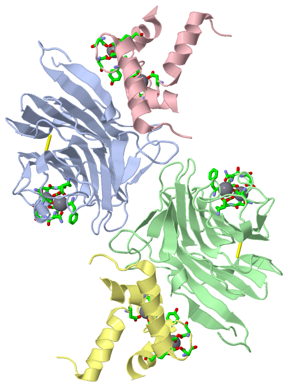
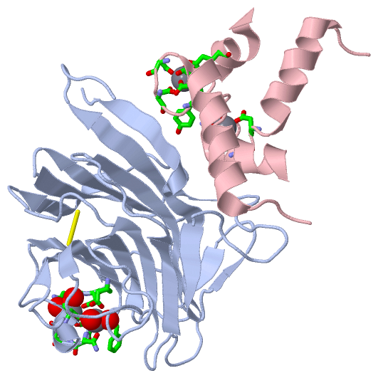
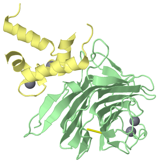
Asymmetric Unit(hide GO term definitions)
Chain A,B   (LMAN1_HUMAN | P49257)
molecular function
    GO:0030246    carbohydrate binding    Interacting selectively and non-covalently with any carbohydrate, which includes monosaccharides, oligosaccharides and polysaccharides as well as substances derived from monosaccharides by reduction of the carbonyl group (alditols), by oxidation of one or more hydroxy groups to afford the corresponding aldehydes, ketones, or carboxylic acids, or by replacement of one or more hydroxy group(s) by a hydrogen atom. Cyclitols are generally not regarded as carbohydrates.
    GO:0005537    mannose binding    Interacting selectively and non-covalently with mannose, a monosaccharide hexose, stereoisomeric with glucose, that occurs naturally only in polymerized forms called mannans.
    GO:0046872    metal ion binding    Interacting selectively and non-covalently with any metal ion.
    GO:0005515    protein binding    Interacting selectively and non-covalently with any protein or protein complex (a complex of two or more proteins that may include other nonprotein molecules).
    GO:0051082    unfolded protein binding    Interacting selectively and non-covalently with an unfolded protein.
biological process
    GO:0048208    COPII vesicle coating    The addition of COPII proteins and adaptor proteins to ER membranes during the formation of transport vesicles, forming a vesicle coat.
    GO:0006888    ER to Golgi vesicle-mediated transport    The directed movement of substances from the endoplasmic reticulum (ER) to the Golgi, mediated by COP II vesicles. Small COP II coated vesicles form from the ER and then fuse directly with the cis-Golgi. Larger structures are transported along microtubules to the cis-Golgi.
    GO:0007030    Golgi organization    A process that is carried out at the cellular level which results in the assembly, arrangement of constituent parts, or disassembly of the Golgi apparatus.
    GO:0007596    blood coagulation    The sequential process in which the multiple coagulation factors of the blood interact, ultimately resulting in the formation of an insoluble fibrin clot; it may be divided into three stages: stage 1, the formation of intrinsic and extrinsic prothrombin converting principle; stage 2, the formation of thrombin; stage 3, the formation of stable fibrin polymers.
    GO:0034498    early endosome to Golgi transport    The directed movement of substances from early endosomes to the Golgi.
    GO:0007029    endoplasmic reticulum organization    A process that is carried out at the cellular level which results in the assembly, arrangement of constituent parts, or disassembly of the endoplasmic reticulum.
    GO:0010638    positive regulation of organelle organization    Any process that increases the frequency, rate or extent of a process involved in the formation, arrangement of constituent parts, or disassembly of an organelle.
    GO:0018279    protein N-linked glycosylation via asparagine    The glycosylation of protein via the N4 atom of peptidyl-asparagine forming N4-glycosyl-L-asparagine; the most common form is N-acetylglucosaminyl asparagine; N-acetylgalactosaminyl asparagine and N4 glucosyl asparagine also occur. This modification typically occurs in extracellular peptides with an N-X-(ST) motif. Partial modification has been observed to occur with cysteine, rather than serine or threonine, in the third position; secondary structure features are important, and proline in the second or fourth positions inhibits modification.
    GO:0032527    protein exit from endoplasmic reticulum    The directed movement of proteins from the endoplasmic reticulum.
    GO:0006457    protein folding    The process of assisting in the covalent and noncovalent assembly of single chain polypeptides or multisubunit complexes into the correct tertiary structure.
    GO:0015031    protein transport    The directed movement of proteins into, out of or within a cell, or between cells, by means of some agent such as a transporter or pore.
    GO:0006810    transport    The directed movement of substances (such as macromolecules, small molecules, ions) or cellular components (such as complexes and organelles) into, out of or within a cell, or between cells, or within a multicellular organism by means of some agent such as a transporter, pore or motor protein.
    GO:0016192    vesicle-mediated transport    A cellular transport process in which transported substances are moved in membrane-bounded vesicles; transported substances are enclosed in the vesicle lumen or located in the vesicle membrane. The process begins with a step that directs a substance to the forming vesicle, and includes vesicle budding and coating. Vesicles are then targeted to, and fuse with, an acceptor membrane.
cellular component
    GO:0030134    COPII-coated ER to Golgi transport vesicle    A vesicle with a coat formed of the COPII coat complex proteins. The COPII coat complex is formed by the Sec23p/Sec24p and the Sec13p/Sec31p heterodimers. COPII-associated vesicles transport proteins from the rough endoplasmic reticulum to the Golgi apparatus (anterograde transport).
    GO:0012507    ER to Golgi transport vesicle membrane    The lipid bilayer surrounding a vesicle transporting substances from the endoplasmic reticulum to the Golgi.
    GO:0005794    Golgi apparatus    A compound membranous cytoplasmic organelle of eukaryotic cells, consisting of flattened, ribosome-free vesicles arranged in a more or less regular stack. The Golgi apparatus differs from the endoplasmic reticulum in often having slightly thicker membranes, appearing in sections as a characteristic shallow semicircle so that the convex side (cis or entry face) abuts the endoplasmic reticulum, secretory vesicles emerging from the concave side (trans or exit face). In vertebrate cells there is usually one such organelle, while in invertebrates and plants, where they are known usually as dictyosomes, there may be several scattered in the cytoplasm. The Golgi apparatus processes proteins produced on the ribosomes of the rough endoplasmic reticulum; such processing includes modification of the core oligosaccharides of glycoproteins, and the sorting and packaging of proteins for transport to a variety of cellular locations. Three different regions of the Golgi are now recognized both in terms of structure and function: cis, in the vicinity of the cis face, trans, in the vicinity of the trans face, and medial, lying between the cis and trans regions.
    GO:0000139    Golgi membrane    The lipid bilayer surrounding any of the compartments of the Golgi apparatus.
    GO:0005783    endoplasmic reticulum    The irregular network of unit membranes, visible only by electron microscopy, that occurs in the cytoplasm of many eukaryotic cells. The membranes form a complex meshwork of tubular channels, which are often expanded into slitlike cavities called cisternae. The ER takes two forms, rough (or granular), with ribosomes adhering to the outer surface, and smooth (with no ribosomes attached).
    GO:0005789    endoplasmic reticulum membrane    The lipid bilayer surrounding the endoplasmic reticulum.
    GO:0005793    endoplasmic reticulum-Golgi intermediate compartment    A complex system of membrane-bounded compartments located between endoplasmic reticulum (ER) and the Golgi complex, with a distinctive membrane protein composition; involved in ER-to-Golgi and Golgi-to-ER transport.
    GO:0033116    endoplasmic reticulum-Golgi intermediate compartment membrane    The lipid bilayer surrounding any of the compartments of the endoplasmic reticulum (ER)-Golgi intermediate compartment system.
    GO:0070062    extracellular exosome    A vesicle that is released into the extracellular region by fusion of the limiting endosomal membrane of a multivesicular body with the plasma membrane. Extracellular exosomes, also simply called exosomes, have a diameter of about 40-100 nm.
    GO:0044220    host cell perinuclear region of cytoplasm    The host cell cytoplasm situated near, or occurring around, the host nucleus.
    GO:0016021    integral component of membrane    The component of a membrane consisting of the gene products and protein complexes having at least some part of their peptide sequence embedded in the hydrophobic region of the membrane.
    GO:0016020    membrane    A lipid bilayer along with all the proteins and protein complexes embedded in it an attached to it.
    GO:0030017    sarcomere    The repeating unit of a myofibril in a muscle cell, composed of an array of overlapping thick and thin filaments between two adjacent Z discs.

Chain C,D   (MCFD2_HUMAN | Q8NI22)
molecular function
    GO:0005509    calcium ion binding    Interacting selectively and non-covalently with calcium ions (Ca2+).
    GO:0046872    metal ion binding    Interacting selectively and non-covalently with any metal ion.
biological process
    GO:0048208    COPII vesicle coating    The addition of COPII proteins and adaptor proteins to ER membranes during the formation of transport vesicles, forming a vesicle coat.
    GO:0006888    ER to Golgi vesicle-mediated transport    The directed movement of substances from the endoplasmic reticulum (ER) to the Golgi, mediated by COP II vesicles. Small COP II coated vesicles form from the ER and then fuse directly with the cis-Golgi. Larger structures are transported along microtubules to the cis-Golgi.
    GO:0018279    protein N-linked glycosylation via asparagine    The glycosylation of protein via the N4 atom of peptidyl-asparagine forming N4-glycosyl-L-asparagine; the most common form is N-acetylglucosaminyl asparagine; N-acetylgalactosaminyl asparagine and N4 glucosyl asparagine also occur. This modification typically occurs in extracellular peptides with an N-X-(ST) motif. Partial modification has been observed to occur with cysteine, rather than serine or threonine, in the third position; secondary structure features are important, and proline in the second or fourth positions inhibits modification.
    GO:0015031    protein transport    The directed movement of proteins into, out of or within a cell, or between cells, by means of some agent such as a transporter or pore.
    GO:0006810    transport    The directed movement of substances (such as macromolecules, small molecules, ions) or cellular components (such as complexes and organelles) into, out of or within a cell, or between cells, or within a multicellular organism by means of some agent such as a transporter, pore or motor protein.
    GO:0016192    vesicle-mediated transport    A cellular transport process in which transported substances are moved in membrane-bounded vesicles; transported substances are enclosed in the vesicle lumen or located in the vesicle membrane. The process begins with a step that directs a substance to the forming vesicle, and includes vesicle budding and coating. Vesicles are then targeted to, and fuse with, an acceptor membrane.
cellular component
    GO:0012507    ER to Golgi transport vesicle membrane    The lipid bilayer surrounding a vesicle transporting substances from the endoplasmic reticulum to the Golgi.
    GO:0005794    Golgi apparatus    A compound membranous cytoplasmic organelle of eukaryotic cells, consisting of flattened, ribosome-free vesicles arranged in a more or less regular stack. The Golgi apparatus differs from the endoplasmic reticulum in often having slightly thicker membranes, appearing in sections as a characteristic shallow semicircle so that the convex side (cis or entry face) abuts the endoplasmic reticulum, secretory vesicles emerging from the concave side (trans or exit face). In vertebrate cells there is usually one such organelle, while in invertebrates and plants, where they are known usually as dictyosomes, there may be several scattered in the cytoplasm. The Golgi apparatus processes proteins produced on the ribosomes of the rough endoplasmic reticulum; such processing includes modification of the core oligosaccharides of glycoproteins, and the sorting and packaging of proteins for transport to a variety of cellular locations. Three different regions of the Golgi are now recognized both in terms of structure and function: cis, in the vicinity of the cis face, trans, in the vicinity of the trans face, and medial, lying between the cis and trans regions.
    GO:0000139    Golgi membrane    The lipid bilayer surrounding any of the compartments of the Golgi apparatus.
    GO:0005783    endoplasmic reticulum    The irregular network of unit membranes, visible only by electron microscopy, that occurs in the cytoplasm of many eukaryotic cells. The membranes form a complex meshwork of tubular channels, which are often expanded into slitlike cavities called cisternae. The ER takes two forms, rough (or granular), with ribosomes adhering to the outer surface, and smooth (with no ribosomes attached).
    GO:0005789    endoplasmic reticulum membrane    The lipid bilayer surrounding the endoplasmic reticulum.
    GO:0005793    endoplasmic reticulum-Golgi intermediate compartment    A complex system of membrane-bounded compartments located between endoplasmic reticulum (ER) and the Golgi complex, with a distinctive membrane protein composition; involved in ER-to-Golgi and Golgi-to-ER transport.
    GO:0033116    endoplasmic reticulum-Golgi intermediate compartment membrane    The lipid bilayer surrounding any of the compartments of the endoplasmic reticulum (ER)-Golgi intermediate compartment system.

 Visualization

(-) Interactive Views

Asymmetric Unit
  Complete Structure
    Jena3D(integrated viewing of ligand, site, SAP, PROSITE, SCOP information)
    WebMol | AstexViewer[tm]@PDBe
(Java Applets, require no local installation except for Java; loading may be slow)
    STRAP
(Java WebStart application, automatic local installation, requires Java; full application with system access!)
    RasMol
(require local installation)
    Molscript (VRML)
(requires installation of a VRML viewer; select preferred view via VRML and generate a mono or stereo PDF format file)
 
  Ligands, Modified Residues, Ions
    CA  [ RasMol | Jena3D ]  +environment [ RasMol | Jena3D ]
 
  Sites
    AC1  [ RasMol ]  +environment [ RasMol ]
    AC2  [ RasMol ]  +environment [ RasMol ]
    AC3  [ RasMol ]  +environment [ RasMol ]
    AC4  [ RasMol ]  +environment [ RasMol ]
    AC5  [ RasMol ]  +environment [ RasMol ]
    AC6  [ RasMol ]  +environment [ RasMol ]
    AC7  [ RasMol ]  +environment [ RasMol ]
    AC8  [ RasMol ]  +environment [ RasMol ]
 
  Cis Peptide Bonds
    Ala A:120 - Asp A:121   [ RasMol ]  
    Ala B:120 - Asp B:121   [ RasMol ]  
    Asn A:162 - Pro A:163   [ RasMol ]  
    Asn B:162 - Pro B:163   [ RasMol ]  
    Gly A:54 - Pro A:55   [ RasMol ]  
    Gly B:54 - Pro B:55   [ RasMol ]  
 
Biological Units
  Complete Structure
    Biological Unit 1  [ Jena3D ]
    Biological Unit 2  [ Jena3D ]

(-) Still Images

Jmol
  protein: cartoon or spacefill or dots and stick; nucleic acid: cartoon and stick; ligands: spacefill; active site: stick
Molscript
  protein, nucleic acid: cartoon; ligands: spacefill; active site: ball and stick

 Databases and Analysis Tools

(-) Databases

Access by PDB/NDB ID
  3lcp
    Family and Domain InformationProDom | SYSTERS
    General Structural InformationGlycoscienceDB | MMDB | NDB | OCA | PDB | PDBe | PDBj | PDBsum | PDBWiki | PQS | PROTEOPEDIA
    Orientation in MembranesOPM
    Protein SurfaceSURFACE
    Secondary StructureDSSP (structure derived) | HSSP (homology derived)
    Structural GenomicsGeneCensus
    Structural NeighboursCE | VAST
    Structure ClassificationCATH | Dali | SCOP
    Validation and Original DataBMRB Data View | BMRB Restraints Grid | EDS | PROCHECK | RECOORD | WHAT_CHECK
 
Access by UniProt ID/Accession number
  LMAN1_HUMAN | P49257
    Comparative Protein Structure ModelsModBase
    Genomic InformationEnsembl
    Protein-protein InteractionDIP
    Sequence, Family and Domain InformationInterPro | Pfam | SMART | UniProtKB/SwissProt
  MCFD2_HUMAN | Q8NI22
    Comparative Protein Structure ModelsModBase
    Genomic InformationEnsembl
    Protein-protein InteractionDIP
    Sequence, Family and Domain InformationInterPro | Pfam | SMART | UniProtKB/SwissProt
 
Access by Enzyme Classificator   (EC Number)
  (no 'Enzyme Classificator' available)
    General Enzyme InformationBRENDA | EC-PDB | Enzyme | IntEnz
    PathwayKEGG | MetaCyc
 
Access by Disease Identifier   (MIM ID)
  227300
    Disease InformationOMIM
  613625
    Disease InformationOMIM
 
Access by GenAge ID
  (no 'GenAge ID' available)
    Age Related InformationGenAge

(-) Analysis Tools

Access by PDB/NDB ID
    Domain InformationXDom
    Interatomic Contacts of Structural UnitsCSU
    Ligand-protein ContactsLPC
    Protein CavitiescastP
    Sequence and Secondary StructurePDBCartoon
    Structure AlignmentSTRAP(Java WebStart application, automatic local installation, requires Java; full application with system access!)
    Structure and Sequence BrowserSTING
 
Access by UniProt ID/Accession number
  LMAN1_HUMAN | P49257
    Protein Disorder PredictionDisEMBL | FoldIndex | GLOBPLOT (for more information see DisProt)
  MCFD2_HUMAN | Q8NI22
    Protein Disorder PredictionDisEMBL | FoldIndex | GLOBPLOT (for more information see DisProt)

 Related Entries

(-) Entries Sharing at Least One Protein Chain (UniProt ID)

UniProtKB/Swiss-Prot
        LMAN1_HUMAN | P492573a4u 3wht 3whu 3wnx 4gkx 4gky 4ygb 4ygc 4ygd 4yge
        MCFD2_HUMAN | Q8NI222vrg 3a4u 3wht 3whu 3wnx 4ygb 4ygc 4ygd 4yge

(-) Related Entries Specified in the PDB File

(no "Related Entries Specified in the PDB File" available for 3LCP)