|
|
|
|
 Description
|
|
Compounds
|
||||||||||||||||||||||||||||||||||||||||||||||||||||||||||||
Chains, Units
Summary Information (see also Sequences/Alignments below) |
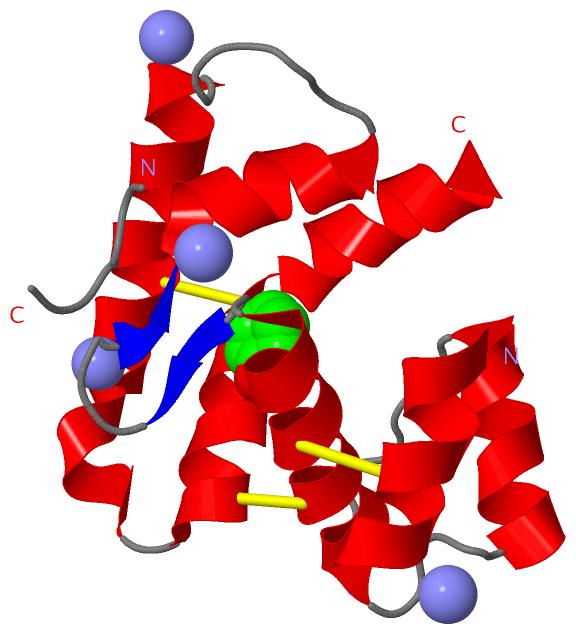
Ligands, Modified Residues, Ions (2, 5)| Asymmetric/Biological Unit (2, 5) |
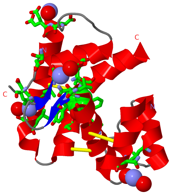
Sites (5, 5)
Asymmetric Unit (5, 5)
|
SS Bonds (3, 3)
Asymmetric/Biological Unit
|
||||||||||||||||
Cis Peptide Bonds (0, 0)| (no "Cis Peptide Bond" information available for 3QME) |
SAPs(SNPs)/Variants (0, 0)| (no "SAP(SNP)/Variant" information available for 3QME) |
PROSITE Motifs (0, 0)| (no "PROSITE Motif" information available for 3QME) |
Exons (0, 0)| (no "Exon" information available for 3QME) |
Sequences/Alignments
Asymmetric/Biological UnitChain A from PDB Type:PROTEIN Length:119 aligned with Q7PGA3_ANOGA | Q7PGA3 from UniProtKB/TrEMBL Length:144 Alignment length:120 144 35 45 55 65 75 85 95 105 115 125 135 |- Q7PGA3_ANOGA 26 ESVIESCSNAVQGAANDELKVHYRANEFPDDPVTHCFVRCIGLELNLYDDKYGVDLQANWENLGNSDDADEEFVAKHRACLEAKNLETIEDLCERAYSAFQCLREDYEMYQNNNNATSE- - SCOP domains ------------------------------------------------------------------------------------------------------------------------ SCOP domains CATH domains ------------------------------------------------------------------------------------------------------------------------ CATH domains Pfam domains PBP_GOBP-3qmeA01 A:26-133 ------------ Pfam domains SAPs(SNPs) ------------------------------------------------------------------------------------------------------------------------ SAPs(SNPs) PROSITE ------------------------------------------------------------------------------------------------------------------------ PROSITE Transcript ------------------------------------------------------------------------------------------------------------------------ Transcript 3qme A 26 ESVIESCSNAVQGAANDELKVHYRANEFPDDPVTHCFVRCIGLELNLYDDKYGVDLQANWENLGNSDDADEEFVAKHRACLEAKNLETIEDLCERAYSAFQCLREDYEMYQNN-ELWSHP 158 35 45 55 65 75 85 95 105 115 125 135 | | 158 138 | 153
|
||||||||||||||||||||
SCOP Domains (0, 0)| (no "SCOP Domain" information available for 3QME) |
CATH Domains (0, 0)| (no "CATH Domain" information available for 3QME) |
Pfam Domains (1, 1)
Asymmetric/Biological Unit
|
Gene Ontology (1, 1)|
Asymmetric/Biological Unit(hide GO term definitions) Chain A (Q7PGA3_ANOGA | Q7PGA3)
|
||||||||||||
Interactive Views
|
|||||||||||||||||||||||||||||||||||||||||||||||||||||||||||||||||||||||||||||||||||||||||||||||||||||||||||||||||||||||||||||||||||||||||||||||||||||||||
Still Images
|
||||||||||||||||
Databases
|
||||||||||||||||||||||||||||||||||||||||||||||||||||||||||||||||||||||||||||||||||||||||||||||||||||||||||||||||||||||||||||||||||||||||||||||||||||||||||||||||
Analysis Tools
|
|||||||||||||||||||||||||||||||||||||||||||||||||||||||||||||
Entries Sharing at Least One Protein Chain (UniProt ID)
Related Entries Specified in the PDB File
|
|