| molecular function |
|---|
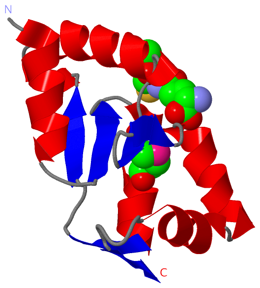
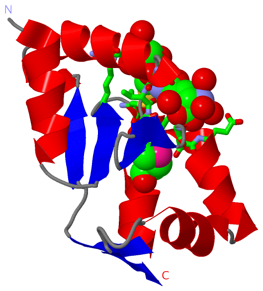
| | GO:0051537 | | 2 iron, 2 sulfur cluster binding | | Interacting selectively and non-covalently with a 2 iron, 2 sulfur (2Fe-2S) cluster; this cluster consists of two iron atoms, with two inorganic sulfur atoms found between the irons and acting as bridging ligands. |
| | GO:0009055 | | electron carrier activity | | Any molecular entity that serves as an electron acceptor and electron donor in an electron transport chain. An electron transport chain is a process in which a series of electron carriers operate together to transfer electrons from donors to any of several different terminal electron acceptors to generate a transmembrane electrochemical gradient. |
| | GO:0004362 | | glutathione-disulfide reductase activity | | Catalysis of the reaction: 2 glutathione + NADP+ = glutathione disulfide + NADPH + H+. |
| | GO:0005506 | | iron ion binding | | Interacting selectively and non-covalently with iron (Fe) ions. |
| | GO:0051536 | | iron-sulfur cluster binding | | Interacting selectively and non-covalently with an iron-sulfur cluster, a combination of iron and sulfur atoms. |
| | GO:0046872 | | metal ion binding | | Interacting selectively and non-covalently with any metal ion. |
| | GO:0015035 | | protein disulfide oxidoreductase activity | | Catalysis of the reaction: a protein with reduced sulfide groups = a protein with oxidized disulfide bonds. |
| | GO:0042803 | | protein homodimerization activity | | Interacting selectively and non-covalently with an identical protein to form a homodimer. |
| biological process |
|---|
| | GO:0045454 | | cell redox homeostasis | | Any process that maintains the redox environment of a cell or compartment within a cell. |
| | GO:0098869 | | cellular oxidant detoxification | | Any process carried out at the cellular level that reduces or removes the toxicity superoxide radicals or hydrogen peroxide. |
| | GO:0034599 | | cellular response to oxidative stress | | Any process that results in a change in state or activity of a cell (in terms of movement, secretion, enzyme production, gene expression, etc.) as a result of oxidative stress, a state often resulting from exposure to high levels of reactive oxygen species, e.g. superoxide anions, hydrogen peroxide (H2O2), and hydroxyl radicals. |
| | GO:0055114 | | oxidation-reduction process | | A metabolic process that results in the removal or addition of one or more electrons to or from a substance, with or without the concomitant removal or addition of a proton or protons. |
| cellular component |
|---|
| | GO:0005794 | | Golgi apparatus | | A compound membranous cytoplasmic organelle of eukaryotic cells, consisting of flattened, ribosome-free vesicles arranged in a more or less regular stack. The Golgi apparatus differs from the endoplasmic reticulum in often having slightly thicker membranes, appearing in sections as a characteristic shallow semicircle so that the convex side (cis or entry face) abuts the endoplasmic reticulum, secretory vesicles emerging from the concave side (trans or exit face). In vertebrate cells there is usually one such organelle, while in invertebrates and plants, where they are known usually as dictyosomes, there may be several scattered in the cytoplasm. The Golgi apparatus processes proteins produced on the ribosomes of the rough endoplasmic reticulum; such processing includes modification of the core oligosaccharides of glycoproteins, and the sorting and packaging of proteins for transport to a variety of cellular locations. Three different regions of the Golgi are now recognized both in terms of structure and function: cis, in the vicinity of the cis face, trans, in the vicinity of the trans face, and medial, lying between the cis and trans regions. |
| | GO:0005796 | | Golgi lumen | | The volume enclosed by the membranes of any cisterna or subcompartment of the Golgi apparatus, including the cis- and trans-Golgi networks. |
| | GO:0005801 | | cis-Golgi network | | The network of interconnected tubular and cisternal structures located at the convex side of the Golgi apparatus, which abuts the endoplasmic reticulum. |
| | GO:0005789 | | endoplasmic reticulum membrane | | The lipid bilayer surrounding the endoplasmic reticulum. |
| | GO:0000324 | | fungal-type vacuole | | A vacuole that has both lytic and storage functions. The fungal vacuole is a large, membrane-bounded organelle that functions as a reservoir for the storage of small molecules (including polyphosphate, amino acids, several divalent cations (e.g. calcium), other ions, and other small molecules) as well as being the primary compartment for degradation. It is an acidic compartment, containing an ensemble of acid hydrolases. At least in S. cerevisiae, there are indications that the morphology of the vacuole is variable and correlated with the cell cycle, with logarithmically growing cells having a multilobed, reticulated vacuole, while stationary phase cells contain a single large structure. |
| | GO:0016021 | | integral component of membrane | | The component of a membrane consisting of the gene products and protein complexes having at least some part of their peptide sequence embedded in the hydrophobic region of the membrane. |
| | GO:0005773 | | vacuole | | A closed structure, found only in eukaryotic cells, that is completely surrounded by unit membrane and contains liquid material. Cells contain one or several vacuoles, that may have different functions from each other. Vacuoles have a diverse array of functions. They can act as a storage organelle for nutrients or waste products, as a degradative compartment, as a cost-effective way of increasing cell size, and as a homeostatic regulator controlling both turgor pressure and pH of the cytosol. |
 Description