|
|
|
|
 Description
|
|
Compounds
|
||||||||||||||||||||||||||||||||||||||||||||||||||||||||
Chains, Units
Summary Information (see also Sequences/Alignments below) |
Ligands, Modified Residues, Ions (2, 3)| Asymmetric/Biological Unit (2, 3) |
Sites (3, 3)
Asymmetric Unit (3, 3)
|
SS Bonds (0, 0)| (no "SS Bond" information available for 3KI9) |
Cis Peptide Bonds (3, 3)
Asymmetric/Biological Unit
|
||||||||||||||||
SAPs(SNPs)/Variants (0, 0)| (no "SAP(SNP)/Variant" information available for 3KI9) |
PROSITE Motifs (0, 0)| (no "PROSITE Motif" information available for 3KI9) |
Exons (0, 0)| (no "Exon" information available for 3KI9) |
Sequences/Alignments
Asymmetric/Biological UnitChain A from PDB Type:PROTEIN Length:467 aligned with PEPVL_STAAC | Q5HF23 from UniProtKB/Swiss-Prot Length:469 Alignment length:470 1 | 9 19 29 39 49 59 69 79 89 99 109 119 129 139 149 159 169 179 189 199 209 219 229 239 249 259 269 279 289 299 309 319 329 339 349 359 369 379 389 399 409 419 429 439 449 459 469 PEPVL_STAAC - -MWKEKVQQYEDQIINDLKGLLAIESVRDDAKASEDAPVGPGPRKALDYMYEIAHRDGFTTHDVDHIAGRIEAGKGNDVLGILCHVDVVPAGDGWDSNPFEPVVTEDAIIARGTLDDKGPTIAAYYAIKILEDMNVDWKKRIHMIIGTDEESDWKCTDRYFKTEEMPTLGFAPDAEFPCIHGEKGITTFDLVQNKLTEDQDEPDYELITFKSGERYNMVPDHAEARVLVKENMTDVIQDFEYFLEQNHLQGDSTVDSGILVLTVEGKAVHGMDPSIGVNAGLYLLKFLASLNLDNNAQAFVAFSNRYLFNSDFGEKMGMKFHTDVMGDVTTNIGVITYDNENAGLFGINLRYPEGFEFEKAMDRFANEIQQYGFEVKLGKVQPPHYVDKNDPFVQKLVTAYRNQTNDMTEPYTIGGGTYARNLDKGVAFGAMFSDSEDLMHQKNEYITKKQLFNATSIYLEAIYSLCVEE 469 SCOP domains -------------------------------------------------------------------------------------------------------------------------------------------------------------------------------------------------------------------------------------------------------------------------------------------------------------------------------------------------------------------------------------------------------------------------------------------------------------------------------------- SCOP domains CATH domains -------------------------------------------------------------------------------------------------------------------------------------------------------------------------------------------------------------------------------------------------------------------------------------------------------------------------------------------------------------------------------------------------------------------------------------------------------------------------------------- CATH domains Pfam domains --------------------------------------------------------------------------------Peptidase_M20-3ki9A01 A:80-464 ----- Pfam domains SAPs(SNPs) -------------------------------------------------------------------------------------------------------------------------------------------------------------------------------------------------------------------------------------------------------------------------------------------------------------------------------------------------------------------------------------------------------------------------------------------------------------------------------------- SAPs(SNPs) PROSITE -------------------------------------------------------------------------------------------------------------------------------------------------------------------------------------------------------------------------------------------------------------------------------------------------------------------------------------------------------------------------------------------------------------------------------------------------------------------------------------- PROSITE Transcript -------------------------------------------------------------------------------------------------------------------------------------------------------------------------------------------------------------------------------------------------------------------------------------------------------------------------------------------------------------------------------------------------------------------------------------------------------------------------------------- Transcript 3ki9 A 0 SMWKEKVQQYEDQIINDLKGLLAIESVRDDAKASEDAPVGPGPRKALDYMYEIAHRDGFTTHDVDHIAGRIEAGKGNDVLGILCHVDVVPAGDGWDSNPFEPVVTEDAIIARGTLDDKGPTIAAYYAIKILEDMNVDWKKRIHMIIGTDEESDWKCTDRYFKTEEMPTLGFAPDAEFPCIHGEKGITTFDLVQNKL---QDEPDYELITFKSGERYNMVPDHAEARVLVKENMTDVIQDFEYFLEQNHLQGDSTVDSGILVLTVEGKAVHGMDPSIGVNAGLYLLKFLASLNLDNNAQAFVAFSNRYLFNSDFGEKMGMKFHTDVMGDVTTNIGVITYDNENAGLFGINLRYPEGFEFEKAMDRFANEIQQYGFEVKLGKVQPPHYVDKNDPFVQKLVTAYRNQTNDMTEPYTIGGGTYARNLDKGVAFGAMFSDSEDLMHQKNEYITKKQLFNATSIYLEAIYSLCVEE 469 9 19 29 39 49 59 69 79 89 99 109 119 129 139 149 159 169 179 189 | 199 209 219 229 239 249 259 269 279 289 299 309 319 329 339 349 359 369 379 389 399 409 419 429 439 449 459 469 195 199
|
||||||||||||||||||||
SCOP Domains (0, 0)| (no "SCOP Domain" information available for 3KI9) |
CATH Domains (0, 0)| (no "CATH Domain" information available for 3KI9) |
Pfam Domains (1, 1)
Asymmetric/Biological Unit

|
Gene Ontology (8, 8)|
Asymmetric/Biological Unit(hide GO term definitions) Chain A (PEPVL_STAAC | Q5HF23)
|
||||||||||||||||||||||||||||||||||||||||||||||||||||||||||||
Interactive Views
|
||||||||||||||||||||||||||||||||||||||||||||||||||||||||||||||||||||||||||||||||||||||||||||||||||||||||||||||||||||||||||||||||||||||||||||||||||||||||||
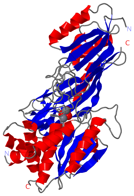
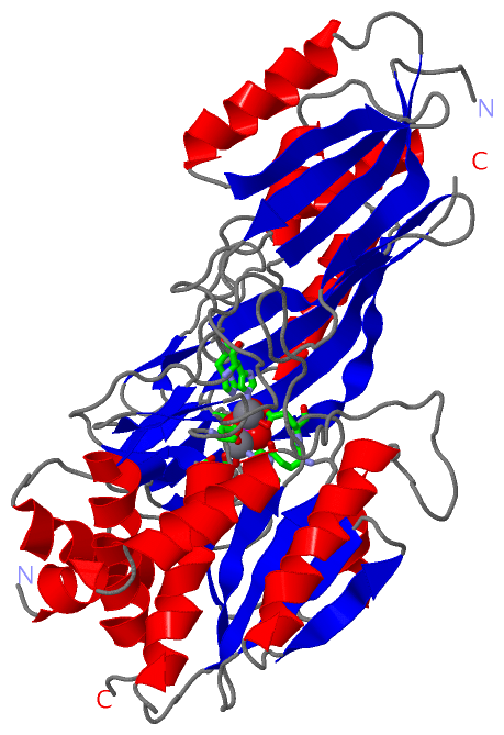
Still Images
|
||||||||||||||||
Databases
|
||||||||||||||||||||||||||||||||||||||||||||||||||||||||||||||||||||||||||||||||||||||||||||||||||||||||||||||||||||||||||||||||||||||||||||||||||||||||||||||||
Analysis Tools
|
|||||||||||||||||||||||||||||||||||||||||||||||||||||||||||||
Entries Sharing at Least One Protein Chain (UniProt ID)
Related Entries Specified in the PDB File
|
|