Show PDB file:   
         Plain Text   HTML   (compressed file size)
QuickSearch:   
by PDB,NDB,UniProt,PROSITE Code or Search Term(s)  
(-)Asymmetric Unit
(-)Asym. Unit - sites
(-)Biological Unit 1
(-)Biol. Unit 1 - sites
(-)Biological Unit 2
collapse expand < >
Image Asymmetric Unit
Asymmetric Unit  (Jmol Viewer)
Image Asym. Unit - sites
Asym. Unit - sites  (Jmol Viewer)
Image Biological Unit 1
Biological Unit 1  (Jmol Viewer)
Image Biol. Unit 1 - sites
Biol. Unit 1 - sites  (Jmol Viewer)
Image Biological Unit 2
Biological Unit 2  (Jmol Viewer)

(-) Description

Title :  CRYSTAL STRUCTURE OF SCD-MPR MUTANT E19Q/K137M PH 7.0
 
Authors :  L. J. Olson, G. Sun, R. N. Bohnsack, F. C. Peterson, N. M. Dahms, J. J. P. Ki
Date :  05 Oct 09  (Deposition) - 24 Nov 09  (Release) - 13 Jul 11  (Revision)
Method :  X-RAY DIFFRACTION
Resolution :  2.30
Chains :  Asym. Unit :  A,B
Biol. Unit 1:  A,B  (1x)
Biol. Unit 2:  A (1x),B (1x)
Keywords :  Transport, Lysosome, Mannose, Receptor, Sugar Binding, Glycoprotein, Membrane, Phosphoprotein, Transmembrane, Protein Transport, Sugar Binding Protein (Keyword Search: [Gene Ontology, PubMed, Web (Google))
 
Reference :  L. J. Olson, G. Sun, R. N. Bohnsack, F. C. Peterson, N. M. Dahms, J. J. Kim
Intermonomer Interactions Are Essential For Lysosomal Enzym Binding By The Cation-Dependent Mannose 6-Phosphate Receptor.
Biochemistry V. 49 236 2010
PubMed-ID: 19928875  |  Reference-DOI: 10.1021/BI901725X

(-) Compounds

Molecule 1 - CATION-DEPENDENT MANNOSE-6-PHOSPHATE RECEPTOR
    ChainsA, B
    EngineeredYES
    Expression SystemPICHIA PASTORIS
    Expression System PlasmidPPICZALPHAA
    Expression System StrainX-33
    Expression System Taxid4922
    FragmentUNP RESIDUES 29-182
    GeneM6PR
    MutationYES
    Organism CommonBOVINE,COW,DOMESTIC CATTLE,DOMESTIC COW
    Organism ScientificBOS TAURUS
    Organism Taxid9913
    SynonymCD MAN-6-P RECEPTOR, CD-MPR, 46 KDA MANNOSE 6-PHOSPHATE RECEPTOR, MPR 46

 Structural Features

(-) Chains, Units

  12
Asymmetric Unit AB
Biological Unit 1 (1x)AB
Biological Unit 2 (1x)A (1x)B (1x)

Summary Information (see also Sequences/Alignments below)

(-) Ligands, Modified Residues, Ions  (3, 12)

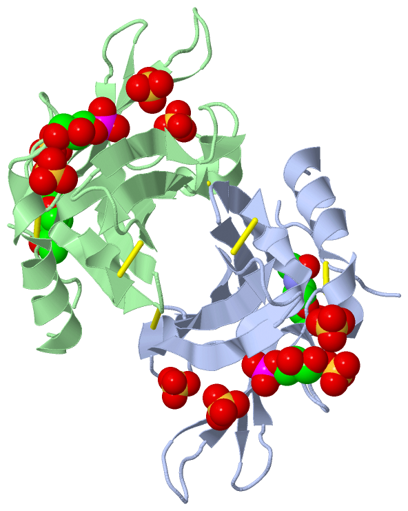
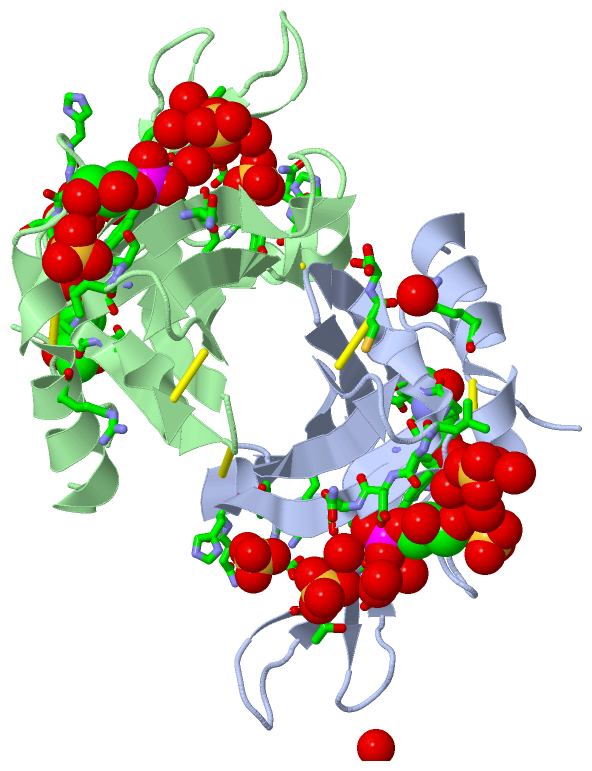
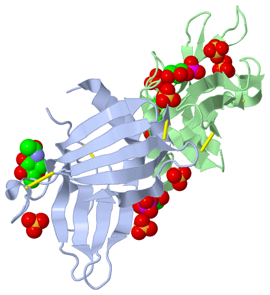
Asymmetric Unit (3, 12)
No.NameCountTypeFull Name
11GP2Ligand/IonSN-GLYCEROL-1-PHOSPHATE
2NAG2Ligand/IonN-ACETYL-D-GLUCOSAMINE
3SO48Ligand/IonSULFATE ION
Biological Unit 1 (3, 12)
No.NameCountTypeFull Name
11GP2Ligand/IonSN-GLYCEROL-1-PHOSPHATE
2NAG2Ligand/IonN-ACETYL-D-GLUCOSAMINE
3SO48Ligand/IonSULFATE ION
Biological Unit 2 (3, 6)
No.NameCountTypeFull Name
11GP1Ligand/IonSN-GLYCEROL-1-PHOSPHATE
2NAG1Ligand/IonN-ACETYL-D-GLUCOSAMINE
3SO44Ligand/IonSULFATE ION

(-) Sites  (12, 12)

Asymmetric Unit (12, 12)
No.NameEvidenceResiduesDescription
01AC1SOFTWAREARG A:79 , ASN A:81 , GLU A:82 , HOH A:592BINDING SITE FOR RESIDUE NAG A 3001
02AC2SOFTWARESER A:33 , PHE A:34 , GLU A:35 , ALA A:125 , ASP A:126 , ASN A:127 , PHE A:128 , HOH A:645 , HOH A:665 , HOH A:690 , HOH A:698 , SO4 A:6002BINDING SITE FOR RESIDUE 1GP A 7000
03AC3SOFTWARETYR A:102 , ASP A:103 , ASN A:104 , HIS A:105 , ARG A:111BINDING SITE FOR RESIDUE SO4 A 6000
04AC4SOFTWARELYS A:32 , SER A:33 , 1GP A:7000BINDING SITE FOR RESIDUE SO4 A 6002
05AC5SOFTWARELYS A:32 , LEU A:124 , HOH A:547 , HOH A:662 , ARG B:25BINDING SITE FOR RESIDUE SO4 A 6003
06AC6SOFTWAREGLU A:35 , SER A:36 , THR A:37 , HOH A:613 , HOH A:645BINDING SITE FOR RESIDUE SO4 A 6007
07AC7SOFTWAREASN B:81 , GLU B:82 , HOH B:609 , HOH B:634BINDING SITE FOR RESIDUE NAG B 4001
08AC8SOFTWAREPHE B:34 , GLU B:35 , ALA B:125 , ASN B:127 , PHE B:128 , HOH B:648BINDING SITE FOR RESIDUE 1GP B 7001
09AC9SOFTWARETYR B:102 , ASP B:103 , ASN B:104 , HIS B:105 , ARG B:111BINDING SITE FOR RESIDUE SO4 B 6001
10BC1SOFTWAREARG A:25 , CYS A:153 , SER A:154 , HOH A:653 , LEU B:124BINDING SITE FOR RESIDUE SO4 B 6004
11BC2SOFTWARESER B:36 , THR B:37 , HOH B:583 , HOH B:623 , HOH B:654BINDING SITE FOR RESIDUE SO4 B 6005
12BC3SOFTWAREHOH A:606 , HIS B:58 , SER B:59 , SER B:60 , ARG B:79 , HOH B:628BINDING SITE FOR RESIDUE SO4 B 6006

(-) SS Bonds  (6, 6)

Asymmetric Unit
No.Residues
1A:6 -A:52
2A:106 -A:141
3A:119 -A:153
4B:6 -B:52
5B:106 -B:141
6B:119 -B:153

(-) Cis Peptide Bonds  (1, 1)

Asymmetric Unit
No.Residues
1Ser A:41 -Pro A:42

 Sequence-Structure Mapping

(-) SAPs(SNPs)/Variants  (0, 0)

(no "SAP(SNP)/Variant" information available for 3K42)

(-) PROSITE Motifs  (0, 0)

(no "PROSITE Motif" information available for 3K42)

(-) Exons   (4, 8)

Asymmetric Unit (4, 8)
 ENSEMBLUniProtKBPDB
No.Transcript IDExonExon IDGenome LocationLengthIDLocationLengthCountLocationLength
1.1ENSBTAT000000242331ENSBTAE00000295015chr5:108383511-108383668158MPRD_BOVIN1-110--
1.2ENSBTAT000000242332ENSBTAE00000197230chr5:108390145-108390324180MPRD_BOVIN1-61612A:3-33 (gaps)
B:5-33 (gaps)
31
29
1.3ENSBTAT000000242333ENSBTAE00000197232chr5:108391558-108391724167MPRD_BOVIN61-117572A:33-89
B:33-89
57
57
1.4ENSBTAT000000242334ENSBTAE00000295014chr5:108392731-108392840110MPRD_BOVIN117-153372A:89-125
B:89-125
37
37
1.5ENSBTAT000000242335ENSBTAE00000197235chr5:108393267-108393397131MPRD_BOVIN154-197442A:126-154 (gaps)
B:126-154
29
29
1.6ENSBTAT000000242336ENSBTAE00000197236chr5:108393940-108394066127MPRD_BOVIN197-239430--
1.7ENSBTAT000000242337ENSBTAE00000197238chr5:108394495-1083960421548MPRD_BOVIN240-279400--

(-) Sequences/Alignments

Asymmetric Unit
   Reformat: Number of residues per line =  ('0' or empty: single-line sequence representation)
  Number of residues per labelling interval =   
  UniProt sequence: complete  aligned part    
   Show mapping: SCOP domains CATH domains Pfam domains Secondary structure (by author)
SAPs(SNPs) PROSITE motifs Exons
(details for a mapped element are shown in a popup box when the mouse pointer rests over it)
Chain A from PDB  Type:PROTEIN  Length:141
 aligned with MPRD_BOVIN | P11456 from UniProtKB/Swiss-Prot  Length:279

    Alignment length:152
                                    40        50        60        70        80        90       100       110       120       130       140       150       160       170       180  
           MPRD_BOVIN    31 EKTCDLVGEKGKESEKELALLKRLTPLFNKSFESTVGQSPDMYSYVFRVCREAGNHSSGAGLVQINKSNGKETVVGRFNETQIFNGSNWIMLIYKGGDEYDNHCGREQRRAVVMISCNRHTLADNFNPVSEERGKVQDCFYLFEMDSSLACS 182
               SCOP domains d3k42a_     A: Cation-dependent mannose 6-phosphate receptor, extracytoplasmic domain                                                                    SCOP domains
               CATH domains 3k42A00     A:3-154 Cation-dependent Mannose-6-phosphate Receptor; Chain A                                                                               CATH domains
               Pfam domains -------------------------------------------------------------------------------------------------------------------------------------------------------- Pfam domains
         Sec.struct. author ........----.hhhhhhhhhhhhhhhh..eeeee......eeeee............eeeeeee.....eeeeeeeeeeeeeee..eeeeeeee......hhhhh...eeeeeee......eeeeeee.-------..eeeeeeehhhh. Sec.struct. author
                 SAPs(SNPs) -------------------------------------------------------------------------------------------------------------------------------------------------------- SAPs(SNPs)
                    PROSITE -------------------------------------------------------------------------------------------------------------------------------------------------------- PROSITE
           Transcript 1 (1) Exon 1.2  PDB: A:3-33 (gaps)   -------------------------------------------------------Exon 1.4  PDB: A:89-125              Exon 1.5 UniProt: 154-197     Transcript 1 (1)
           Transcript 1 (2) ------------------------------Exon 1.3  PDB: A:33-89 UniProt: 61-117                   ----------------------------------------------------------------- Transcript 1 (2)
                 3k42 A   3 EKTCDLVG----ESEKQLALLKRLTPLFQKSFESTVGQSPDMYSYVFRVCREAGQHSSGAGLVQIQKSNGKETVVGRFNETQIFQGSNWIMLIYKGGDEYDNHCGREQRRAVVMISCNRHTLADNFNPVSE-------CFYLFEMDSSLACS 154
                                   | -  |     22        32        42        52        62        72        82        92       102       112       122       132|      142       152  
                                  10   15                                                                                                                   133     141             

Chain B from PDB  Type:PROTEIN  Length:146
 aligned with MPRD_BOVIN | P11456 from UniProtKB/Swiss-Prot  Length:279

    Alignment length:150
                                    42        52        62        72        82        92       102       112       122       132       142       152       162       172       182
           MPRD_BOVIN    33 TCDLVGEKGKESEKELALLKRLTPLFNKSFESTVGQSPDMYSYVFRVCREAGNHSSGAGLVQINKSNGKETVVGRFNETQIFNGSNWIMLIYKGGDEYDNHCGREQRRAVVMISCNRHTLADNFNPVSEERGKVQDCFYLFEMDSSLACS 182
               SCOP domains d3k42b    _ B: Cation-dependent mannose 6-phosphate receptor, extracytoplasmic domain                                                                  SCOP domains
               CATH domains 3k42B0    0 B:5-154 Cation-dependent Mannose-6-phosphate Receptor; Chain A                                                                             CATH domains
           Pfam domains (1) Man-6-    P_recep-3k42B01 B:5-154                                                                                                                      Pfam domains (1)
           Pfam domains (2) Man-6-    P_recep-3k42B02 B:5-154                                                                                                                      Pfam domains (2)
         Sec.struct. author ......----.hhhhhhhhhhhhhhh...eeeee......eeeee............eeeeeee.....eeeeeeeeeeeeee...eeeeeeee......hhhhh...eeeeeee......eeeee.....hhhhhh.eeeeeeehhhh. Sec.struct. author
                 SAPs(SNPs) ------------------------------------------------------------------------------------------------------------------------------------------------------ SAPs(SNPs)
                    PROSITE ------------------------------------------------------------------------------------------------------------------------------------------------------ PROSITE
           Transcript 1 (1) Exon 1.2  PDB: B:5-33 (gaps) -------------------------------------------------------Exon 1.4  PDB: B:89-125              Exon 1.5  PDB: B:126-154      Transcript 1 (1)
           Transcript 1 (2) ----------------------------Exon 1.3  PDB: B:33-89 UniProt: 61-117                   ----------------------------------------------------------------- Transcript 1 (2)
                 3k42 B   5 TCDLVG----ESEKQLALLKRLTPLFQKSFESTVGQSPDMYSYVFRVCREAGQHSSGAGLVQIQKSNGKETVVGRFNETQIFQGSNWIMLIYKGGDEYDNHCGREQRRAVVMISCNRHTLADNFNPVSEERGMVQDCFYLFEMDSSLACS 154
                                 |   -|       24        34        44        54        64        74        84        94       104       114       124       134       144       154
                                10   15                                                                                                                                           

   Legend:   → Mismatch (orange background)
  - → Gap (green background, '-', border residues have a numbering label)
    → Modified Residue (blue background, lower-case, 'x' indicates undefined single-letter code, labelled with number + name)
  x → Chemical Group (purple background, 'x', labelled with number + name, e.g. ACE or NH2)
  extra numbering lines below/above indicate numbering irregularities and modified residue names etc., number ends below/above '|'

 Classification and Annotation

(-) SCOP Domains  (1, 2)

Asymmetric Unit

(-) CATH Domains  (1, 2)

Asymmetric Unit
(-)
Class: Mainly Beta (13760)

(-) Pfam Domains  (1, 2)

Asymmetric Unit
(-)
Clan: M6PR (17)

(-) Gene Ontology  (14, 14)

Asymmetric Unit(hide GO term definitions)
Chain A,B   (MPRD_BOVIN | P11456)
molecular function
    GO:0005537    mannose binding    Interacting selectively and non-covalently with mannose, a monosaccharide hexose, stereoisomeric with glucose, that occurs naturally only in polymerized forms called mannans.
    GO:0015578    mannose transmembrane transporter activity    Enables the transfer of mannose from one side of the membrane to the other. Mannose is the aldohexose manno-hexose, the C-2 epimer of glucose. The D-(+)-form is widely distributed in mannans and hemicelluloses and is of major importance in the core oligosaccharide of N-linked oligosaccharides of glycoproteins.
biological process
    GO:0015761    mannose transport    The directed movement of mannose into, out of or within a cell, or between cells, by means of some agent such as a transporter or pore. Mannose is the aldohexose manno-hexose, the C-2 epimer of glucose. The D-(+)-form is widely distributed in mannans and hemicelluloses and is of major importance in the core oligosaccharide of N-linked oligosaccharides of glycoproteins.
    GO:0006810    transport    The directed movement of substances (such as macromolecules, small molecules, ions) or cellular components (such as complexes and organelles) into, out of or within a cell, or between cells, or within a multicellular organism by means of some agent such as a transporter, pore or motor protein.
cellular component
    GO:0005794    Golgi apparatus    A compound membranous cytoplasmic organelle of eukaryotic cells, consisting of flattened, ribosome-free vesicles arranged in a more or less regular stack. The Golgi apparatus differs from the endoplasmic reticulum in often having slightly thicker membranes, appearing in sections as a characteristic shallow semicircle so that the convex side (cis or entry face) abuts the endoplasmic reticulum, secretory vesicles emerging from the concave side (trans or exit face). In vertebrate cells there is usually one such organelle, while in invertebrates and plants, where they are known usually as dictyosomes, there may be several scattered in the cytoplasm. The Golgi apparatus processes proteins produced on the ribosomes of the rough endoplasmic reticulum; such processing includes modification of the core oligosaccharides of glycoproteins, and the sorting and packaging of proteins for transport to a variety of cellular locations. Three different regions of the Golgi are now recognized both in terms of structure and function: cis, in the vicinity of the cis face, trans, in the vicinity of the trans face, and medial, lying between the cis and trans regions.
    GO:0005768    endosome    A vacuole to which materials ingested by endocytosis are delivered.
    GO:0016021    integral component of membrane    The component of a membrane consisting of the gene products and protein complexes having at least some part of their peptide sequence embedded in the hydrophobic region of the membrane.
    GO:0005770    late endosome    A prelysosomal endocytic organelle differentiated from early endosomes by lower lumenal pH and different protein composition. Late endosomes are more spherical than early endosomes and are mostly juxtanuclear, being concentrated near the microtubule organizing center.
    GO:0005765    lysosomal membrane    The lipid bilayer surrounding the lysosome and separating its contents from the cell cytoplasm.
    GO:0005764    lysosome    A small lytic vacuole that has cell cycle-independent morphology and is found in most animal cells and that contains a variety of hydrolases, most of which have their maximal activities in the pH range 5-6. The contained enzymes display latency if properly isolated. About 40 different lysosomal hydrolases are known and lysosomes have a great variety of morphologies and functions.
    GO:0000323    lytic vacuole    A vacuole that is maintained at an acidic pH and which contains degradative enzymes, including a wide variety of acid hydrolases.
    GO:0016020    membrane    A lipid bilayer along with all the proteins and protein complexes embedded in it an attached to it.
    GO:0048471    perinuclear region of cytoplasm    Cytoplasm situated near, or occurring around, the nucleus.
    GO:0005802    trans-Golgi network    The network of interconnected tubular and cisternal structures located within the Golgi apparatus on the side distal to the endoplasmic reticulum, from which secretory vesicles emerge. The trans-Golgi network is important in the later stages of protein secretion where it is thought to play a key role in the sorting and targeting of secreted proteins to the correct destination.

 Visualization

(-) Interactive Views

Asymmetric Unit
  Complete Structure
    Jena3D(integrated viewing of ligand, site, SAP, PROSITE, SCOP information)
    WebMol | AstexViewer[tm]@PDBe
(Java Applets, require no local installation except for Java; loading may be slow)
    STRAP
(Java WebStart application, automatic local installation, requires Java; full application with system access!)
    RasMol
(require local installation)
    Molscript (VRML)
(requires installation of a VRML viewer; select preferred view via VRML and generate a mono or stereo PDF format file)
 
  Ligands, Modified Residues, Ions
    1GP  [ RasMol | Jena3D ]  +environment [ RasMol | Jena3D ]
    NAG  [ RasMol | Jena3D ]  +environment [ RasMol | Jena3D ]
    SO4  [ RasMol | Jena3D ]  +environment [ RasMol | Jena3D ]
 
  Sites
    AC1  [ RasMol ]  +environment [ RasMol ]
    AC2  [ RasMol ]  +environment [ RasMol ]
    AC3  [ RasMol ]  +environment [ RasMol ]
    AC4  [ RasMol ]  +environment [ RasMol ]
    AC5  [ RasMol ]  +environment [ RasMol ]
    AC6  [ RasMol ]  +environment [ RasMol ]
    AC7  [ RasMol ]  +environment [ RasMol ]
    AC8  [ RasMol ]  +environment [ RasMol ]
    AC9  [ RasMol ]  +environment [ RasMol ]
    BC1  [ RasMol ]  +environment [ RasMol ]
    BC2  [ RasMol ]  +environment [ RasMol ]
    BC3  [ RasMol ]  +environment [ RasMol ]
 
  Cis Peptide Bonds
    Ser A:41 - Pro A:42   [ RasMol ]  
 
Biological Units
  Complete Structure
    Biological Unit 1  [ Jena3D ]
    Biological Unit 2  [ Jena3D ]

(-) Still Images

Jmol
  protein: cartoon or spacefill or dots and stick; nucleic acid: cartoon and stick; ligands: spacefill; active site: stick
Molscript
  protein, nucleic acid: cartoon; ligands: spacefill; active site: ball and stick

 Databases and Analysis Tools

(-) Databases

Access by PDB/NDB ID
  3k42
    Family and Domain InformationProDom | SYSTERS
    General Structural InformationGlycoscienceDB | MMDB | NDB | OCA | PDB | PDBe | PDBj | PDBsum | PDBWiki | PQS | PROTEOPEDIA
    Orientation in MembranesOPM
    Protein SurfaceSURFACE
    Secondary StructureDSSP (structure derived) | HSSP (homology derived)
    Structural GenomicsGeneCensus
    Structural NeighboursCE | VAST
    Structure ClassificationCATH | Dali | SCOP
    Validation and Original DataBMRB Data View | BMRB Restraints Grid | EDS | PROCHECK | RECOORD | WHAT_CHECK
 
Access by UniProt ID/Accession number
  MPRD_BOVIN | P11456
    Comparative Protein Structure ModelsModBase
    Genomic InformationEnsembl
    Protein-protein InteractionDIP
    Sequence, Family and Domain InformationInterPro | Pfam | SMART | UniProtKB/SwissProt
 
Access by Enzyme Classificator   (EC Number)
  (no 'Enzyme Classificator' available)
    General Enzyme InformationBRENDA | EC-PDB | Enzyme | IntEnz
    PathwayKEGG | MetaCyc
 
Access by Disease Identifier   (MIM ID)
  (no 'MIM ID' available)
    Disease InformationOMIM
 
Access by GenAge ID
  (no 'GenAge ID' available)
    Age Related InformationGenAge

(-) Analysis Tools

Access by PDB/NDB ID
    Domain InformationXDom
    Interatomic Contacts of Structural UnitsCSU
    Ligand-protein ContactsLPC
    Protein CavitiescastP
    Sequence and Secondary StructurePDBCartoon
    Structure AlignmentSTRAP(Java WebStart application, automatic local installation, requires Java; full application with system access!)
    Structure and Sequence BrowserSTING
 
Access by UniProt ID/Accession number
  MPRD_BOVIN | P11456
    Protein Disorder PredictionDisEMBL | FoldIndex | GLOBPLOT (for more information see DisProt)

 Related Entries

(-) Entries Sharing at Least One Protein Chain (UniProt ID)

UniProtKB/Swiss-Prot
        MPRD_BOVIN | P114561c39 1keo 1m6p 2rl7 2rl8 2rl9 2rlb 3cy4 3k41 3k43

(-) Related Entries Specified in the PDB File

3k41 3k43