|
|
|
|
 Description
|
|
Compounds
|
||||||||||||||||||||||||||||||||||||||||||||||||||||
Chains, Units
Summary Information (see also Sequences/Alignments below) |
Ligands, Modified Residues, Ions (0, 0)| (no "Ligand,Modified Residues,Ions" information available for 3I42) |
Sites (0, 0)| (no "Site" information available for 3I42) |
SS Bonds (0, 0)| (no "SS Bond" information available for 3I42) |
Cis Peptide Bonds (1, 1)
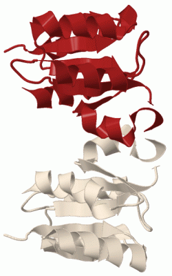
Asymmetric Unit
|
||||||||
SAPs(SNPs)/Variants (0, 0)| (no "SAP(SNP)/Variant" information available for 3I42) |
PROSITE Motifs (0, 0)| (no "PROSITE Motif" information available for 3I42) |
Exons (0, 0)| (no "Exon" information available for 3I42) |
Sequences/Alignments
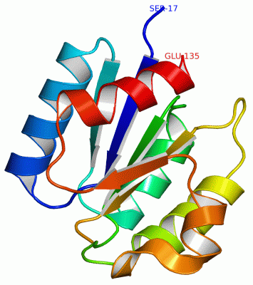
Asymmetric UnitChain A from PDB Type:PROTEIN Length:118 aligned with Q1H0I3_METFK | Q1H0I3 from UniProtKB/TrEMBL Length:134 Alignment length:119 134 26 36 46 56 66 76 86 96 106 116 126 | Q1H0I3_METFK 17 QAQQALIVEDYQAAAETFKELLEMLGFQADYVMSGTDALHAMSTRGYDAVFIDLNLPDTSGLALVKQLRALPMEKTSKFVAVSGFAKNDLGKEACELFDFYLEKPIDIASLEPILQSI- - SCOP domains d3i42a_ A: automated matches SCOP domains CATH domains ----------------------------------------------------------------------------------------------------------------------- CATH domains Pfam domains ----------------------------------------------------------------------------------------------------------------------- Pfam domains SAPs(SNPs) ----------------------------------------------------------------------------------------------------------------------- SAPs(SNPs) PROSITE ----------------------------------------------------------------------------------------------------------------------- PROSITE Transcript ----------------------------------------------------------------------------------------------------------------------- Transcript 3i42 A 17 SLQQALIVEDYQAAAETFKELLEMLGFQADYVMSGTDALHAMSTRGYDAVFIDLNLPDTSGLALVKQLRALPMEKTSKFVAVSGF-KNDLGKEACELFDFYLEKPIDIASLEPILQSIE 135 26 36 46 56 66 76 86 96 | |106 116 126 101 | 103
|
||||||||||||||||||||
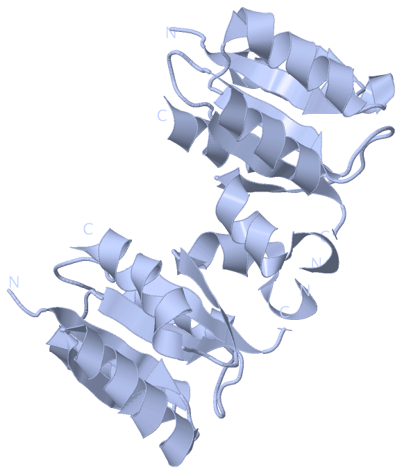
SCOP Domains (1, 1)
Asymmetric Unit
 |
CATH Domains (0, 0)| (no "CATH Domain" information available for 3I42) |
Pfam Domains (0, 0)| (no "Pfam Domain" information available for 3I42) |
Gene Ontology (2, 2)|
Asymmetric Unit(hide GO term definitions) Chain A (Q1H0I3_METFK | Q1H0I3)
|
||||||||||||||||||||||||
Interactive Views
|
|||||||||||||||||||||||||||||||||||||||||||||||||||||||||||||||||||||||||||||||||||||||||||||||||||||||||||||||||||||||||||||||||||||||
Still Images
|
||||||||||||||||
Databases
|
||||||||||||||||||||||||||||||||||||||||||||||||||||||||||||||||||||||||||||||||||||||||||||||||||||||||||||||||||||||||||||||||||||||||||||||||||||||||||||||||
Analysis Tools
|
|||||||||||||||||||||||||||||||||||||||||||||||||||||||||||||
Entries Sharing at Least One Protein Chain (UniProt ID)
Related Entries Specified in the PDB File
|
|