|
|
|
|
 Description
|
|
Compounds
|
||||||||||||||||||||||||||||||||||||||||||||||||||||
Chains, Units
Summary Information (see also Sequences/Alignments below) |
Ligands, Modified Residues, Ions (1, 2)
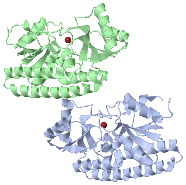
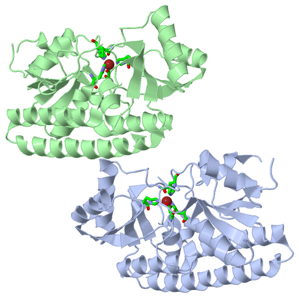
Asymmetric Unit (1, 2)
|
Sites (2, 2)
Asymmetric Unit (2, 2)
|
SS Bonds (0, 0)| (no "SS Bond" information available for 3HJT) |
Cis Peptide Bonds (0, 0)| (no "Cis Peptide Bond" information available for 3HJT) |
SAPs(SNPs)/Variants (0, 0)| (no "SAP(SNP)/Variant" information available for 3HJT) |
PROSITE Motifs (0, 0)| (no "PROSITE Motif" information available for 3HJT) |
Exons (0, 0)| (no "Exon" information available for 3HJT) |
Sequences/Alignments
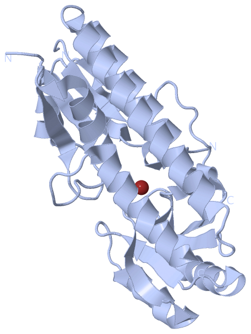
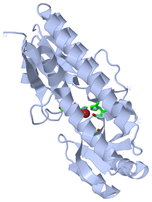
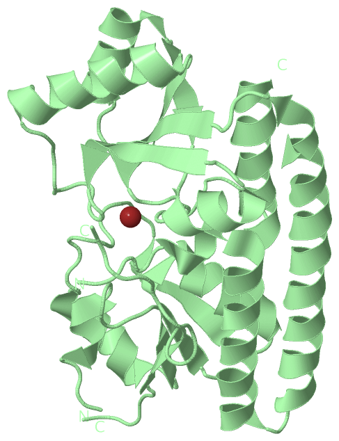
Asymmetric UnitChain A from PDB Type:PROTEIN Length:260 aligned with Q9ZHG8_STRAG | Q9ZHG8 from UniProtKB/TrEMBL Length:306 Alignment length:276 40 50 60 70 80 90 100 110 120 130 140 150 160 170 180 190 200 210 220 230 240 250 260 270 280 290 300 Q9ZHG8_STRAG 31 QGMSVVTSFYPMYAMTKEVSGDLNDVRMIQSGAGIHSFEPSVNDVAAIYDADLFVYHSHTLEAWARDLDPNLKKSKVNVFEASKPLTLDRVKGLEDMEVTQGIDPATLYDPHTWTDPVLAGEEAVNIAKELGHLDPKHKDSYTKKAKAFKKEAEQLTEEYTQKFKKVRSKTFVTQHTAFSYLAKRFGLKQLGISGISPEQEPSPRQLKEIQDFVKEYNVKTIFAEDNVNPKIAHAIAKSTGAKVKTLSPLEAAPSGNKTYLENLRANLEVLYQQLK 306 SCOP domains d3hjta_ A: automated matches SCOP domains CATH domains 3hjtA01 A:31-197 Nitrogenase molybdenum iron protein domain 3hjtA02 A:198-306 Nitrogenase molybdenum iron protein domain CATH domains Pfam domains ------------------------------------------------------------------------------------------------------------------------------------------------------------------------------------------------------------------------------------------------------------------------------------ Pfam domains SAPs(SNPs) ------------------------------------------------------------------------------------------------------------------------------------------------------------------------------------------------------------------------------------------------------------------------------------ SAPs(SNPs) PROSITE ------------------------------------------------------------------------------------------------------------------------------------------------------------------------------------------------------------------------------------------------------------------------------------ PROSITE Transcript ------------------------------------------------------------------------------------------------------------------------------------------------------------------------------------------------------------------------------------------------------------------------------------ Transcript 3hjt A 31 QGMSVVTSFYPMYAMTKEVSGDLNDVRMIQSGAGIHSFEPSVNDVAAIYDADLFVYHSHTLEAWARDLDP---KSKVNVFEASKPLTLDRVKG-------------TLYDPHTWTDPVLAGEEAVNIAKELGHLDPKHKDSYTKKAKAFKKEAEQLTEEYTQKFKKVRSKTFVTQHTAFSYLAKRFGLKQLGISGISPEQEPSPRQLKEIQDFVKEYNVKTIFAEDNVNPKIAHAIAKSTGAKVKTLSPLEAAPSGNKTYLENLRANLEVLYQQLK 306 40 50 60 70 80 90 100 | 110 120 | - |140 150 160 170 180 190 200 210 220 230 240 250 260 270 280 290 300 100 104 123 137 Chain B from PDB Type:PROTEIN Length:257 aligned with Q9ZHG8_STRAG | Q9ZHG8 from UniProtKB/TrEMBL Length:306 Alignment length:275 41 51 61 71 81 91 101 111 121 131 141 151 161 171 181 191 201 211 221 231 241 251 261 271 281 291 301 Q9ZHG8_STRAG 32 GMSVVTSFYPMYAMTKEVSGDLNDVRMIQSGAGIHSFEPSVNDVAAIYDADLFVYHSHTLEAWARDLDPNLKKSKVNVFEASKPLTLDRVKGLEDMEVTQGIDPATLYDPHTWTDPVLAGEEAVNIAKELGHLDPKHKDSYTKKAKAFKKEAEQLTEEYTQKFKKVRSKTFVTQHTAFSYLAKRFGLKQLGISGISPEQEPSPRQLKEIQDFVKEYNVKTIFAEDNVNPKIAHAIAKSTGAKVKTLSPLEAAPSGNKTYLENLRANLEVLYQQLK 306 SCOP domains d3hjtb_ B: automated matches SCOP domains CATH domains 3hjtB01 B:32-197 Nitrogenase molybdenum iron protein domain 3hjtB02 B:198-306 Nitrogenase molybdenum iron protein domain CATH domains Pfam domains ----------------------------------------------------------------------------------------------------------------------------------------------------------------------------------------------------------------------------------------------------------------------------------- Pfam domains SAPs(SNPs) ----------------------------------------------------------------------------------------------------------------------------------------------------------------------------------------------------------------------------------------------------------------------------------- SAPs(SNPs) PROSITE ----------------------------------------------------------------------------------------------------------------------------------------------------------------------------------------------------------------------------------------------------------------------------------- PROSITE Transcript ----------------------------------------------------------------------------------------------------------------------------------------------------------------------------------------------------------------------------------------------------------------------------------- Transcript 3hjt B 32 GMSVVTSFYPMYAMTKEVSGDLNDVRMIQSGAGIHSFEPSVNDVAAIYDADLFVYHSHTLEAWARDLDP---KSKVNVFEASKPLTLDRVK---------------LYDPHTWTDPVLAGEEAVNIAKELGHLDPKHKDSYTKKAKAFKKEAEQLTEEYTQKFKKVRSKTFVTQHTAFSYLAKRFGLKQLGISGISPEQEPSPRQLKEIQDFVKEYNVKTIFAEDNVNPKIAHAIAKSTGAKVKTLSPLEAAPSGNKTYLENLRANLEVLYQQLK 306 41 51 61 71 81 91 |- | 111 121| - |141 151 161 171 181 191 201 211 221 231 241 251 261 271 281 291 301 100 104 122 138
|
||||||||||||||||||||
SCOP Domains (1, 2)
Asymmetric Unit
 |
CATH Domains (1, 4)
Asymmetric Unit
|
Pfam Domains (0, 0)| (no "Pfam Domain" information available for 3HJT) |
Gene Ontology (4, 4)|
Asymmetric Unit(hide GO term definitions) Chain A,B (Q9ZHG8_STRAG | Q9ZHG8)
|
||||||||||||||||||||||||||||||||||||
Interactive Views
|
||||||||||||||||||||||||||||||||||||||||||||||||||||||||||||||||||||||||||||||||||||||||||||||||||||||||||||||||||||||||||||||||||||||||||||||||||||
Still Images
|
||||||||||||||||
Databases
|
||||||||||||||||||||||||||||||||||||||||||||||||||||||||||||||||||||||||||||||||||||||||||||||||||||||||||||||||||||||||||||||||||||||||||||||||||||||||||||||||
Analysis Tools
|
|||||||||||||||||||||||||||||||||||||||||||||||||||||||||||||
Entries Sharing at Least One Protein Chain (UniProt ID)
Related Entries Specified in the PDB File
|
|