Show PDB file:   
         Plain Text   HTML   (compressed file size)
QuickSearch:   
by PDB,NDB,UniProt,PROSITE Code or Search Term(s)  
(-)Asymmetric Unit
(-)Asym. Unit - sites
(-)Biological Unit 1
(-)Biol. Unit 1 - sites
(-)Biological Unit 2
collapse expand < >
Image Asymmetric Unit
Asymmetric Unit  (Jmol Viewer)
Image Asym. Unit - sites
Asym. Unit - sites  (Jmol Viewer)
Image Biological Unit 1
Biological Unit 1  (Jmol Viewer)
Image Biol. Unit 1 - sites
Biol. Unit 1 - sites  (Jmol Viewer)
Image Biological Unit 2
Biological Unit 2  (Jmol Viewer)

(-) Description

Title :  CRYSTAL STRUCTURE OF 4-METHYLMUCONOLACTONE METHYLISOMERASE (H52A) IN COMPLEX WITH 4-METHYLMUCONOLACTONE
 
Authors :  M. Marin, D. W. Heinz, D. H. Pieper, B. U. Klink
Date :  12 May 09  (Deposition) - 29 Sep 09  (Release) - 22 Jan 14  (Revision)
Method :  X-RAY DIFFRACTION
Resolution :  1.90
Chains :  Asym. Unit :  A,B,C,D
Biol. Unit 1:  A,D  (1x)
Biol. Unit 2:  B,C  (1x)
Keywords :  Ferredoxin, Ferredoxin-Like Fold, Beta-Barrel, Isomerase, 4- Methylmuconolactone Methylisomerase, H52A, Biodegradation, Ortho- Cleavage (Keyword Search: [Gene Ontology, PubMed, Web (Google))
 
Reference :  M. Marin, D. W. Heinz, D. H. Pieper, B. U. Klink
Crystal Structure And Catalytic Mechanism Of 4-Methylmuconolactone Methylisomerase
J. Biol. Chem. V. 284 32709 2009
PubMed-ID: 19801657  |  Reference-DOI: 10.1074/JBC.M109.024604

(-) Compounds

Molecule 1 - 4-METHYLMUCONOLACTONE METHYLISOMERASE
    ChainsA, B, C, D
    EC Number5.4.99.14
    EngineeredYES
    Expression SystemESCHERICHIA COLI
    Expression System PlasmidPASKIBAMMLI
    Expression System StrainJM109
    Expression System Taxid562
    Expression System Vector TypePLASMID
    GeneMMLI
    MutationYES
    Organism ScientificPSEUDOMONAS REINEKEI
    Organism Taxid395598
    StrainMT1

 Structural Features

(-) Chains, Units

  1234
Asymmetric Unit ABCD
Biological Unit 1 (1x)A  D
Biological Unit 2 (1x) BC 

Summary Information (see also Sequences/Alignments below)

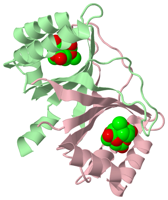
(-) Ligands, Modified Residues, Ions  (1, 7)

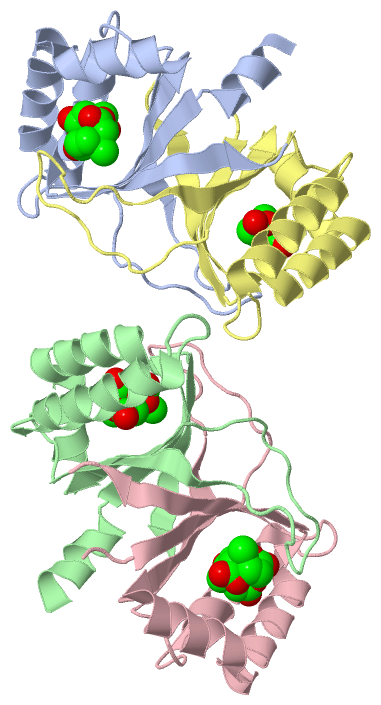
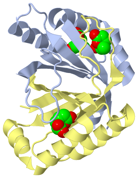
Asymmetric Unit (1, 7)
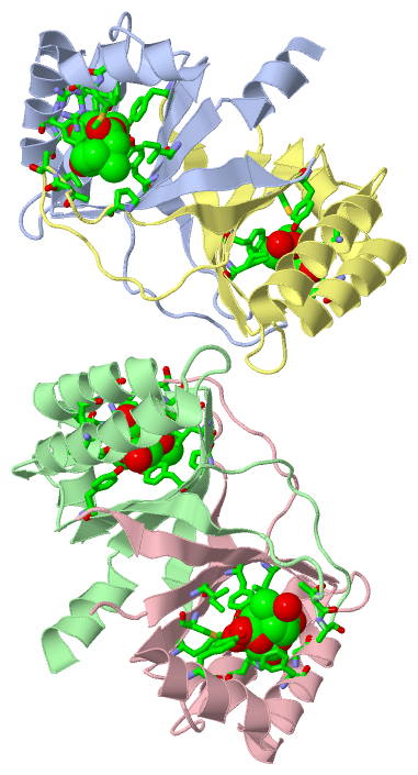
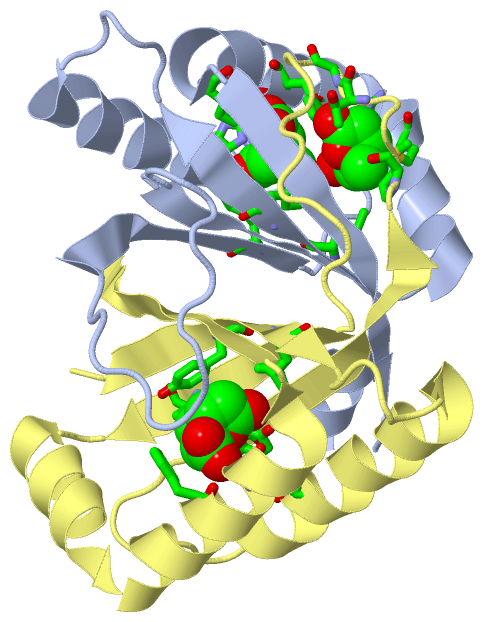
No.NameCountTypeFull Name
14ML7Ligand/Ion[(2S)-2-METHYL-5-OXO-2,5-DIHYDROFURAN-2-YL]ACETIC ACID
Biological Unit 1 (1, 3)
No.NameCountTypeFull Name
14ML3Ligand/Ion[(2S)-2-METHYL-5-OXO-2,5-DIHYDROFURAN-2-YL]ACETIC ACID
Biological Unit 2 (1, 4)
No.NameCountTypeFull Name
14ML4Ligand/Ion[(2S)-2-METHYL-5-OXO-2,5-DIHYDROFURAN-2-YL]ACETIC ACID

(-) Sites  (7, 7)

Asymmetric Unit (7, 7)
No.NameEvidenceResiduesDescription
1AC1SOFTWARETYR A:6 , HIS A:26 , TYR A:39 , TYR A:78 , TRP A:87 , PHE A:88 , GLY A:91 , ILE A:95 , 4ML A:202BINDING SITE FOR RESIDUE 4ML A 201
2AC2SOFTWAREILE A:4 , TYR A:6 , TYR A:78 , MET A:79 , ARG A:84 , PHE A:88 , 4ML A:201 , PRO D:48 , THR D:51 , ALA D:52BINDING SITE FOR RESIDUE 4ML A 202
3AC3SOFTWARETYR B:6 , HIS B:26 , TYR B:39 , TYR B:78 , TRP B:87 , PHE B:88 , GLY B:91 , ILE B:95 , 4ML B:202BINDING SITE FOR RESIDUE 4ML B 201
4AC4SOFTWARETYR B:6 , TYR B:78 , MET B:79 , ARG B:84 , PHE B:88 , LEU B:98 , 4ML B:201 , PRO C:48 , THR C:51 , ALA C:52BINDING SITE FOR RESIDUE 4ML B 202
5AC5SOFTWARETYR C:6 , LEU C:8 , HIS C:26 , TYR C:39 , CYS C:67 , PHE C:88 , GLY C:91BINDING SITE FOR RESIDUE 4ML C 201
6AC6SOFTWARETHR B:51 , ALA B:52 , HOH B:482 , ILE C:4 , TYR C:6 , TYR C:75 , PHE C:88 , HOH C:123BINDING SITE FOR RESIDUE 4ML C 202
7AC7SOFTWARETYR D:6 , LEU D:8 , HIS D:26 , TYR D:39 , CYS D:67 , PHE D:88 , GLY D:91BINDING SITE FOR RESIDUE 4ML D 201

(-) SS Bonds  (0, 0)

(no "SS Bond" information available for 3HFK)

(-) Cis Peptide Bonds  (0, 0)

(no "Cis Peptide Bond" information available for 3HFK)

 Sequence-Structure Mapping

(-) SAPs(SNPs)/Variants  (0, 0)

(no "SAP(SNP)/Variant" information available for 3HFK)

(-) PROSITE Motifs  (0, 0)

(no "PROSITE Motif" information available for 3HFK)

(-) Exons   (0, 0)

(no "Exon" information available for 3HFK)

(-) Sequences/Alignments

Asymmetric Unit
   Reformat: Number of residues per line =  ('0' or empty: single-line sequence representation)
  Number of residues per labelling interval =   
  UniProt sequence: complete  aligned part    
   Show mapping: SCOP domains CATH domains Pfam domains Secondary structure (by author)
SAPs(SNPs) PROSITE motifs Exons
(details for a mapped element are shown in a popup box when the mouse pointer rests over it)
Chain A from PDB  Type:PROTEIN  Length:115
 aligned with C5MR76_9PSED | C5MR76 from UniProtKB/TrEMBL  Length:107

    Alignment length:115
                                    1                                                                                                          
                                    |2        12        22        32        42        52        62        72        82        92       102     
         C5MR76_9PSED     - --------MIRILYLLVKPESMSHEQFRKECVVHFQMSAGMPGLHKYEVRLVAGNPTDTHVPYLDVGRIDAIGECWFASEEQYQVYMESDIRKAWFEHGKYFIGQLKPFVTEELV 107
               SCOP domains d3hfka_ A: automated matches                                                                                        SCOP domains
               CATH domains ------------------------------------------------------------------------------------------------------------------- CATH domains
               Pfam domains ------------------------------------------------------------------------------------------------------------------- Pfam domains
         Sec.struct. author hhhhhhhh.eeeeeeee.....hhhhhhhhhhhhhhh........eeeeeeeee................eeeeeee.hhhhhhhhhhhhhhhhhhhhhhhhh.eeeeeeeee.. Sec.struct. author
                 SAPs(SNPs) ------------------------------------------------------------------------------------------------------------------- SAPs(SNPs)
                    PROSITE ------------------------------------------------------------------------------------------------------------------- PROSITE
                 Transcript ------------------------------------------------------------------------------------------------------------------- Transcript
                 3hfk A  -7 QFEKIEGRMIRILYLLVKPESMSHEQFRKECVVHFQMSAGMPGLHKYEVRLVAGNPTDTAVPYLDVGRIDAIGECWFASEEQYQVYMESDIRKAWFEHGKYFIGQLKPFVTEELV 107
                                     2        12        22        32        42        52        62        72        82        92       102     

Chain B from PDB  Type:PROTEIN  Length:116
 aligned with C5MR76_9PSED | C5MR76 from UniProtKB/TrEMBL  Length:107

    Alignment length:116
                                     1                                                                                                          
                                     1        11        21        31        41        51        61        71        81        91       101      
         C5MR76_9PSED     - ---------MIRILYLLVKPESMSHEQFRKECVVHFQMSAGMPGLHKYEVRLVAGNPTDTHVPYLDVGRIDAIGECWFASEEQYQVYMESDIRKAWFEHGKYFIGQLKPFVTEELV 107
               SCOP domains d3hfkb_ B: automated matches                                                                                         SCOP domains
               CATH domains -------------------------------------------------------------------------------------------------------------------- CATH domains
               Pfam domains -------------------------------------------------------------------------------------------------------------------- Pfam domains
         Sec.struct. author hhhhhhhhh.eeeeeeee.....hhhhhhhhhhhhhhhhh......eeeeeeeee................eeeeeee.hhhhhhhhhhhhhhhhhhhhhhhhh.eeeeeeeee.. Sec.struct. author
                 SAPs(SNPs) -------------------------------------------------------------------------------------------------------------------- SAPs(SNPs)
                    PROSITE -------------------------------------------------------------------------------------------------------------------- PROSITE
                 Transcript -------------------------------------------------------------------------------------------------------------------- Transcript
                 3hfk B  -8 PQFEKIEGRMIRILYLLVKPESMSHEQFRKECVVHFQMSAGMPGLHKYEVRLVAGNPTDTAVPYLDVGRIDAIGECWFASEEQYQVYMESDIRKAWFEHGKYFIGQLKPFVTEELV 107
                                     1        11        21        31        41        51        61        71        81        91       101      

Chain C from PDB  Type:PROTEIN  Length:109
 aligned with C5MR76_9PSED | C5MR76 from UniProtKB/TrEMBL  Length:107

    Alignment length:109
                              1                                                                                                          
                              |      8        18        28        38        48        58        68        78        88        98         
         C5MR76_9PSED     - --MIRILYLLVKPESMSHEQFRKECVVHFQMSAGMPGLHKYEVRLVAGNPTDTHVPYLDVGRIDAIGECWFASEEQYQVYMESDIRKAWFEHGKYFIGQLKPFVTEELV 107
               SCOP domains d3hfkc_ C: automated matches                                                                                  SCOP domains
               CATH domains ------------------------------------------------------------------------------------------------------------- CATH domains
               Pfam domains ------------------------------------------------------------------------------------------------------------- Pfam domains
         Sec.struct. author ...eeeeeeee.....hhhhhhhhhhhhhhhh.......eeeeeeeee................eeeeeee.hhhhhhhhhhhhhhhhhhhhhhhhh.eeeeeeeee.. Sec.struct. author
                 SAPs(SNPs) ------------------------------------------------------------------------------------------------------------- SAPs(SNPs)
                    PROSITE ------------------------------------------------------------------------------------------------------------- PROSITE
                 Transcript ------------------------------------------------------------------------------------------------------------- Transcript
                 3hfk C  -1 GRMIRILYLLVKPESMSHEQFRKECVVHFQMSAGMPGLHKYEVRLVAGNPTDTAVPYLDVGRIDAIGECWFASEEQYQVYMESDIRKAWFEHGKYFIGQLKPFVTEELV 107
                                     8        18        28        38        48        58        68        78        88        98         

Chain D from PDB  Type:PROTEIN  Length:108
 aligned with C5MR76_9PSED | C5MR76 from UniProtKB/TrEMBL  Length:107

    Alignment length:108
                             1                                                                                                          
                             |       9        19        29        39        49        59        69        79        89        99        
         C5MR76_9PSED     - -MIRILYLLVKPESMSHEQFRKECVVHFQMSAGMPGLHKYEVRLVAGNPTDTHVPYLDVGRIDAIGECWFASEEQYQVYMESDIRKAWFEHGKYFIGQLKPFVTEELV 107
               SCOP domains d3hfkd_ D: automated matches                                                                                 SCOP domains
               CATH domains ------------------------------------------------------------------------------------------------------------ CATH domains
               Pfam domains ------------------------------------------------------------------------------------------------------------ Pfam domains
         Sec.struct. author ..eeeeeeee.....hhhhhhhhhhhhhhhh.......eeeeeeeee................eeeeeee.hhhhhhhhhhhhhhhhhhhhhhhhh.eeeeeeeee.. Sec.struct. author
                 SAPs(SNPs) ------------------------------------------------------------------------------------------------------------ SAPs(SNPs)
                    PROSITE ------------------------------------------------------------------------------------------------------------ PROSITE
                 Transcript ------------------------------------------------------------------------------------------------------------ Transcript
                 3hfk D   0 RMIRILYLLVKPESMSHEQFRKECVVHFQMSAGMPGLHKYEVRLVAGNPTDTAVPYLDVGRIDAIGECWFASEEQYQVYMESDIRKAWFEHGKYFIGQLKPFVTEELV 107
                                     9        19        29        39        49        59        69        79        89        99        

   Legend:   → Mismatch (orange background)
  - → Gap (green background, '-', border residues have a numbering label)
    → Modified Residue (blue background, lower-case, 'x' indicates undefined single-letter code, labelled with number + name)
  x → Chemical Group (purple background, 'x', labelled with number + name, e.g. ACE or NH2)
  extra numbering lines below/above indicate numbering irregularities and modified residue names etc., number ends below/above '|'

 Classification and Annotation

(-) SCOP Domains  (1, 4)

Asymmetric Unit

(-) CATH Domains  (0, 0)

(no "CATH Domain" information available for 3HFK)

(-) Pfam Domains  (0, 0)

(no "Pfam Domain" information available for 3HFK)

(-) Gene Ontology  (1, 1)

Asymmetric Unit(hide GO term definitions)
Chain A,B,C,D   (C5MR76_9PSED | C5MR76)
molecular function
    GO:0016853    isomerase activity    Catalysis of the geometric or structural changes within one molecule. Isomerase is the systematic name for any enzyme of EC class 5.

 Visualization

(-) Interactive Views

Asymmetric Unit
  Complete Structure
    Jena3D(integrated viewing of ligand, site, SAP, PROSITE, SCOP information)
    WebMol | AstexViewer[tm]@PDBe
(Java Applets, require no local installation except for Java; loading may be slow)
    STRAP
(Java WebStart application, automatic local installation, requires Java; full application with system access!)
    RasMol
(require local installation)
    Molscript (VRML)
(requires installation of a VRML viewer; select preferred view via VRML and generate a mono or stereo PDF format file)
 
  Ligands, Modified Residues, Ions
    4ML  [ RasMol | Jena3D ]  +environment [ RasMol | Jena3D ]
 
  Sites
    AC1  [ RasMol ]  +environment [ RasMol ]
    AC2  [ RasMol ]  +environment [ RasMol ]
    AC3  [ RasMol ]  +environment [ RasMol ]
    AC4  [ RasMol ]  +environment [ RasMol ]
    AC5  [ RasMol ]  +environment [ RasMol ]
    AC6  [ RasMol ]  +environment [ RasMol ]
    AC7  [ RasMol ]  +environment [ RasMol ]
 
  Cis Peptide Bonds
(no "Cis Peptide Bonds" information available for 3hfk)
 
Biological Units
  Complete Structure
    Biological Unit 1  [ Jena3D ]
    Biological Unit 2  [ Jena3D ]

(-) Still Images

Jmol
  protein: cartoon or spacefill or dots and stick; nucleic acid: cartoon and stick; ligands: spacefill; active site: stick
Molscript
  protein, nucleic acid: cartoon; ligands: spacefill; active site: ball and stick

 Databases and Analysis Tools

(-) Databases

Access by PDB/NDB ID
  3hfk
    Family and Domain InformationProDom | SYSTERS
    General Structural InformationGlycoscienceDB | MMDB | NDB | OCA | PDB | PDBe | PDBj | PDBsum | PDBWiki | PQS | PROTEOPEDIA
    Orientation in MembranesOPM
    Protein SurfaceSURFACE
    Secondary StructureDSSP (structure derived) | HSSP (homology derived)
    Structural GenomicsGeneCensus
    Structural NeighboursCE | VAST
    Structure ClassificationCATH | Dali | SCOP
    Validation and Original DataBMRB Data View | BMRB Restraints Grid | EDS | PROCHECK | RECOORD | WHAT_CHECK
 
Access by UniProt ID/Accession number
  C5MR76_9PSED | C5MR76
    Comparative Protein Structure ModelsModBase
    Genomic InformationEnsembl
    Protein-protein InteractionDIP
    Sequence, Family and Domain InformationInterPro | Pfam | SMART | UniProtKB/TrEMBL
 
Access by Enzyme Classificator   (EC Number)
  5.4.99.14
    General Enzyme InformationBRENDA | EC-PDB | Enzyme | IntEnz
    PathwayKEGG | MetaCyc
 
Access by Disease Identifier   (MIM ID)
  (no 'MIM ID' available)
    Disease InformationOMIM
 
Access by GenAge ID
  (no 'GenAge ID' available)
    Age Related InformationGenAge

(-) Analysis Tools

Access by PDB/NDB ID
    Domain InformationXDom
    Interatomic Contacts of Structural UnitsCSU
    Ligand-protein ContactsLPC
    Protein CavitiescastP
    Sequence and Secondary StructurePDBCartoon
    Structure AlignmentSTRAP(Java WebStart application, automatic local installation, requires Java; full application with system access!)
    Structure and Sequence BrowserSTING
 
Access by UniProt ID/Accession number
  C5MR76_9PSED | C5MR76
    Protein Disorder PredictionDisEMBL | FoldIndex | GLOBPLOT (for more information see DisProt)

 Related Entries

(-) Entries Sharing at Least One Protein Chain (UniProt ID)

UniProtKB/TrEMBL
        C5MR76_9PSED | C5MR763hds 3hf5

(-) Related Entries Specified in the PDB File

3hds THE WILDTYPE PROTEIN COMPLEXED WITH A MES BUFFER MOLECULE
3hf5 THE WILDTYPE PROTEIN COMPLEXED WITH 3-METHYLMUCONOLACTONE