|
|
|
|
 Description
|
|
Compounds
|
||||||||||||||||||||||||||||||||||||||||||||||||
Chains, Units
Summary Information (see also Sequences/Alignments below) |
Ligands, Modified Residues, Ions (0, 0)| (no "Ligand,Modified Residues,Ions" information available for 3HFI) |
Sites (0, 0)| (no "Site" information available for 3HFI) |
SS Bonds (0, 0)| (no "SS Bond" information available for 3HFI) |
Cis Peptide Bonds (0, 0)| (no "Cis Peptide Bond" information available for 3HFI) |
SAPs(SNPs)/Variants (0, 0)| (no "SAP(SNP)/Variant" information available for 3HFI) |
PROSITE Motifs (0, 0)| (no "PROSITE Motif" information available for 3HFI) |
Exons (0, 0)| (no "Exon" information available for 3HFI) |
Sequences/Alignments
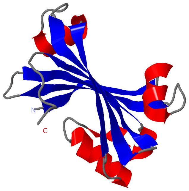
Asymmetric UnitChain A from PDB Type:PROTEIN Length:137 aligned with A0A0H2VBL3_E | A0A0H2VBL3 from UniProtKB/TrEMBL Length:251 Alignment length:137 115 125 135 145 155 165 175 185 195 205 215 225 235 A0A0H2VBL3_E 106 TTEVITSRIEPANRYVAEKLRITPGQDILYLERLRSIGDEKAMLIENRINIELCPGIVEIDFNQHNLFPTIESLSKRKIRYSESRYAARLIGNERGHFLDISEDAPVLHLEQLVFFSRELPVEFGNVWLKGNKYYLG 242 SCOP domains d3hfia_ A: automated matches SCOP domains CATH domains ----------------------------------------------------------------------------------------------------------------------------------------- CATH domains Pfam domains ----------------------------------------------------------------------------------------------------------------------------------------- Pfam domains SAPs(SNPs) ----------------------------------------------------------------------------------------------------------------------------------------- SAPs(SNPs) PROSITE ----------------------------------------------------------------------------------------------------------------------------------------- PROSITE Transcript ----------------------------------------------------------------------------------------------------------------------------------------- Transcript 3hfi A 25 TTEVITSRIEPANRYVAEKLRITPGQDILYLERLRSIGDEKAMLIENRINIELCPGIVEIDFNQHNLFPTIESLSKRKIRYSESRYAARLIGNERGHFLDISEDAPVLHLEQLVFFSRELPVEFGNVWLKGNKYYLG 161 34 44 54 64 74 84 94 104 114 124 134 144 154
|
||||||||||||||||||||
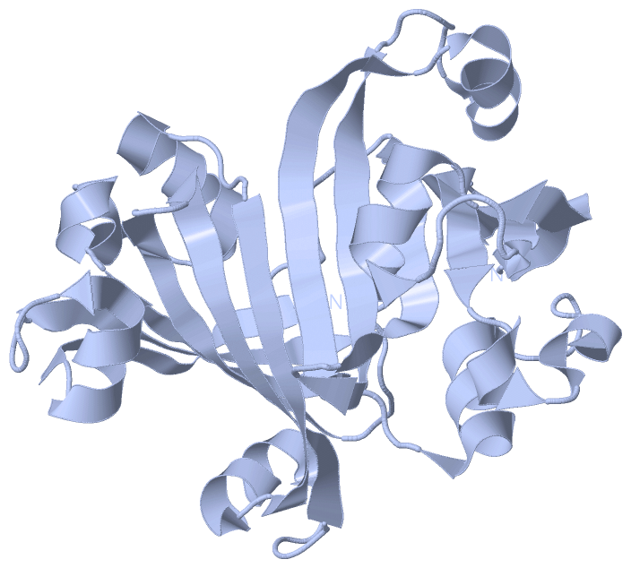
SCOP Domains (1, 1)
Asymmetric Unit
 |
CATH Domains (0, 0)| (no "CATH Domain" information available for 3HFI) |
Pfam Domains (0, 0)| (no "Pfam Domain" information available for 3HFI) |
Gene Ontology (0, 0)|
Asymmetric Unit(hide GO term definitions)
(no "Gene Ontology" information available for 3HFI)
|
Interactive Views
|
||||||||||||||||||||||||||||||||||||||||||||||||||||||||||||||||||||||||||||||||||||||||||||||||||||||||||||||||||||||||||||||||||||||
Still Images
|
||||||||||||||||
Databases
|
||||||||||||||||||||||||||||||||||||||||||||||||||||||||||||||||||||||||||||||||||||||||||||||||||||||||||||||||||||||||||||||||||||||||||||||||||||||||||||||||
Analysis Tools
|
|||||||||||||||||||||||||||||||||||||||||||||||||||||||||||||
Entries Sharing at Least One Protein Chain (UniProt ID)
Related Entries Specified in the PDB File
|
|