|
|
|
|
 Description
|
|
Compounds
|
||||||||||||||||||||||||||||||||||||||||
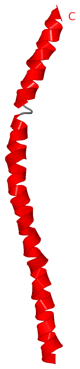
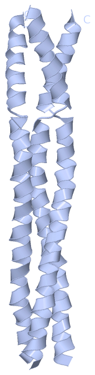
Chains, Units
Summary Information (see also Sequences/Alignments below) |
Ligands, Modified Residues, Ions (0, 0)| (no "Ligand,Modified Residues,Ions" information available for 3H7Z) |
Sites (0, 0)| (no "Site" information available for 3H7Z) |
SS Bonds (0, 0)| (no "SS Bond" information available for 3H7Z) |
Cis Peptide Bonds (0, 0)| (no "Cis Peptide Bond" information available for 3H7Z) |
SAPs(SNPs)/Variants (0, 0)| (no "SAP(SNP)/Variant" information available for 3H7Z) |
PROSITE Motifs (0, 0)| (no "PROSITE Motif" information available for 3H7Z) |
Exons (0, 0)| (no "Exon" information available for 3H7Z) |
Sequences/Alignments
Asymmetric UnitChain A from PDB Type:PROTEIN Length:61 aligned with YADA2_YEREN | P0C2W0 from UniProtKB/Swiss-Prot Length:422 Alignment length:61 312 322 332 342 352 362 YADA2_YEREN 303 HTLKTANSYTDVTVSNSTKKAIRESNQYTDHKFHQLDNRLDKLDTRVDKGLASSAALNSLF 363 SCOP domains ------------------------------------------------------------- SCOP domains CATH domains ------------------------------------------------------------- CATH domains Pfam domains ------------------------------------------------------------- Pfam domains SAPs(SNPs) ------------------------------------------------------------- SAPs(SNPs) PROSITE ------------------------------------------------------------- PROSITE Transcript ------------------------------------------------------------- Transcript 3h7z A 303 HTLKTANSYTDVTVSNSTKKAIRESNQYTDHKFHQLDNRLDKLDTRLLKLLASSAALNSLL 363 312 322 332 342 352 362
|
||||||||||||||||||||
SCOP Domains (0, 0)| (no "SCOP Domain" information available for 3H7Z) |
CATH Domains (0, 0)| (no "CATH Domain" information available for 3H7Z) |
Pfam Domains (0, 0)| (no "Pfam Domain" information available for 3H7Z) |
Gene Ontology (7, 7)|
Asymmetric Unit(hide GO term definitions) Chain A (YADA2_YEREN | P0C2W0)
|
||||||||||||||||||||||||||||||||||||||||||||||||||||||||||||
Interactive Views
|
||||||||||||||||||||||||||||||||||||||||||||||||||||||||||||||||||||||||||||||||||||||||||||||||||||||||||||||||||||||||||||||||||||||
Still Images
|
||||||||||||||||
Databases
|
||||||||||||||||||||||||||||||||||||||||||||||||||||||||||||||||||||||||||||||||||||||||||||||||||||||||||||||||||||||||||||||||||||||||||||||||||||||||||||||||
Analysis Tools
|
|||||||||||||||||||||||||||||||||||||||||||||||||||||||||||||
Entries Sharing at Least One Protein Chain (UniProt ID)
Related Entries Specified in the PDB File
|
|