Show PDB file:   
         Plain Text   HTML   (compressed file size)
QuickSearch:   
by PDB,NDB,UniProt,PROSITE Code or Search Term(s)  
(-)Asymmetric Unit
(-)Asym. Unit - sites
(-)Biological Unit 1
(-)Biol. Unit 1 - sites
(-)Biological Unit 2
(-)Biological Unit 3
(-)Biological Unit 4
collapse expand < >
Image Asymmetric Unit
Asymmetric Unit  (Jmol Viewer)
Image Asym. Unit - sites
Asym. Unit - sites  (Jmol Viewer)
Image Biological Unit 1
Biological Unit 1  (Jmol Viewer)
Image Biol. Unit 1 - sites
Biol. Unit 1 - sites  (Jmol Viewer)
Image Biological Unit 2
Biological Unit 2  (Jmol Viewer)
Image Biological Unit 3
Biological Unit 3  (Jmol Viewer)
Image Biological Unit 4
Biological Unit 4  (Jmol Viewer)

(-) Description

Title :  THE CRYSTAL STRUCTURE OF THE CATHEPSIN K VARIANT M5 IN COMPLEX WITH CHONDROITIN-4-SULFATE
 
Authors :  M. M. Cherney, M. Kienetz, D. Bromme, M. N. G. James
Date :  24 Apr 09  (Deposition) - 28 Apr 10  (Release) - 15 Jan 14  (Revision)
Method :  X-RAY DIFFRACTION
Resolution :  2.24
Chains :  Asym. Unit :  A,E
Biol. Unit 1:  A  (1x)
Biol. Unit 2:  E  (1x)
Biol. Unit 3:  A  (2x)
Biol. Unit 4:  E  (2x)
Keywords :  Glycosaminoglycan, Sulfhydryl Peptidase, Cathepsin K Mutant, Ternary Complex, Disease Mutation, Disulfide Bond, Glycoprotein, Hydrolase, Lysosome, Protease, Thiol Protease, Zymogen (Keyword Search: [Gene Ontology, PubMed, Web (Google))
 
Reference :  M. M. Cherney, F. Lecaille, M. Kienitz, F. S. Nallaseth, Z. Li, M. N. James, D. Bromme
Structure-Activity Analysis Of Cathepsin K/Chondroitin 4-Sulfate Interactions.
J. Biol. Chem. V. 286 8988 2011
PubMed-ID: 21193413  |  Reference-DOI: 10.1074/JBC.M110.126706

(-) Compounds

Molecule 1 - CATHEPSIN K
    ChainsA, E
    EC Number3.4.22.38
    EngineeredYES
    Expression SystemESCHERICHIA COLI
    Expression System Taxid562
    FragmentUNP RESIDUES 115-329
    GeneCTSK, CTSO, CTSO2
    MutationYES
    Organism CommonHUMAN
    Organism ScientificHOMO SAPIENS
    Organism Taxid9606
    SynonymCATHEPSIN O, CATHEPSIN X, CATHEPSIN O2

 Structural Features

(-) Chains, Units

  12
Asymmetric Unit AE
Biological Unit 1 (1x)A 
Biological Unit 2 (1x) E
Biological Unit 3 (2x)A 
Biological Unit 4 (2x) E

Summary Information (see also Sequences/Alignments below)

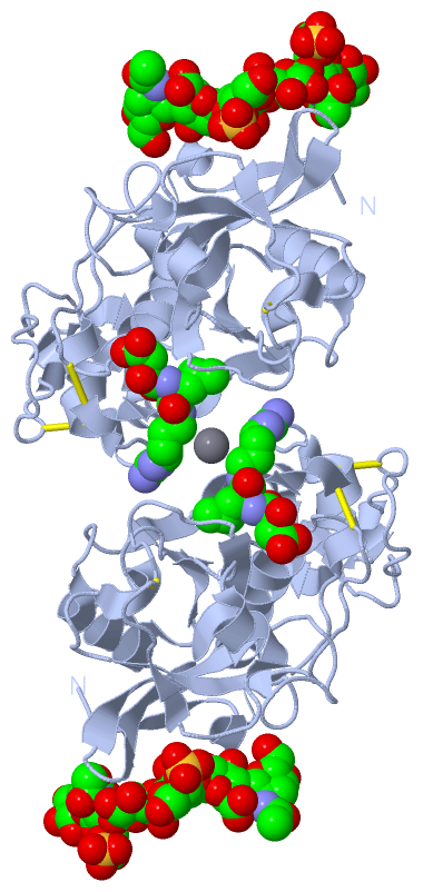
(-) Ligands, Modified Residues, Ions  (4, 10)

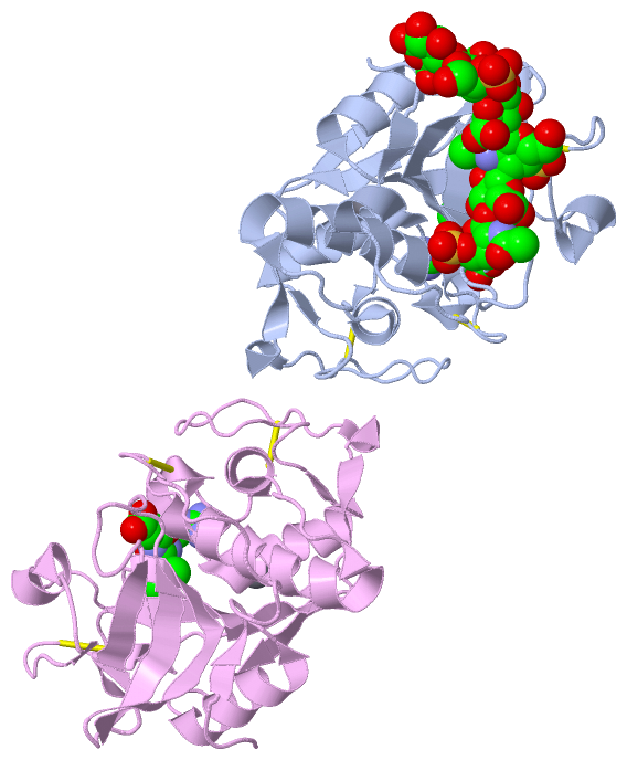
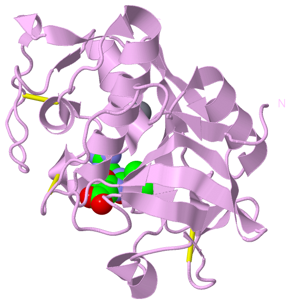
Asymmetric Unit (4, 10)
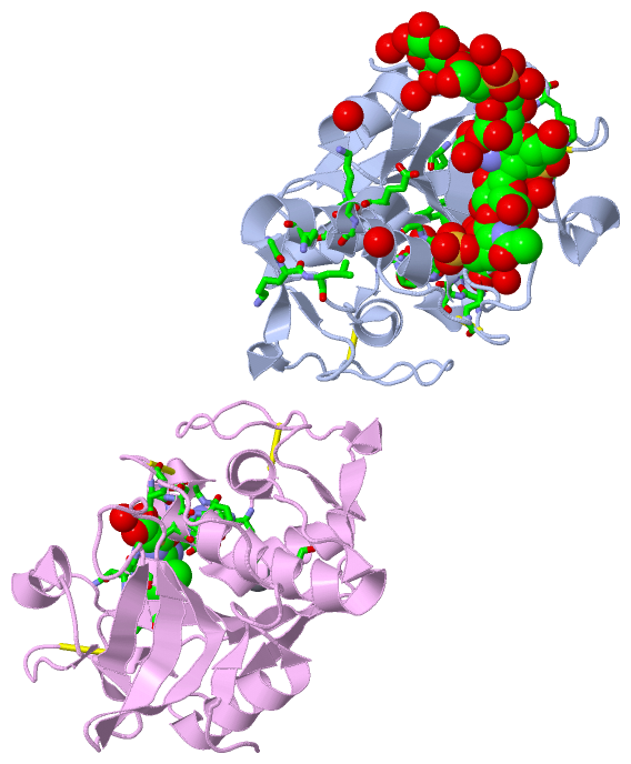
No.NameCountTypeFull Name
1ASG3Ligand/Ion2-DEOXY-2-ACETAMIDO-BETA-D-GALACTOSE-4-SULFATE
2BDP3Ligand/IonBETA-D-GLUCOPYRANURONIC ACID
3CA2Ligand/IonCALCIUM ION
4E642Ligand/IonN-[N-[1-HYDROXYCARBOXYETHYL-CARBONYL]LEUCYLAMINO-BUTYL]-GUANIDINE
Biological Unit 1 (3, 7)
No.NameCountTypeFull Name
1ASG3Ligand/Ion2-DEOXY-2-ACETAMIDO-BETA-D-GALACTOSE-4-SULFATE
2BDP3Ligand/IonBETA-D-GLUCOPYRANURONIC ACID
3CA-1Ligand/IonCALCIUM ION
4E641Ligand/IonN-[N-[1-HYDROXYCARBOXYETHYL-CARBONYL]LEUCYLAMINO-BUTYL]-GUANIDINE
Biological Unit 2 (1, 1)
No.NameCountTypeFull Name
1ASG-1Ligand/Ion2-DEOXY-2-ACETAMIDO-BETA-D-GALACTOSE-4-SULFATE
2BDP-1Ligand/IonBETA-D-GLUCOPYRANURONIC ACID
3CA-1Ligand/IonCALCIUM ION
4E641Ligand/IonN-[N-[1-HYDROXYCARBOXYETHYL-CARBONYL]LEUCYLAMINO-BUTYL]-GUANIDINE
Biological Unit 3 (3, 14)
No.NameCountTypeFull Name
1ASG6Ligand/Ion2-DEOXY-2-ACETAMIDO-BETA-D-GALACTOSE-4-SULFATE
2BDP6Ligand/IonBETA-D-GLUCOPYRANURONIC ACID
3CA-1Ligand/IonCALCIUM ION
4E642Ligand/IonN-[N-[1-HYDROXYCARBOXYETHYL-CARBONYL]LEUCYLAMINO-BUTYL]-GUANIDINE
Biological Unit 4 (1, 2)
No.NameCountTypeFull Name
1ASG-1Ligand/Ion2-DEOXY-2-ACETAMIDO-BETA-D-GALACTOSE-4-SULFATE
2BDP-1Ligand/IonBETA-D-GLUCOPYRANURONIC ACID
3CA-1Ligand/IonCALCIUM ION
4E642Ligand/IonN-[N-[1-HYDROXYCARBOXYETHYL-CARBONYL]LEUCYLAMINO-BUTYL]-GUANIDINE

(-) Sites  (10, 10)

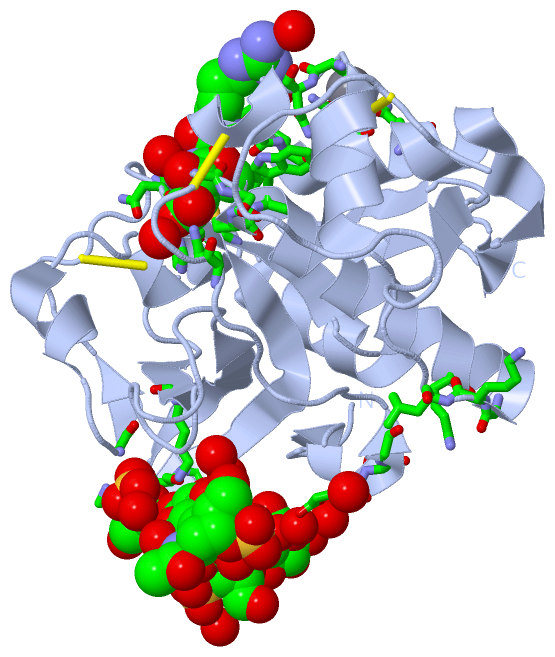
Asymmetric Unit (10, 10)
No.NameEvidenceResiduesDescription
01AC1SOFTWAREGLU A:59 , ASN A:70 , GLN A:73BINDING SITE FOR RESIDUE CA A 300
02AC2SOFTWAREASG A:217 , HOH A:231 , HOH A:281BINDING SITE FOR RESIDUE BDP A 216
03AC3SOFTWARELYS A:39 , GLY A:43 , LYS A:44 , LEU A:45 , GLU A:171 , BDP A:216 , BDP A:218 , HOH A:239 , HOH A:244BINDING SITE FOR RESIDUE ASG A 217
04AC4SOFTWARELYS A:39 , LYS A:173 , ASG A:217 , ASG A:219 , HOH A:226 , HOH A:241BINDING SITE FOR RESIDUE BDP A 218
05AC5SOFTWAREGLU A:9 , GLY A:11 , GLY A:148 , SER A:172 , LYS A:173 , LYS A:195 , BDP A:218 , BDP A:220 , HOH A:356 , HOH A:365 , HOH A:372BINDING SITE FOR RESIDUE ASG A 219
06AC6SOFTWARELYS A:10 , LYS A:195 , ASG A:219 , ASG A:221BINDING SITE FOR RESIDUE BDP A 220
07AC7SOFTWARELYS A:10 , BDP A:220 , HOH A:240 , HOH A:361BINDING SITE FOR RESIDUE ASG A 221
08AC8SOFTWAREGLN A:19 , GLY A:23 , SER A:24 , CYS A:25 , TRP A:26 , GLU A:59 , ASN A:60 , GLY A:65 , GLY A:66 , TYR A:67 , ALA A:134 , LEU A:160 , ASN A:161 , HIS A:162 , LEU A:209 , HOH A:261 , HOH A:289 , HOH A:292 , HOH A:302BINDING SITE FOR RESIDUE E64 A 222
09AC9SOFTWAREGLU E:59 , ASN E:70 , GLN E:73BINDING SITE FOR RESIDUE CA E 300
10BC1SOFTWAREGLN E:19 , GLY E:23 , SER E:24 , CYS E:25 , TRP E:26 , GLU E:59 , ASN E:60 , ASP E:61 , GLY E:65 , GLY E:66 , ALA E:134 , LEU E:160 , ASN E:161 , HIS E:162 , HOH E:243BINDING SITE FOR RESIDUE E64 E 216

(-) SS Bonds  (6, 6)

Asymmetric Unit
No.Residues
1A:22 -A:63
2A:56 -A:96
3A:155 -A:204
4E:22 -E:63
5E:56 -E:96
6E:155 -E:204

(-) Cis Peptide Bonds  (0, 0)

(no "Cis Peptide Bond" information available for 3H7D)

 Sequence-Structure Mapping

(-) SAPs(SNPs)/Variants  (5, 10)

Asymmetric Unit (5, 10)
  dbSNPPDB
No.SourceVariant IDVariantUniProt IDStatusIDChainVariant
1UniProtVAR_074023R122PCATK_HUMANDisease (PKND)  ---A/ER8P
2UniProtVAR_006725G146RCATK_HUMANDisease (PKND)74315302A/EG32R
3UniProtVAR_015739A277VCATK_HUMANDisease (PKND)74315304A/EA163V
4UniProtVAR_074024Y283CCATK_HUMANDisease (PKND)  ---A/EY169C
5UniProtVAR_006726L309PCATK_HUMANDisease (PKND)29001685A/EK195P

  SNP/SAP Summary Statistics (UniProtKB/Swiss-Prot)
Biological Unit 1 (5, 5)
  dbSNPPDB
No.SourceVariant IDVariantUniProt IDStatusIDChainVariant
1UniProtVAR_074023R122PCATK_HUMANDisease (PKND)  ---AR8P
2UniProtVAR_006725G146RCATK_HUMANDisease (PKND)74315302AG32R
3UniProtVAR_015739A277VCATK_HUMANDisease (PKND)74315304AA163V
4UniProtVAR_074024Y283CCATK_HUMANDisease (PKND)  ---AY169C
5UniProtVAR_006726L309PCATK_HUMANDisease (PKND)29001685AK195P

  SNP/SAP Summary Statistics (UniProtKB/Swiss-Prot)
Biological Unit 2 (5, 5)
  dbSNPPDB
No.SourceVariant IDVariantUniProt IDStatusIDChainVariant
1UniProtVAR_074023R122PCATK_HUMANDisease (PKND)  ---ER8P
2UniProtVAR_006725G146RCATK_HUMANDisease (PKND)74315302EG32R
3UniProtVAR_015739A277VCATK_HUMANDisease (PKND)74315304EA163V
4UniProtVAR_074024Y283CCATK_HUMANDisease (PKND)  ---EY169C
5UniProtVAR_006726L309PCATK_HUMANDisease (PKND)29001685EK195P

  SNP/SAP Summary Statistics (UniProtKB/Swiss-Prot)
Biological Unit 3 (5, 10)
  dbSNPPDB
No.SourceVariant IDVariantUniProt IDStatusIDChainVariant
1UniProtVAR_074023R122PCATK_HUMANDisease (PKND)  ---AR8P
2UniProtVAR_006725G146RCATK_HUMANDisease (PKND)74315302AG32R
3UniProtVAR_015739A277VCATK_HUMANDisease (PKND)74315304AA163V
4UniProtVAR_074024Y283CCATK_HUMANDisease (PKND)  ---AY169C
5UniProtVAR_006726L309PCATK_HUMANDisease (PKND)29001685AK195P

  SNP/SAP Summary Statistics (UniProtKB/Swiss-Prot)
Biological Unit 4 (5, 10)
  dbSNPPDB
No.SourceVariant IDVariantUniProt IDStatusIDChainVariant
1UniProtVAR_074023R122PCATK_HUMANDisease (PKND)  ---ER8P
2UniProtVAR_006725G146RCATK_HUMANDisease (PKND)74315302EG32R
3UniProtVAR_015739A277VCATK_HUMANDisease (PKND)74315304EA163V
4UniProtVAR_074024Y283CCATK_HUMANDisease (PKND)  ---EY169C
5UniProtVAR_006726L309PCATK_HUMANDisease (PKND)29001685EK195P

  SNP/SAP Summary Statistics (UniProtKB/Swiss-Prot)

(-) PROSITE Motifs  (3, 6)

Asymmetric Unit (3, 6)
 PROSITEUniProtKBPDB
No.IDACDescriptionIDLocationCountLocation
1THIOL_PROTEASE_CYSPS00139 Eukaryotic thiol (cysteine) proteases cysteine active site.CATK_HUMAN133-144
 
  2A:19-30
E:19-30
2THIOL_PROTEASE_HISPS00639 Eukaryotic thiol (cysteine) proteases histidine active site.CATK_HUMAN274-284
 
  2A:160-170
E:160-170
3THIOL_PROTEASE_ASNPS00640 Eukaryotic thiol (cysteine) proteases asparagine active site.CATK_HUMAN291-310
 
  2A:177-196
E:177-196
Biological Unit 1 (3, 3)
 PROSITEUniProtKBPDB
No.IDACDescriptionIDLocationCountLocation
1THIOL_PROTEASE_CYSPS00139 Eukaryotic thiol (cysteine) proteases cysteine active site.CATK_HUMAN133-144
 
  1A:19-30
-
2THIOL_PROTEASE_HISPS00639 Eukaryotic thiol (cysteine) proteases histidine active site.CATK_HUMAN274-284
 
  1A:160-170
-
3THIOL_PROTEASE_ASNPS00640 Eukaryotic thiol (cysteine) proteases asparagine active site.CATK_HUMAN291-310
 
  1A:177-196
-
Biological Unit 2 (3, 3)
 PROSITEUniProtKBPDB
No.IDACDescriptionIDLocationCountLocation
1THIOL_PROTEASE_CYSPS00139 Eukaryotic thiol (cysteine) proteases cysteine active site.CATK_HUMAN133-144
 
  1-
E:19-30
2THIOL_PROTEASE_HISPS00639 Eukaryotic thiol (cysteine) proteases histidine active site.CATK_HUMAN274-284
 
  1-
E:160-170
3THIOL_PROTEASE_ASNPS00640 Eukaryotic thiol (cysteine) proteases asparagine active site.CATK_HUMAN291-310
 
  1-
E:177-196
Biological Unit 3 (3, 6)
 PROSITEUniProtKBPDB
No.IDACDescriptionIDLocationCountLocation
1THIOL_PROTEASE_CYSPS00139 Eukaryotic thiol (cysteine) proteases cysteine active site.CATK_HUMAN133-144
 
  2A:19-30
-
2THIOL_PROTEASE_HISPS00639 Eukaryotic thiol (cysteine) proteases histidine active site.CATK_HUMAN274-284
 
  2A:160-170
-
3THIOL_PROTEASE_ASNPS00640 Eukaryotic thiol (cysteine) proteases asparagine active site.CATK_HUMAN291-310
 
  2A:177-196
-
Biological Unit 4 (3, 6)
 PROSITEUniProtKBPDB
No.IDACDescriptionIDLocationCountLocation
1THIOL_PROTEASE_CYSPS00139 Eukaryotic thiol (cysteine) proteases cysteine active site.CATK_HUMAN133-144
 
  2-
E:19-30
2THIOL_PROTEASE_HISPS00639 Eukaryotic thiol (cysteine) proteases histidine active site.CATK_HUMAN274-284
 
  2-
E:160-170
3THIOL_PROTEASE_ASNPS00640 Eukaryotic thiol (cysteine) proteases asparagine active site.CATK_HUMAN291-310
 
  2-
E:177-196

(-) Exons   (5, 10)

Asymmetric Unit (5, 10)
 ENSEMBLUniProtKBPDB
No.Transcript IDExonExon IDGenome LocationLengthIDLocationLengthCountLocationLength
1.1ENST000002716511ENSE00001343333chr1:150780799-150780690110CATK_HUMAN-00--
1.3aENST000002716513aENSE00001691906chr1:150779282-150779162121CATK_HUMAN1-40400--
1.4ENST000002716514ENSE00001044450chr1:150778700-150778578123CATK_HUMAN41-81410--
1.5ENST000002716515ENSE00001044471chr1:150778492-150778337156CATK_HUMAN82-133522A:1-19
E:1-19
19
19
1.6bENST000002716516bENSE00002193797chr1:150776715-150776497219CATK_HUMAN134-206732A:20-92
E:20-92
73
73
1.7ENST000002716517ENSE00001610479chr1:150772185-150772020166CATK_HUMAN207-262562A:93-148
E:93-148
56
56
1.8ENST000002716518ENSE00001602868chr1:150771749-150771644106CATK_HUMAN262-297362A:148-183
E:148-183
36
36
1.9ENST000002716519ENSE00000959757chr1:150769374-150768684691CATK_HUMAN297-329332A:183-215
E:183-215
33
33

(-) Sequences/Alignments

Asymmetric Unit
   Reformat: Number of residues per line =  ('0' or empty: single-line sequence representation)
  Number of residues per labelling interval =   
  UniProt sequence: complete  aligned part    
   Show mapping: SCOP domains CATH domains Pfam domains Secondary structure (by author)
SAPs(SNPs) PROSITE motifs Exons
(details for a mapped element are shown in a popup box when the mouse pointer rests over it)
Chain A from PDB  Type:PROTEIN  Length:215
 aligned with CATK_HUMAN | P43235 from UniProtKB/Swiss-Prot  Length:329

    Alignment length:215
                                   124       134       144       154       164       174       184       194       204       214       224       234       244       254       264       274       284       294       304       314       324     
           CATK_HUMAN   115 APDSVDYRKKGYVTPVKNQGQCGSCWAFSSVGALEGQLKKKTGKLLNLSPQNLVDCVSENDGCGGGYMTNAFQYVQKNRGIDSEDAYPYVGQEESCMYNPTGKAAKCRGYREIPEGNEKALKRAVARVGPVSVAIDASLTSFQFYSKGVYYDESCNSDNLNHAVLAVGYGIQKGNKHWIIKNSWGENWGNKGYILMARNKNNACGIANLASFPKM 329
               SCOP domains ----------------------------------------------------------------------------------------------------------------------------------------------------------------------------------------------------------------------- SCOP domains
               CATH domains 3h7dA00 A:1-215 Cysteine proteinases                                                                                                                                                                                    CATH domains
               Pfam domains ----------------------------------------------------------------------------------------------------------------------------------------------------------------------------------------------------------------------- Pfam domains
         Sec.struct. author ....ee.hhhh............hhhhhhhhhhhhhhhhhhhh.....hhhhhhhhh...hhhhh.hhhhhhhhhhhhh.ee................hhh.eee....eee....hhhhhhhhhhhh..eeeee...hhhhhh....ee...........eeeeeeeeeee..eeeeeee...........eeeee....hhhhh....eee.. Sec.struct. author
                 SAPs(SNPs) -------P-----------------------R----------------------------------------------------------------------------------------------------------------------------------V-----C-------------------------P-------------------- SAPs(SNPs)
                    PROSITE ------------------THIOL_PROTEA---------------------------------------------------------------------------------------------------------------------------------THIOL_PROTE------THIOL_PROTEASE_ASN  ------------------- PROSITE
           Transcript 1 (1) Exon 1.5           Exon 1.6b  PDB: A:20-92 UniProt: 134-206                                 Exon 1.7  PDB: A:93-148 UniProt: 207-262                ----------------------------------Exon 1.9  PDB: A:183-215          Transcript 1 (1)
           Transcript 1 (2) ---------------------------------------------------------------------------------------------------------------------------------------------------Exon 1.8  PDB: A:148-183            -------------------------------- Transcript 1 (2)
                 3h7d A   1 APDSVDYREKGYVTPVKNQGQCGSCWAFSSVGALEGQLKKKTGKLLNLSPQNLVDCVSENDGCGGGYMTNAFQYVQKNRGIDSEDAYPYVGQEESCMYNPTGKAAKCRGYREIPEGNEKALKRAVARVGPVSVAIDASLTSFQFYSKGVYYDESCNSDNLNHAVLAVGYGESKGNKHWIIKNSWGENWGMGGYIKMARNKNNACGIANLASFPKM 215
                                    10        20        30        40        50        60        70        80        90       100       110       120       130       140       150       160       170       180       190       200       210     

Chain E from PDB  Type:PROTEIN  Length:215
 aligned with CATK_HUMAN | P43235 from UniProtKB/Swiss-Prot  Length:329

    Alignment length:215
                                   124       134       144       154       164       174       184       194       204       214       224       234       244       254       264       274       284       294       304       314       324     
           CATK_HUMAN   115 APDSVDYRKKGYVTPVKNQGQCGSCWAFSSVGALEGQLKKKTGKLLNLSPQNLVDCVSENDGCGGGYMTNAFQYVQKNRGIDSEDAYPYVGQEESCMYNPTGKAAKCRGYREIPEGNEKALKRAVARVGPVSVAIDASLTSFQFYSKGVYYDESCNSDNLNHAVLAVGYGIQKGNKHWIIKNSWGENWGNKGYILMARNKNNACGIANLASFPKM 329
               SCOP domains ----------------------------------------------------------------------------------------------------------------------------------------------------------------------------------------------------------------------- SCOP domains
               CATH domains 3h7dE00 E:1-215 Cysteine proteinases                                                                                                                                                                                    CATH domains
               Pfam domains ----------------------------------------------------------------------------------------------------------------------------------------------------------------------------------------------------------------------- Pfam domains
         Sec.struct. author ....ee.................hhhhhhhhhhhhhhhhhhhh.....hhhhhhhhh...hhhhh.hhhhhhhhhhhh..ee................hhh.eee...eeee....hhhhhhhhhhhh..eeeee...hhhhhh....ee...........eeeeeeeeeee..eeeeeee...........eeeee....hhhhh....eeee. Sec.struct. author
                 SAPs(SNPs) -------P-----------------------R----------------------------------------------------------------------------------------------------------------------------------V-----C-------------------------P-------------------- SAPs(SNPs)
                    PROSITE ------------------THIOL_PROTEA---------------------------------------------------------------------------------------------------------------------------------THIOL_PROTE------THIOL_PROTEASE_ASN  ------------------- PROSITE
           Transcript 1 (1) Exon 1.5           Exon 1.6b  PDB: E:20-92 UniProt: 134-206                                 Exon 1.7  PDB: E:93-148 UniProt: 207-262                ----------------------------------Exon 1.9  PDB: E:183-215          Transcript 1 (1)
           Transcript 1 (2) ---------------------------------------------------------------------------------------------------------------------------------------------------Exon 1.8  PDB: E:148-183            -------------------------------- Transcript 1 (2)
                 3h7d E   1 APDSVDYREKGYVTPVKNQGQCGSCWAFSSVGALEGQLKKKTGKLLNLSPQNLVDCVSENDGCGGGYMTNAFQYVQKNRGIDSEDAYPYVGQEESCMYNPTGKAAKCRGYREIPEGNEKALKRAVARVGPVSVAIDASLTSFQFYSKGVYYDESCNSDNLNHAVLAVGYGESKGNKHWIIKNSWGENWGMGGYIKMARNKNNACGIANLASFPKM 215
                                    10        20        30        40        50        60        70        80        90       100       110       120       130       140       150       160       170       180       190       200       210     

   Legend:   → Mismatch (orange background)
  - → Gap (green background, '-', border residues have a numbering label)
    → Modified Residue (blue background, lower-case, 'x' indicates undefined single-letter code, labelled with number + name)
  x → Chemical Group (purple background, 'x', labelled with number + name, e.g. ACE or NH2)
  extra numbering lines below/above indicate numbering irregularities and modified residue names etc., number ends below/above '|'

 Classification and Annotation

(-) SCOP Domains  (0, 0)

(no "SCOP Domain" information available for 3H7D)

(-) CATH Domains  (1, 2)

Asymmetric Unit
(-)
Class: Alpha Beta (26913)

(-) Pfam Domains  (0, 0)

(no "Pfam Domain" information available for 3H7D)

(-) Gene Ontology  (23, 23)

Asymmetric Unit(hide GO term definitions)
Chain A,E   (CATK_HUMAN | P43235)
molecular function
    GO:0005518    collagen binding    Interacting selectively and non-covalently with collagen, a group of fibrous proteins of very high tensile strength that form the main component of connective tissue in animals. Collagen is highly enriched in glycine (some regions are 33% glycine) and proline, occurring predominantly as 3-hydroxyproline (about 20%).
    GO:0004197    cysteine-type endopeptidase activity    Catalysis of the hydrolysis of internal, alpha-peptide bonds in a polypeptide chain by a mechanism in which the sulfhydryl group of a cysteine residue at the active center acts as a nucleophile.
    GO:0008234    cysteine-type peptidase activity    Catalysis of the hydrolysis of peptide bonds in a polypeptide chain by a mechanism in which the sulfhydryl group of a cysteine residue at the active center acts as a nucleophile.
    GO:0001968    fibronectin binding    Interacting selectively and non-covalently with a fibronectin, a group of related adhesive glycoproteins of high molecular weight found on the surface of animal cells, connective tissue matrices, and in extracellular fluids.
    GO:0016787    hydrolase activity    Catalysis of the hydrolysis of various bonds, e.g. C-O, C-N, C-C, phosphoric anhydride bonds, etc. Hydrolase is the systematic name for any enzyme of EC class 3.
    GO:0008233    peptidase activity    Catalysis of the hydrolysis of a peptide bond. A peptide bond is a covalent bond formed when the carbon atom from the carboxyl group of one amino acid shares electrons with the nitrogen atom from the amino group of a second amino acid.
    GO:0005515    protein binding    Interacting selectively and non-covalently with any protein or protein complex (a complex of two or more proteins that may include other nonprotein molecules).
    GO:0043394    proteoglycan binding    Interacting selectively and non-covalently with a proteoglycan, any glycoprotein in which the carbohydrate units are glycosaminoglycans.
    GO:0004252    serine-type endopeptidase activity    Catalysis of the hydrolysis of internal, alpha-peptide bonds in a polypeptide chain by a catalytic mechanism that involves a catalytic triad consisting of a serine nucleophile that is activated by a proton relay involving an acidic residue (e.g. aspartate or glutamate) and a basic residue (usually histidine).
biological process
    GO:0045453    bone resorption    The process in which specialized cells known as osteoclasts degrade the organic and inorganic portions of bone, and endocytose and transport the degradation products.
    GO:0030574    collagen catabolic process    The proteolytic chemical reactions and pathways resulting in the breakdown of collagen in the extracellular matrix, usually carried out by proteases secreted by nearby cells.
    GO:0022617    extracellular matrix disassembly    A process that results in the breakdown of the extracellular matrix.
    GO:0001957    intramembranous ossification    Direct ossification that occurs within mesenchyme or an accumulation of relatively unspecialized cells.
    GO:1903955    positive regulation of protein targeting to mitochondrion    Any process that activates or increases the frequency, rate or extent of protein targeting to mitochondrion.
    GO:0006508    proteolysis    The hydrolysis of proteins into smaller polypeptides and/or amino acids by cleavage of their peptide bonds.
    GO:0051603    proteolysis involved in cellular protein catabolic process    The hydrolysis of a peptide bond or bonds within a protein as part of the chemical reactions and pathways resulting in the breakdown of a protein by individual cells.
    GO:1903146    regulation of mitophagy    Any process that modulates the frequency, rate or extent of mitochondrion degradation (mitophagy).
    GO:0002224    toll-like receptor signaling pathway    Any series of molecular signals generated as a consequence of binding to a toll-like receptor. Toll-like receptors directly bind pattern motifs from a variety of microbial sources to initiate innate immune response.
cellular component
    GO:0005737    cytoplasm    All of the contents of a cell excluding the plasma membrane and nucleus, but including other subcellular structures.
    GO:0036021    endolysosome lumen    The volume enclosed by the membrane of an endolysosome. An endolysosome is a transient hybrid organelle formed by fusion of a late endosome with a lysosome.
    GO:0005576    extracellular region    The space external to the outermost structure of a cell. For cells without external protective or external encapsulating structures this refers to space outside of the plasma membrane. This term covers the host cell environment outside an intracellular parasite.
    GO:0005615    extracellular space    That part of a multicellular organism outside the cells proper, usually taken to be outside the plasma membranes, and occupied by fluid.
    GO:0005764    lysosome    A small lytic vacuole that has cell cycle-independent morphology and is found in most animal cells and that contains a variety of hydrolases, most of which have their maximal activities in the pH range 5-6. The contained enzymes display latency if properly isolated. About 40 different lysosomal hydrolases are known and lysosomes have a great variety of morphologies and functions.

 Visualization

(-) Interactive Views

Asymmetric Unit
  Complete Structure
    Jena3D(integrated viewing of ligand, site, SAP, PROSITE, SCOP information)
    WebMol | AstexViewer[tm]@PDBe
(Java Applets, require no local installation except for Java; loading may be slow)
    STRAP
(Java WebStart application, automatic local installation, requires Java; full application with system access!)
    RasMol
(require local installation)
    Molscript (VRML)
(requires installation of a VRML viewer; select preferred view via VRML and generate a mono or stereo PDF format file)
 
  Ligands, Modified Residues, Ions
    ASG  [ RasMol | Jena3D ]  +environment [ RasMol | Jena3D ]
    BDP  [ RasMol | Jena3D ]  +environment [ RasMol | Jena3D ]
    CA  [ RasMol | Jena3D ]  +environment [ RasMol | Jena3D ]
    E64  [ RasMol | Jena3D ]  +environment [ RasMol | Jena3D ]
 
  Sites
    AC1  [ RasMol ]  +environment [ RasMol ]
    AC2  [ RasMol ]  +environment [ RasMol ]
    AC3  [ RasMol ]  +environment [ RasMol ]
    AC4  [ RasMol ]  +environment [ RasMol ]
    AC5  [ RasMol ]  +environment [ RasMol ]
    AC6  [ RasMol ]  +environment [ RasMol ]
    AC7  [ RasMol ]  +environment [ RasMol ]
    AC8  [ RasMol ]  +environment [ RasMol ]
    AC9  [ RasMol ]  +environment [ RasMol ]
    BC1  [ RasMol ]  +environment [ RasMol ]
 
  Cis Peptide Bonds
(no "Cis Peptide Bonds" information available for 3h7d)
 
Biological Units
  Complete Structure
    Biological Unit 1  [ Jena3D ]
    Biological Unit 2  [ Jena3D ]
    Biological Unit 3  [ Jena3D ]
    Biological Unit 4  [ Jena3D ]

(-) Still Images

Jmol
  protein: cartoon or spacefill or dots and stick; nucleic acid: cartoon and stick; ligands: spacefill; active site: stick
Molscript
  protein, nucleic acid: cartoon; ligands: spacefill; active site: ball and stick

 Databases and Analysis Tools

(-) Databases

Access by PDB/NDB ID
  3h7d
    Family and Domain InformationProDom | SYSTERS
    General Structural InformationGlycoscienceDB | MMDB | NDB | OCA | PDB | PDBe | PDBj | PDBsum | PDBWiki | PQS | PROTEOPEDIA
    Orientation in MembranesOPM
    Protein SurfaceSURFACE
    Secondary StructureDSSP (structure derived) | HSSP (homology derived)
    Structural GenomicsGeneCensus
    Structural NeighboursCE | VAST
    Structure ClassificationCATH | Dali | SCOP
    Validation and Original DataBMRB Data View | BMRB Restraints Grid | EDS | PROCHECK | RECOORD | WHAT_CHECK
 
Access by UniProt ID/Accession number
  CATK_HUMAN | P43235
    Comparative Protein Structure ModelsModBase
    Genomic InformationEnsembl
    Protein-protein InteractionDIP
    Sequence, Family and Domain InformationInterPro | Pfam | SMART | UniProtKB/SwissProt
 
Access by Enzyme Classificator   (EC Number)
  3.4.22.38
    General Enzyme InformationBRENDA | EC-PDB | Enzyme | IntEnz
    PathwayKEGG | MetaCyc
 
Access by Disease Identifier   (MIM ID)
  265800
    Disease InformationOMIM
 
Access by GenAge ID
  (no 'GenAge ID' available)
    Age Related InformationGenAge

(-) Analysis Tools

Access by PDB/NDB ID
    Domain InformationXDom
    Interatomic Contacts of Structural UnitsCSU
    Ligand-protein ContactsLPC
    Protein CavitiescastP
    Sequence and Secondary StructurePDBCartoon
    Structure AlignmentSTRAP(Java WebStart application, automatic local installation, requires Java; full application with system access!)
    Structure and Sequence BrowserSTING
 
Access by UniProt ID/Accession number
  CATK_HUMAN | P43235
    Protein Disorder PredictionDisEMBL | FoldIndex | GLOBPLOT (for more information see DisProt)

 Related Entries

(-) Entries Sharing at Least One Protein Chain (UniProt ID)

UniProtKB/Swiss-Prot
        CATK_HUMAN | P432351atk 1au0 1au2 1au3 1au4 1ayu 1ayv 1ayw 1bgo 1by8 1mem 1nl6 1nlj 1q6k 1snk 1tu6 1u9v 1u9w 1u9x 1vsn 1yk7 1yk8 1yt7 2ato 2aux 2auz 2bdl 2r6n 3c9e 3kw9 3kwb 3kwz 3kx1 3o0u 3o1g 3ovz 4dmx 4dmy 4n79 4n8w 4x6h 4x6i 4x6j 4yv8 4yva 5j94 5ja7 5jh3 5tdi 5tun 7pck

(-) Related Entries Specified in the PDB File

3c9e