|
|
|
|
 Description
|
|
Compounds
|
||||||||||||||||||||||||||||||||||||||||
Chains, Units
Summary Information (see also Sequences/Alignments below) |
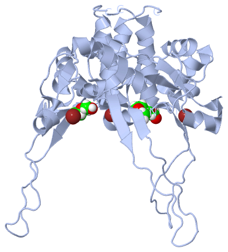
Ligands, Modified Residues, Ions (4, 4)
Asymmetric Unit (4, 4)
|
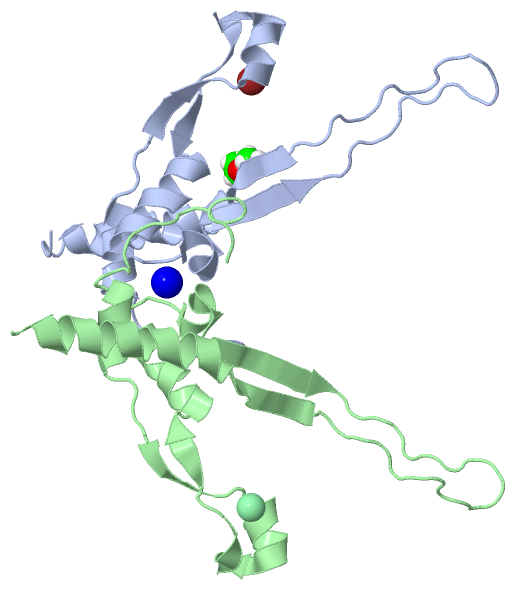
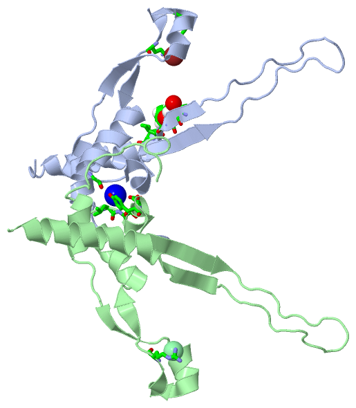
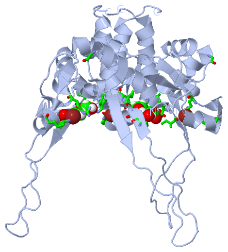
Sites (4, 4)
Asymmetric Unit (4, 4)
|
SS Bonds (0, 0)| (no "SS Bond" information available for 3GUD) |
Cis Peptide Bonds (2, 2)
Asymmetric Unit
|
||||||||||||
SAPs(SNPs)/Variants (0, 0)| (no "SAP(SNP)/Variant" information available for 3GUD) |
PROSITE Motifs (0, 0)| (no "PROSITE Motif" information available for 3GUD) |
Exons (0, 0)| (no "Exon" information available for 3GUD) |
Sequences/Alignments
Asymmetric UnitChain A from PDB Type:PROTEIN Length:119 aligned with Q9FZW3_BPGA1 | Q9FZW3 from UniProtKB/TrEMBL Length:740 Alignment length:119 631 641 651 661 671 681 691 701 711 721 731 Q9FZW3_BPGA1 622 ERHKTDIAPISDKVLDAWEKVKFYQYKFKDAVDEKGEEARYHFGVIAQQIVKVFEDEGLSAFDYGLVGYDEWEATEDEYDSEGNLVEKGREAGNIYSIRPTECQWLEMACMRRKLERLS 740 SCOP domains ----------------------------------------------------------------------------------------------------------------------- SCOP domains CATH domains ----------------------------------------------------------------------------------------------------------------------- CATH domains Pfam domains ----------------------------------------------------------------------------------------------------------------------- Pfam domains SAPs(SNPs) ----------------------------------------------------------------------------------------------------------------------- SAPs(SNPs) PROSITE ----------------------------------------------------------------------------------------------------------------------- PROSITE Transcript ----------------------------------------------------------------------------------------------------------------------- Transcript 3gud A 622 ERHKTDIAPISDKVLDAWEKVKFYQYKFKDAVDEKGEEARYHFGVIAQQIVKVFEDEGLSAFDYGLVGYDEWEATEDEYDSEGNLVEKGREAGNIYSIRPTECQWLEMACMRRKLERLS 740 631 641 651 661 671 681 691 701 711 721 731 Chain B from PDB Type:PROTEIN Length:120 aligned with Q9FZW3_BPGA1 | Q9FZW3 from UniProtKB/TrEMBL Length:740 Alignment length:120 629 639 649 659 669 679 689 699 709 719 729 739 Q9FZW3_BPGA1 620 SDERHKTDIAPISDKVLDAWEKVKFYQYKFKDAVDEKGEEARYHFGVIAQQIVKVFEDEGLSAFDYGLVGYDEWEATEDEYDSEGNLVEKGREAGNIYSIRPTECQWLEMACMRRKLERL 739 SCOP domains ------------------------------------------------------------------------------------------------------------------------ SCOP domains CATH domains ------------------------------------------------------------------------------------------------------------------------ CATH domains Pfam domains ------------------------------------------------------------------------------------------------------------------------ Pfam domains SAPs(SNPs) ------------------------------------------------------------------------------------------------------------------------ SAPs(SNPs) PROSITE ------------------------------------------------------------------------------------------------------------------------ PROSITE Transcript ------------------------------------------------------------------------------------------------------------------------ Transcript 3gud B 620 SDERHKTDIAPISDKVLDAWEKVKFYQYKFKDAVDEKGEEARYHFGVIAQQIVKVFEDEGLSAFDYGLVGYDEWEATEDEYDSEGNLVEKGREAGNIYSIRPTECQWLEMACMRRKLERL 739 629 639 649 659 669 679 689 699 709 719 729 739
|
||||||||||||||||||||
SCOP Domains (0, 0)| (no "SCOP Domain" information available for 3GUD) |
CATH Domains (0, 0)| (no "CATH Domain" information available for 3GUD) |
Pfam Domains (0, 0)| (no "Pfam Domain" information available for 3GUD) |
Gene Ontology (0, 0)|
Asymmetric Unit(hide GO term definitions)
(no "Gene Ontology" information available for 3GUD)
|
Interactive Views
|
|||||||||||||||||||||||||||||||||||||||||||||||||||||||||||||||||||||||||||||||||||||||||||||||||||||||||||||||||||||||||||||||||||||||||||||||||||||||||||||||||||||||||||||||||||||||||||||||
Still Images
|
||||||||||||||||
Databases
|
||||||||||||||||||||||||||||||||||||||||||||||||||||||||||||||||||||||||||||||||||||||||||||||||||||||||||||||||||||||||||||||||||||||||||||||||||||||||||||||||
Analysis Tools
|
|||||||||||||||||||||||||||||||||||||||||||||||||||||||||||||
Entries Sharing at Least One Protein Chain (UniProt ID)
Related Entries Specified in the PDB File
|
|