| molecular function |
|---|
| | GO:0005515 | | protein binding | | Interacting selectively and non-covalently with any protein or protein complex (a complex of two or more proteins that may include other nonprotein molecules). |
| | GO:0031386 | | protein tag | | A molecular function exhibited by a protein that is covalently attached (AKA tagged or conjugated) to another protein where it acts as a marker, recognized by the cellular apparatus to target the tagged protein for some cellular process such as modification, sequestration, transport or degradation. |
| biological process |
|---|
| | GO:0006310 | | DNA recombination | | Any process in which a new genotype is formed by reassortment of genes resulting in gene combinations different from those that were present in the parents. In eukaryotes genetic recombination can occur by chromosome assortment, intrachromosomal recombination, or nonreciprocal interchromosomal recombination. Intrachromosomal recombination occurs by crossing over. In bacteria it may occur by genetic transformation, conjugation, transduction, or F-duction. |
| | GO:0006281 | | DNA repair | | The process of restoring DNA after damage. Genomes are subject to damage by chemical and physical agents in the environment (e.g. UV and ionizing radiations, chemical mutagens, fungal and bacterial toxins, etc.) and by free radicals or alkylating agents endogenously generated in metabolism. DNA is also damaged because of errors during its replication. A variety of different DNA repair pathways have been reported that include direct reversal, base excision repair, nucleotide excision repair, photoreactivation, bypass, double-strand break repair pathway, and mismatch repair pathway. |
| | GO:0006974 | | cellular response to DNA damage stimulus | | Any process that results in a change in state or activity of a cell (in terms of movement, secretion, enzyme production, gene expression, etc.) as a result of a stimulus indicating damage to its DNA from environmental insults or errors during metabolism. |
| | GO:0000724 | | double-strand break repair via homologous recombination | | The error-free repair of a double-strand break in DNA in which the broken DNA molecule is repaired using homologous sequences. A strand in the broken DNA searches for a homologous region in an intact chromosome to serve as the template for DNA synthesis. The restoration of two intact DNA molecules results in the exchange, reciprocal or nonreciprocal, of genetic material between the intact DNA molecule and the broken DNA molecule. |
| | GO:0016925 | | protein sumoylation | | The process in which a SUMO protein (small ubiquitin-related modifier) is conjugated to a target protein via an isopeptide bond between the carboxyl terminus of SUMO with an epsilon-amino group of a lysine residue of the target protein. |
| | GO:0031297 | | replication fork processing | | The process in which a DNA replication fork that has stalled is restored to a functional state and replication is restarted. The stalling may be due to DNA damage, DNA secondary structure, bound proteins, dNTP shortage, or other causes. |
| cellular component |
|---|
| | GO:0005634 | | nucleus | | A membrane-bounded organelle of eukaryotic cells in which chromosomes are housed and replicated. In most cells, the nucleus contains all of the cell's chromosomes except the organellar chromosomes, and is the site of RNA synthesis and processing. In some species, or in specialized cell types, RNA metabolism or DNA replication may be absent. |
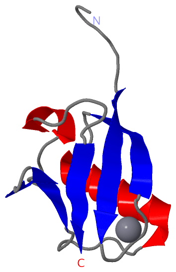
 Description
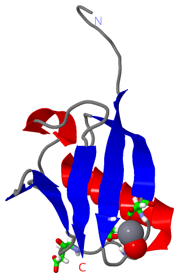