|
|
|
|
 Description
|
|
Compounds
|
||||||||||||||||||||||||||||
Chains, Units
Summary Information (see also Sequences/Alignments below) |
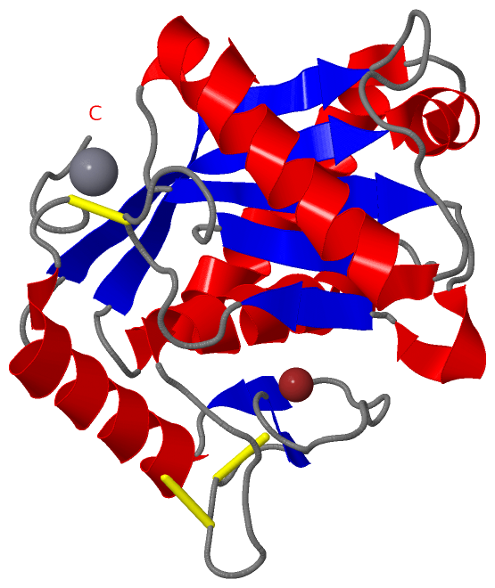
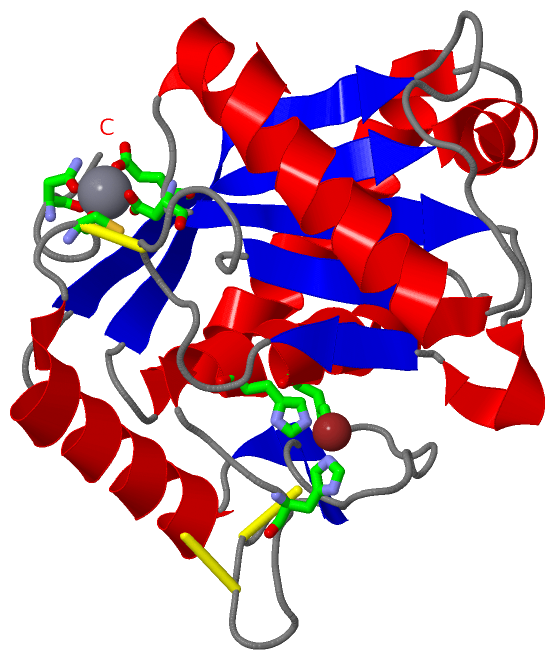
Ligands, Modified Residues, Ions (2, 2)| Asymmetric/Biological Unit (2, 2) |
Sites (2, 2)
Asymmetric Unit (2, 2)
|
SS Bonds (3, 3)
Asymmetric/Biological Unit
|
||||||||||||||||
Cis Peptide Bonds (0, 0)| (no "Cis Peptide Bond" information available for 3GBO) |
SAPs(SNPs)/Variants (0, 0)| (no "SAP(SNP)/Variant" information available for 3GBO) |
PROSITE Motifs (2, 2)
Asymmetric/Biological Unit (2, 2)
|
||||||||||||||||||||||||||||||||
Exons (0, 0)| (no "Exon" information available for 3GBO) |
Sequences/Alignments
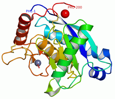
Asymmetric/Biological UnitChain A from PDB Type:PROTEIN Length:200 aligned with VM1BI_BOTMO | P85314 from UniProtKB/Swiss-Prot Length:205 Alignment length:200 14 24 34 44 54 64 74 84 94 104 114 124 134 144 154 164 174 184 194 204 VM1BI_BOTMO 5 FSPRYIELVVVADHGMFKKYNSNLNTIRKWVHEMVNSMNGFYRSVDVTASLANLEVWSKKDLINVQKDSRETLKSFGEWRERDLLPRISHDNAQLLTAIVFDGHTIGRAYTGGMCDPRHSVGVVMDHSPKNLQVAVTMAHELGHNLGMHHDGNQCHCDAASCIMADSLSVVLSYEFSDCSQNQYQTYLTKHNPQCILNEP 204 SCOP domains d3gboa_ A: automated matches SCOP domains CATH domains 3gboA00 A:1-200 Collagenase (Catalytic Domain) CATH domains Pfam domains -------------------------------------------------------------------------------------------------------------------------------------------------------------------------------------------------------- Pfam domains SAPs(SNPs) -------------------------------------------------------------------------------------------------------------------------------------------------------------------------------------------------------- SAPs(SNPs) PROSITE (1) ---ADAM_MEPRO PDB: A:4-200 UniProt: 8-204 PROSITE (1) PROSITE (2) ----------------------------------------------------------------------------------------------------------------------------------------ZINC_PROTE------------------------------------------------------ PROSITE (2) Transcript -------------------------------------------------------------------------------------------------------------------------------------------------------------------------------------------------------- Transcript 3gbo A 1 FSPRHIELVVVADHGMFKKYNSNLNTIRKWVHEMVNSMNGFYRSVDVTASLANLEVWSKKDLINVQKDSRETLKSFGEWRERDLLPRISHDNAQLLTTIVFDGHVIGRAFTGGMCDPRHSVGVVMDHSPKNLQVAVTMAHELGHNLGMHHDGNQCHCDAASCIMADSLSQVLSYEFSDCSQNQYQTYLTKHNPQCILNEP 200 10 20 30 40 50 60 70 80 90 100 110 120 130 140 150 160 170 180 190 200
|
||||||||||||||||||||
SCOP Domains (1, 1)
Asymmetric/Biological Unit
 |
CATH Domains (1, 1)
Asymmetric/Biological Unit
|
Pfam Domains (0, 0)| (no "Pfam Domain" information available for 3GBO) |
Gene Ontology (7, 7)|
Asymmetric/Biological Unit(hide GO term definitions) Chain A (VM1BI_BOTMO | P85314)
|
||||||||||||||||||||||||||||||||||||||||||||||||||||||||||||
Interactive Views
|
||||||||||||||||||||||||||||||||||||||||||||||||||||||||||||||||||||||||||||||||||||||||||||||||||||||||||||||||||||||||||||||||||||
Still Images
|
||||||||||||||||
Databases
|
||||||||||||||||||||||||||||||||||||||||||||||||||||||||||||||||||||||||||||||||||||||||||||||||||||||||||||||||||||||||||||||||||||||||||||||||||||||||||||||||
Analysis Tools
|
|||||||||||||||||||||||||||||||||||||||||||||||||||||||||||||
Entries Sharing at Least One Protein Chain (UniProt ID)
Related Entries Specified in the PDB File
|
|