|
|
|
|
 Description
|
|
Compounds
|
||||||||||||||||||||||||||||||||||||||||||||
Chains, Units
Summary Information (see also Sequences/Alignments below) |
Ligands, Modified Residues, Ions (3, 10)
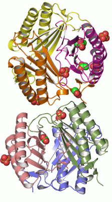
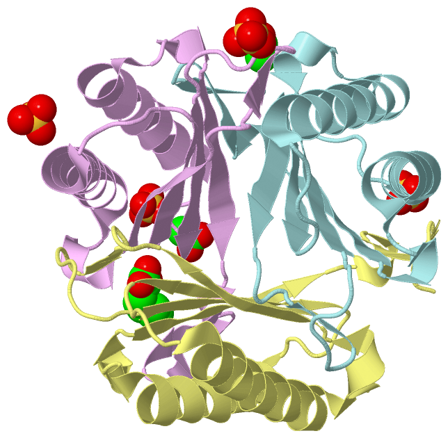
Asymmetric Unit (3, 10)
|
Sites (10, 10)
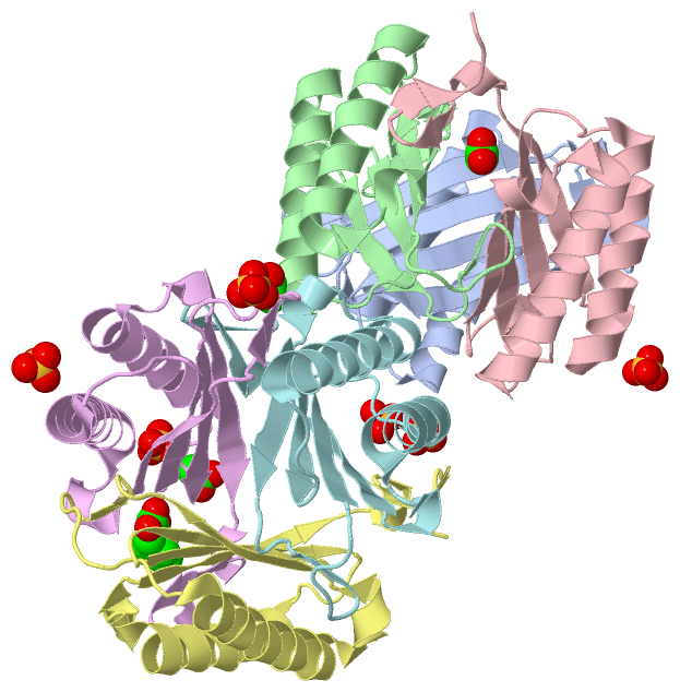
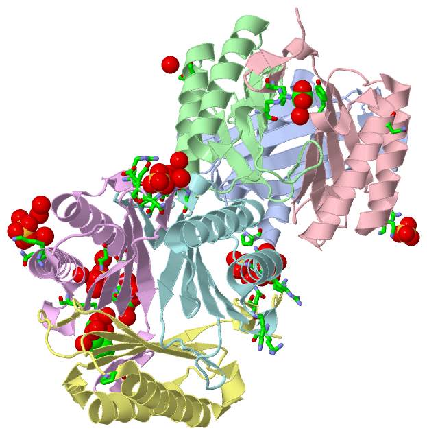
Asymmetric Unit (10, 10)
|
SS Bonds (0, 0)| (no "SS Bond" information available for 3GAC) |
Cis Peptide Bonds (0, 0)| (no "Cis Peptide Bond" information available for 3GAC) |
SAPs(SNPs)/Variants (0, 0)| (no "SAP(SNP)/Variant" information available for 3GAC) |
PROSITE Motifs (0, 0)| (no "PROSITE Motif" information available for 3GAC) |
Exons (0, 0)| (no "Exon" information available for 3GAC) |
Sequences/Alignments
Asymmetric UnitChain A from PDB Type:PROTEIN Length:115 aligned with Q1HEA2_PLAYO | Q1HEA2 from UniProtKB/TrEMBL Length:116 Alignment length:115 11 21 31 41 51 61 71 81 91 101 111 Q1HEA2_PLAYO 2 PCCELITNISIPDDKAQNALSEIEDAISNVLGKPVAYIMSNYDYQKNLRFSGSNEGYCFVRLTSIGGINRSNNSSLADKITKILSNHLGVKPRRVYIEFRDCSAQNFAFSGSLFG 116 SCOP domains d3gaca_ A: Microphage migration inhibition factor (MIF) SCOP domains CATH domains ------------------------------------------------------------------------------------------------------------------- CATH domains Pfam domains ------------------------------------------------------------------------------------------------------------------- Pfam domains SAPs(SNPs) ------------------------------------------------------------------------------------------------------------------- SAPs(SNPs) PROSITE ------------------------------------------------------------------------------------------------------------------- PROSITE Transcript ------------------------------------------------------------------------------------------------------------------- Transcript 3gac A 2 PCCELITNISIPDDKAQNALSEIEDAISNVLGKPVAYIMSNYDYQKNLRFSGSNEGYCFVRLTSIGGINRSNNSSLADKITKILSNHLGVKPRRVYIEFRDCSAQNFAFSGSLFG 116 11 21 31 41 51 61 71 81 91 101 111 Chain B from PDB Type:PROTEIN Length:116 aligned with Q1HEA2_PLAYO | Q1HEA2 from UniProtKB/TrEMBL Length:116 Alignment length:116 116 11 21 31 41 51 61 71 81 91 101 111 | Q1HEA2_PLAYO 2 PCCELITNISIPDDKAQNALSEIEDAISNVLGKPVAYIMSNYDYQKNLRFSGSNEGYCFVRLTSIGGINRSNNSSLADKITKILSNHLGVKPRRVYIEFRDCSAQNFAFSGSLFG- - SCOP domains d3gacb_ B: Microphage migration inhibition factor (MIF) SCOP domains CATH domains -------------------------------------------------------------------------------------------------------------------- CATH domains Pfam domains -------------------------------------------------------------------------------------------------------------------- Pfam domains SAPs(SNPs) -------------------------------------------------------------------------------------------------------------------- SAPs(SNPs) PROSITE -------------------------------------------------------------------------------------------------------------------- PROSITE Transcript -------------------------------------------------------------------------------------------------------------------- Transcript 3gac B 2 PCCELITNISIPDDKAQNALSEIEDAISNVLGKPVAYIMSNYDYQKNLRFSGSNEGYCFVRLTSIGGINRSNNSSLADKITKILSNHLGVKPRRVYIEFRDCSAQNFAFSGSLFGL 117 11 21 31 41 51 61 71 81 91 101 111 Chain C from PDB Type:PROTEIN Length:117 aligned with Q1HEA2_PLAYO | Q1HEA2 from UniProtKB/TrEMBL Length:116 Alignment length:117 116 11 21 31 41 51 61 71 81 91 101 111 | Q1HEA2_PLAYO 2 PCCELITNISIPDDKAQNALSEIEDAISNVLGKPVAYIMSNYDYQKNLRFSGSNEGYCFVRLTSIGGINRSNNSSLADKITKILSNHLGVKPRRVYIEFRDCSAQNFAFSGSLFG-- - SCOP domains d3gacc_ C: Microphage migration inhibition factor (MIF) SCOP domains CATH domains --------------------------------------------------------------------------------------------------------------------- CATH domains Pfam domains --------------------------------------------------------------------------------------------------------------------- Pfam domains SAPs(SNPs) --------------------------------------------------------------------------------------------------------------------- SAPs(SNPs) PROSITE --------------------------------------------------------------------------------------------------------------------- PROSITE Transcript --------------------------------------------------------------------------------------------------------------------- Transcript 3gac C 2 PCCELITNISIPDDKAQNALSEIEDAISNVLGKPVAYIMSNYDYQKNLRFSGSNEGYCFVRLTSIGGINRSNNSSLADKITKILSNHLGVKPRRVYIEFRDCSAQNFAFSGSLFGLE 118 11 21 31 41 51 61 71 81 91 101 111 Chain D from PDB Type:PROTEIN Length:115 aligned with Q1HEA2_PLAYO | Q1HEA2 from UniProtKB/TrEMBL Length:116 Alignment length:115 11 21 31 41 51 61 71 81 91 101 111 Q1HEA2_PLAYO 2 PCCELITNISIPDDKAQNALSEIEDAISNVLGKPVAYIMSNYDYQKNLRFSGSNEGYCFVRLTSIGGINRSNNSSLADKITKILSNHLGVKPRRVYIEFRDCSAQNFAFSGSLFG 116 SCOP domains d3gacd_ D: Microphage migration inhibition factor (MIF) SCOP domains CATH domains ------------------------------------------------------------------------------------------------------------------- CATH domains Pfam domains ------------------------------------------------------------------------------------------------------------------- Pfam domains SAPs(SNPs) ------------------------------------------------------------------------------------------------------------------- SAPs(SNPs) PROSITE ------------------------------------------------------------------------------------------------------------------- PROSITE Transcript ------------------------------------------------------------------------------------------------------------------- Transcript 3gac D 2 PCCELITNISIPDDKAQNALSEIEDAISNVLGKPVAYIMSNYDYQKNLRFSGSNEGYCFVRLTSIGGINRSNNSSLADKITKILSNHLGVKPRRVYIEFRDCSAQNFAFSGSLFG 116 11 21 31 41 51 61 71 81 91 101 111 Chain E from PDB Type:PROTEIN Length:115 aligned with Q1HEA2_PLAYO | Q1HEA2 from UniProtKB/TrEMBL Length:116 Alignment length:115 11 21 31 41 51 61 71 81 91 101 111 Q1HEA2_PLAYO 2 PCCELITNISIPDDKAQNALSEIEDAISNVLGKPVAYIMSNYDYQKNLRFSGSNEGYCFVRLTSIGGINRSNNSSLADKITKILSNHLGVKPRRVYIEFRDCSAQNFAFSGSLFG 116 SCOP domains d3gace_ E: Microphage migration inhibition factor (MIF) SCOP domains CATH domains ------------------------------------------------------------------------------------------------------------------- CATH domains Pfam domains ------------------------------------------------------------------------------------------------------------------- Pfam domains SAPs(SNPs) ------------------------------------------------------------------------------------------------------------------- SAPs(SNPs) PROSITE ------------------------------------------------------------------------------------------------------------------- PROSITE Transcript ------------------------------------------------------------------------------------------------------------------- Transcript 3gac E 2 PCCELITNISIPDDKAQNALSEIEDAISNVLGKPVAYIMSNYDYQKNLRFSGSNEGYCFVRLTSIGGINRSNNSSLADKITKILSNHLGVKPRRVYIEFRDCSAQNFAFSGSLFG 116 11 21 31 41 51 61 71 81 91 101 111 Chain F from PDB Type:PROTEIN Length:116 aligned with Q1HEA2_PLAYO | Q1HEA2 from UniProtKB/TrEMBL Length:116 Alignment length:116 116 11 21 31 41 51 61 71 81 91 101 111 | Q1HEA2_PLAYO 2 PCCELITNISIPDDKAQNALSEIEDAISNVLGKPVAYIMSNYDYQKNLRFSGSNEGYCFVRLTSIGGINRSNNSSLADKITKILSNHLGVKPRRVYIEFRDCSAQNFAFSGSLFG- - SCOP domains d3gacf_ F: Microphage migration inhibition factor (MIF) SCOP domains CATH domains -------------------------------------------------------------------------------------------------------------------- CATH domains Pfam domains -------------------------------------------------------------------------------------------------------------------- Pfam domains SAPs(SNPs) -------------------------------------------------------------------------------------------------------------------- SAPs(SNPs) PROSITE -------------------------------------------------------------------------------------------------------------------- PROSITE Transcript -------------------------------------------------------------------------------------------------------------------- Transcript 3gac F 2 PCCELITNISIPDDKAQNALSEIEDAISNVLGKPVAYIMSNYDYQKNLRFSGSNEGYCFVRLTSIGGINRSNNSSLADKITKILSNHLGVKPRRVYIEFRDCSAQNFAFSGSLFGL 117 11 21 31 41 51 61 71 81 91 101 111
|
||||||||||||||||||||
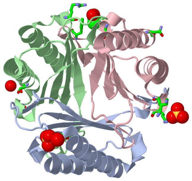
SCOP Domains (1, 6)
Asymmetric Unit
 |
CATH Domains (0, 0)| (no "CATH Domain" information available for 3GAC) |
Pfam Domains (0, 0)| (no "Pfam Domain" information available for 3GAC) |
Gene Ontology (0, 0)|
Asymmetric Unit(hide GO term definitions)
(no "Gene Ontology" information available for 3GAC)
|
Interactive Views
|
||||||||||||||||||||||||||||||||||||||||||||||||||||||||||||||||||||||||||||||||||||||||||||||||||||||||||||||||||||||||||||||||||||||||||||||||||||||||||||||||||||||||||||||||||||||||||||||||||||||||||||||||||||||||||
Still Images
|
||||||||||||||||
Databases
|
||||||||||||||||||||||||||||||||||||||||||||||||||||||||||||||||||||||||||||||||||||||||||||||||||||||||||||||||||||||||||||||||||||||||||||||||||||||||||||||||
Analysis Tools
|
|||||||||||||||||||||||||||||||||||||||||||||||||||||||||||||
Entries Sharing at Least One Protein Chain (UniProt ID)
Related Entries Specified in the PDB File
|
|