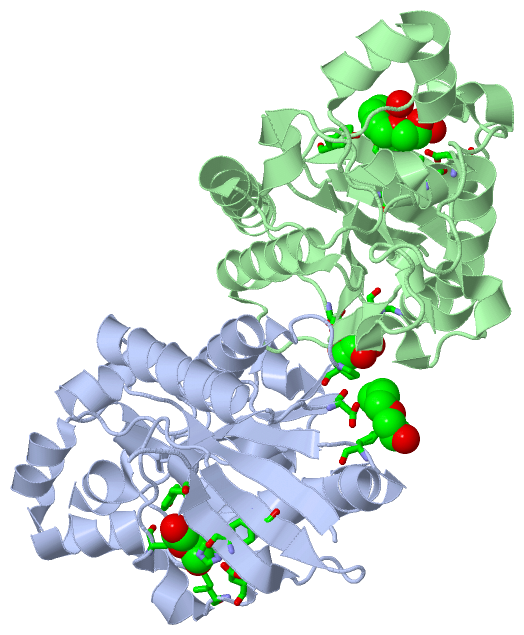
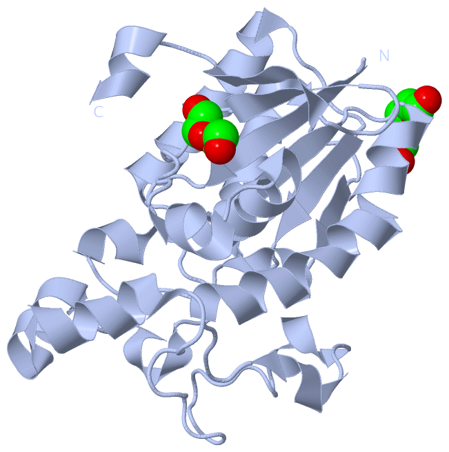
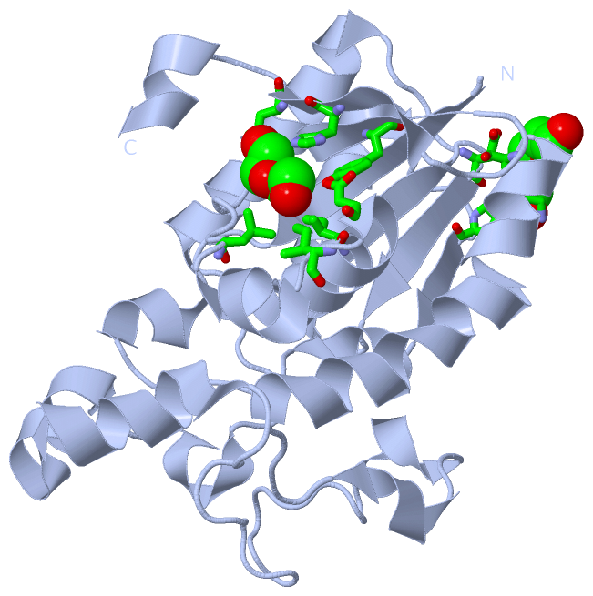
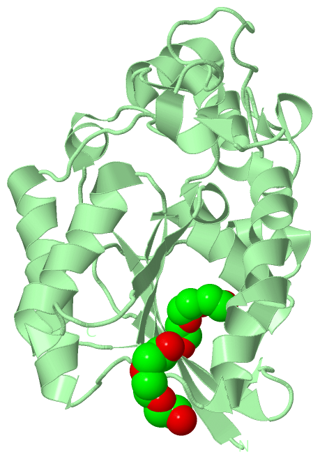
Show PDB file:   
         Plain Text   HTML   (compressed file size)
QuickSearch:   
by PDB,NDB,UniProt,PROSITE Code or Search Term(s)  
(-)Asymmetric Unit
(-)Asym. Unit - sites
(-)Biological Unit 1
(-)Biol. Unit 1 - sites
(-)Biological Unit 2
(-)Biological Unit 3
collapse expand < >
Image Asymmetric Unit
Asymmetric Unit  (Jmol Viewer)
Image Asym. Unit - sites
Asym. Unit - sites  (Jmol Viewer)
Image Biological Unit 1
Biological Unit 1  (Jmol Viewer)
Image Biol. Unit 1 - sites
Biol. Unit 1 - sites  (Jmol Viewer)
Image Biological Unit 2
Biological Unit 2  (Jmol Viewer)
Image Biological Unit 3
Biological Unit 3  (Jmol Viewer)

(-) Description

Title :  CRYSTAL STRUCTURE OF PHOSPHOGLYCEROMUTASE FROM BURKHOLDERIA PSEUDOMALLEI 1710B
 
Authors :  Seattle Structural Genomics Center For Infectious Disease (S
Date :  23 Oct 08  (Deposition) - 11 Nov 08  (Release) - 05 Oct 11  (Revision)
Method :  X-RAY DIFFRACTION
Resolution :  2.10
Chains :  Asym. Unit :  A,B
Biol. Unit 1:  A  (1x)
Biol. Unit 2:  B  (1x)
Biol. Unit 3:  A,B  (1x)
Keywords :  Ssgcid, Phosphoglyceromutase, Burkholderia Pseudomallei, Glycolysis, Isomerase, Structural Genomics, Seattle Structural Genomics Center For Infectious Disease (Keyword Search: [Gene Ontology, PubMed, Web (Google))
 
Reference :  D. R. Davies, B. L. Staker, J. A. Abendroth, T. E. Edwards, R. Hartley, J. Leonard, H. Kim, A. L. Rychel, S. N. Hewitt, P. J. Myler, L. J. Stewart
An Ensemble Of Structures Of Burkholderia Pseudomallei 2, 3-Bisphosphoglycerate-Dependent Phosphoglycerate Mutase.
Acta Crystallogr. , Sect. F V. 67 1044 2011
PubMed-ID: 21904048  |  Reference-DOI: 10.1107/S1744309111030405

(-) Compounds

Molecule 1 - 2,3-BISPHOSPHOGLYCERATE-DEPENDENT PHOSPHOGLYCERATE MUTASE
    ChainsA, B
    EC Number5.4.2.1
    EngineeredYES
    Expression SystemESCHERICHIA COLI
    Expression System PlasmidAVA0421
    Expression System StrainBL21(DE3)
    Expression System Taxid562
    Expression System Vector TypePLASMID
    GeneGPMA, BURPS1710B_0662
    Organism ScientificBURKHOLDERIA PSEUDOMALLEI 1710B
    Organism Taxid320372
    Strain1719B
    SynonymPHOSPHOGLYCEROMUTASE, PGAM, BPG-DEPENDENT PGAM, DPGM

 Structural Features

(-) Chains, Units

  12
Asymmetric Unit AB
Biological Unit 1 (1x)A 
Biological Unit 2 (1x) B
Biological Unit 3 (1x)AB

Summary Information (see also Sequences/Alignments below)

(-) Ligands, Modified Residues, Ions  (1, 4)

Asymmetric Unit (1, 4)
No.NameCountTypeFull Name
1PG44Ligand/IonTETRAETHYLENE GLYCOL
Biological Unit 1 (1, 2)
No.NameCountTypeFull Name
1PG42Ligand/IonTETRAETHYLENE GLYCOL
Biological Unit 2 (1, 2)
No.NameCountTypeFull Name
1PG42Ligand/IonTETRAETHYLENE GLYCOL
Biological Unit 3 (1, 4)
No.NameCountTypeFull Name
1PG44Ligand/IonTETRAETHYLENE GLYCOL

(-) Sites  (4, 4)

Asymmetric Unit (4, 4)
No.NameEvidenceResiduesDescription
1AC1SOFTWAREASP A:51 , PRO A:78 , LYS A:175BINDING SITE FOR RESIDUE PG4 A 250
2AC2SOFTWAREILE A:190 , ASP A:194 , ILE A:196 , LEU A:204 , TYR A:214 , HIS A:225 , TYR A:227BINDING SITE FOR RESIDUE PG4 A 251
3AC3SOFTWAREASP B:51 , GLY B:174 , LYS B:175BINDING SITE FOR RESIDUE PG4 B 250
4AC4SOFTWAREILE B:190 , ASP B:194 , TYR B:214 , HIS B:225 , TYR B:227 , HOH B:357 , HOH B:426BINDING SITE FOR RESIDUE PG4 B 251

(-) SS Bonds  (0, 0)

(no "SS Bond" information available for 3EZN)

(-) Cis Peptide Bonds  (0, 0)

(no "Cis Peptide Bond" information available for 3EZN)

 Sequence-Structure Mapping

(-) SAPs(SNPs)/Variants  (0, 0)

(no "SAP(SNP)/Variant" information available for 3EZN)

(-) PROSITE Motifs  (1, 2)

Asymmetric Unit (1, 2)
 PROSITEUniProtKBPDB
No.IDACDescriptionIDLocationCountLocation
1PG_MUTASEPS00175 Phosphoglycerate mutase family phosphohistidine signature.GPMA_BURP16-15
 
  2A:6-15
B:6-15
Biological Unit 1 (1, 1)
 PROSITEUniProtKBPDB
No.IDACDescriptionIDLocationCountLocation
1PG_MUTASEPS00175 Phosphoglycerate mutase family phosphohistidine signature.GPMA_BURP16-15
 
  1A:6-15
-
Biological Unit 2 (1, 1)
 PROSITEUniProtKBPDB
No.IDACDescriptionIDLocationCountLocation
1PG_MUTASEPS00175 Phosphoglycerate mutase family phosphohistidine signature.GPMA_BURP16-15
 
  1-
B:6-15
Biological Unit 3 (1, 2)
 PROSITEUniProtKBPDB
No.IDACDescriptionIDLocationCountLocation
1PG_MUTASEPS00175 Phosphoglycerate mutase family phosphohistidine signature.GPMA_BURP16-15
 
  2A:6-15
B:6-15

(-) Exons   (0, 0)

(no "Exon" information available for 3EZN)

(-) Sequences/Alignments

Asymmetric Unit
   Reformat: Number of residues per line =  ('0' or empty: single-line sequence representation)
  Number of residues per labelling interval =   
  UniProt sequence: complete  aligned part    
   Show mapping: SCOP domains CATH domains Pfam domains Secondary structure (by author)
SAPs(SNPs) PROSITE motifs Exons
(details for a mapped element are shown in a popup box when the mouse pointer rests over it)
Chain A from PDB  Type:PROTEIN  Length:236
 aligned with GPMA_BURP1 | Q3JWH7 from UniProtKB/Swiss-Prot  Length:249

    Alignment length:236
                                    10        20        30        40        50        60        70        80        90       100       110       120       130       140       150       160       170       180       190       200       210       220       230      
           GPMA_BURP1     1 MYKLVLIRHGESTWNKENRFTGWVDVDLTEQGNREARQAGQLLKEAGYTFDIAYTSVLKRAIRTLWHVQDQMDLMYVPVVHSWRLNERHYGALSGLNKAETAAKYGDEQVLVWRRSYDTPPPALEPGDERAPYADPRYAKVPREQLPLTECLKDTVARVLPLWNESIAPAVKAGKQVLIAAHGNSLRALIKYLDGISDADIVGLNIPNGVPLVYELDESLTPIRHYYLGDQEAIAK 236
               SCOP domains d3ezna_ A: automated matches                                                                                                                                                                                                                 SCOP domains
               CATH domains 3eznA00 A:1-236 Phosphoglycerate mutase-like                                                                                                                                                                                                 CATH domains
               Pfam domains -------------------------------------------------------------------------------------------------------------------------------------------------------------------------------------------------------------------------------------------- Pfam domains
         Sec.struct. author .eeeeeee...hhhhhh...........hhhhhhhhhhhhhhhhhh.....eeee..hhhhhhhhhhhhhhhh.....eee.hhhh...hhhhh..hhhhhhhhhhhhhhhhhhhh..................hhhhh..hhhhh....hhhhhhhhhhhhhhhhhhhhhhh...eeeeehhhhhhhhhhhhh..hhhhhh.........eeeee.....eeeeee..hhhhhhh Sec.struct. author
                 SAPs(SNPs) -------------------------------------------------------------------------------------------------------------------------------------------------------------------------------------------------------------------------------------------- SAPs(SNPs)
                    PROSITE -----PG_MUTASE ----------------------------------------------------------------------------------------------------------------------------------------------------------------------------------------------------------------------------- PROSITE
                 Transcript -------------------------------------------------------------------------------------------------------------------------------------------------------------------------------------------------------------------------------------------- Transcript
                 3ezn A   1 MYKLVLIRHGESTWNKENRFTGWVDVDLTEQGNREARQAGQLLKEAGYTFDIAYTSVLKRAIRTLWHVQDQMDLMYVPVVHSWRLNERHYGALSGLNKAETAAKYGDEQVLVWRRSYDTPPPALEPGDERAPYADPRYAKVPREQLPLTECLKDTVARVLPLWNESIAPAVKAGKQVLIAAHGNSLRALIKYLDGISDADIVGLNIPNGVPLVYELDESLTPIRHYYLGDQEAIAK 236
                                    10        20        30        40        50        60        70        80        90       100       110       120       130       140       150       160       170       180       190       200       210       220       230      

Chain B from PDB  Type:PROTEIN  Length:231
 aligned with GPMA_BURP1 | Q3JWH7 from UniProtKB/Swiss-Prot  Length:249

    Alignment length:231
                             1                                                                                                                                                                                                                                     
                             |       9        19        29        39        49        59        69        79        89        99       109       119       129       139       149       159       169       179       189       199       209       219       229 
           GPMA_BURP1     - -MYKLVLIRHGESTWNKENRFTGWVDVDLTEQGNREARQAGQLLKEAGYTFDIAYTSVLKRAIRTLWHVQDQMDLMYVPVVHSWRLNERHYGALSGLNKAETAAKYGDEQVLVWRRSYDTPPPALEPGDERAPYADPRYAKVPREQLPLTECLKDTVARVLPLWNESIAPAVKAGKQVLIAAHGNSLRALIKYLDGISDADIVGLNIPNGVPLVYELDESLTPIRHYYLGD 230
               SCOP domains d3eznb_ B: automated matches                                                                                                                                                                                                            SCOP domains
               CATH domains 3eznB00 B:0-230 Phosphoglycerate mutase-like                                                                                                                                                                                            CATH domains
               Pfam domains --------------------------------------------------------------------------------------------------------------------------------------------------------------------------------------------------------------------------------------- Pfam domains
         Sec.struct. author .eeeeeeee...hhhhhh...........hhhhhhhhhhhhhhhhhh.....eeee..hhhhhhhhhhhhhhhh.....eee.hhhh...hhhhh..hhhhhhhhhhhhhhhhhhhh..................hhhhh..hhhhh....hhhhhhhhhhhhhhhhhhhhhhh...eeeeehhhhhhhhhhhhh..hhhhhh.........eeeeee....eeeeee... Sec.struct. author
                 SAPs(SNPs) --------------------------------------------------------------------------------------------------------------------------------------------------------------------------------------------------------------------------------------- SAPs(SNPs)
                    PROSITE ------PG_MUTASE ----------------------------------------------------------------------------------------------------------------------------------------------------------------------------------------------------------------------- PROSITE
                 Transcript --------------------------------------------------------------------------------------------------------------------------------------------------------------------------------------------------------------------------------------- Transcript
                 3ezn B   0 HMYKLVLIRHGESTWNKENRFTGWVDVDLTEQGNREARQAGQLLKEAGYTFDIAYTSVLKRAIRTLWHVQDQMDLMYVPVVHSWRLNERHYGALSGLNKAETAAKYGDEQVLVWRRSYDTPPPALEPGDERAPYADPRYAKVPREQLPLTECLKDTVARVLPLWNESIAPAVKAGKQVLIAAHGNSLRALIKYLDGISDADIVGLNIPNGVPLVYELDESLTPIRHYYLGD 230
                                     9        19        29        39        49        59        69        79        89        99       109       119       129       139       149       159       169       179       189       199       209       219       229 

   Legend:   → Mismatch (orange background)
  - → Gap (green background, '-', border residues have a numbering label)
    → Modified Residue (blue background, lower-case, 'x' indicates undefined single-letter code, labelled with number + name)
  x → Chemical Group (purple background, 'x', labelled with number + name, e.g. ACE or NH2)
  extra numbering lines below/above indicate numbering irregularities and modified residue names etc., number ends below/above '|'

 Classification and Annotation

(-) SCOP Domains  (1, 2)

Asymmetric Unit

(-) CATH Domains  (1, 2)

Asymmetric Unit
(-)
Class: Alpha Beta (26913)

(-) Pfam Domains  (0, 0)

(no "Pfam Domain" information available for 3EZN)

(-) Gene Ontology  (8, 8)

Asymmetric Unit(hide GO term definitions)
Chain A,B   (GPMA_BURP1 | Q3JWH7)
molecular function
    GO:0046538    2,3-bisphosphoglycerate-dependent phosphoglycerate mutase activity    Catalysis of the reaction: 2-phospho-D-glycerate = 3-phospho-D-glycerate; this reaction requires the cofactor 2,3-bisphosphoglycerate.
    GO:0003824    catalytic activity    Catalysis of a biochemical reaction at physiological temperatures. In biologically catalyzed reactions, the reactants are known as substrates, and the catalysts are naturally occurring macromolecular substances known as enzymes. Enzymes possess specific binding sites for substrates, and are usually composed wholly or largely of protein, but RNA that has catalytic activity (ribozyme) is often also regarded as enzymatic.
    GO:0016868    intramolecular transferase activity, phosphotransferases    Catalysis of the transfer of a phosphate group from one position to another within a single molecule.
    GO:0016853    isomerase activity    Catalysis of the geometric or structural changes within one molecule. Isomerase is the systematic name for any enzyme of EC class 5.
    GO:0004619    phosphoglycerate mutase activity    Catalysis of the reaction: 2-phospho-D-glycerate = 3-phospho-D-glycerate.
biological process
    GO:0006094    gluconeogenesis    The formation of glucose from noncarbohydrate precursors, such as pyruvate, amino acids and glycerol.
    GO:0006096    glycolytic process    The chemical reactions and pathways resulting in the breakdown of a carbohydrate into pyruvate, with the concomitant production of a small amount of ATP and the reduction of NAD(P) to NAD(P)H. Glycolysis begins with the metabolism of a carbohydrate to generate products that can enter the pathway and ends with the production of pyruvate. Pyruvate may be converted to acetyl-coenzyme A, ethanol, lactate, or other small molecules.
    GO:0008152    metabolic process    The chemical reactions and pathways, including anabolism and catabolism, by which living organisms transform chemical substances. Metabolic processes typically transform small molecules, but also include macromolecular processes such as DNA repair and replication, and protein synthesis and degradation.

 Visualization

(-) Interactive Views

Asymmetric Unit
  Complete Structure
    Jena3D(integrated viewing of ligand, site, SAP, PROSITE, SCOP information)
    WebMol | AstexViewer[tm]@PDBe
(Java Applets, require no local installation except for Java; loading may be slow)
    STRAP
(Java WebStart application, automatic local installation, requires Java; full application with system access!)
    RasMol
(require local installation)
    Molscript (VRML)
(requires installation of a VRML viewer; select preferred view via VRML and generate a mono or stereo PDF format file)
 
  Ligands, Modified Residues, Ions
    PG4  [ RasMol | Jena3D ]  +environment [ RasMol | Jena3D ]
 
  Sites
    AC1  [ RasMol ]  +environment [ RasMol ]
    AC2  [ RasMol ]  +environment [ RasMol ]
    AC3  [ RasMol ]  +environment [ RasMol ]
    AC4  [ RasMol ]  +environment [ RasMol ]
 
  Cis Peptide Bonds
(no "Cis Peptide Bonds" information available for 3ezn)
 
Biological Units
  Complete Structure
    Biological Unit 1  [ Jena3D ]
    Biological Unit 2  [ Jena3D ]
    Biological Unit 3  [ Jena3D ]

(-) Still Images

Jmol
  protein: cartoon or spacefill or dots and stick; nucleic acid: cartoon and stick; ligands: spacefill; active site: stick
Molscript
  protein, nucleic acid: cartoon; ligands: spacefill; active site: ball and stick

 Databases and Analysis Tools

(-) Databases

Access by PDB/NDB ID
  3ezn
    Family and Domain InformationProDom | SYSTERS
    General Structural InformationGlycoscienceDB | MMDB | NDB | OCA | PDB | PDBe | PDBj | PDBsum | PDBWiki | PQS | PROTEOPEDIA
    Orientation in MembranesOPM
    Protein SurfaceSURFACE
    Secondary StructureDSSP (structure derived) | HSSP (homology derived)
    Structural GenomicsGeneCensus
    Structural NeighboursCE | VAST
    Structure ClassificationCATH | Dali | SCOP
    Validation and Original DataBMRB Data View | BMRB Restraints Grid | EDS | PROCHECK | RECOORD | WHAT_CHECK
 
Access by UniProt ID/Accession number
  GPMA_BURP1 | Q3JWH7
    Comparative Protein Structure ModelsModBase
    Genomic InformationEnsembl
    Protein-protein InteractionDIP
    Sequence, Family and Domain InformationInterPro | Pfam | SMART | UniProtKB/SwissProt
 
Access by Enzyme Classificator   (EC Number)
  5.4.2.1
    General Enzyme InformationBRENDA | EC-PDB | Enzyme | IntEnz
    PathwayKEGG | MetaCyc
 
Access by Disease Identifier   (MIM ID)
  (no 'MIM ID' available)
    Disease InformationOMIM
 
Access by GenAge ID
  (no 'GenAge ID' available)
    Age Related InformationGenAge

(-) Analysis Tools

Access by PDB/NDB ID
    Domain InformationXDom
    Interatomic Contacts of Structural UnitsCSU
    Ligand-protein ContactsLPC
    Protein CavitiescastP
    Sequence and Secondary StructurePDBCartoon
    Structure AlignmentSTRAP(Java WebStart application, automatic local installation, requires Java; full application with system access!)
    Structure and Sequence BrowserSTING
 
Access by UniProt ID/Accession number
  GPMA_BURP1 | Q3JWH7
    Protein Disorder PredictionDisEMBL | FoldIndex | GLOBPLOT (for more information see DisProt)

 Related Entries

(-) Entries Sharing at Least One Protein Chain (UniProt ID)

UniProtKB/Swiss-Prot
        GPMA_BURP1 | Q3JWH73fdz 3gp3 3gp5 3gw8 3lnt

(-) Related Entries Specified in the PDB File

3fdz 3gp3 3gp5 3gw8