|
|
|
|
 Description
|
|
Compounds
|
||||||||||||||||||||||||||||||||||||||||||||||||||||
Chains, Units
Summary Information (see also Sequences/Alignments below) |
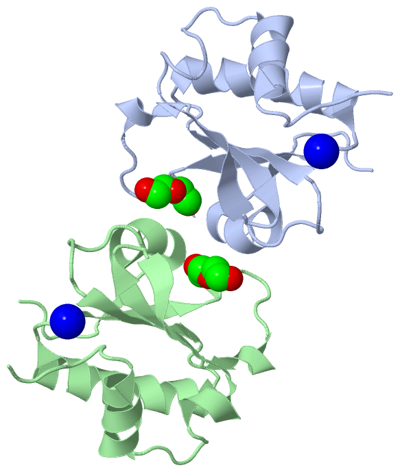
Ligands, Modified Residues, Ions (2, 4)| Asymmetric Unit (2, 4) Biological Unit 1 (1, 2) Biological Unit 2 (1, 1) Biological Unit 3 (1, 1) |
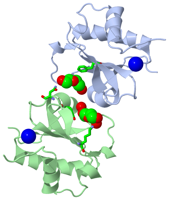
Sites (2, 2)
Asymmetric Unit (2, 2)
|
SS Bonds (0, 0)| (no "SS Bond" information available for 3EVI) |
Cis Peptide Bonds (2, 2)
Asymmetric Unit
|
||||||||||||
SAPs(SNPs)/Variants (0, 0)| (no "SAP(SNP)/Variant" information available for 3EVI) |
PROSITE Motifs (0, 0)| (no "PROSITE Motif" information available for 3EVI) |
Exons (0, 0)| (no "Exon" information available for 3EVI) |
Sequences/Alignments
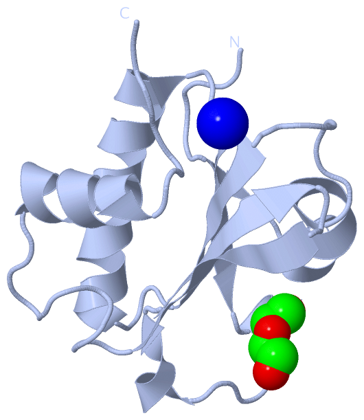
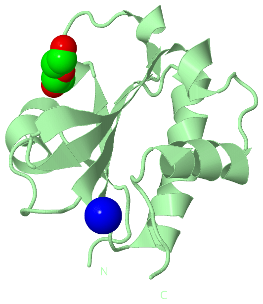
Asymmetric UnitChain A from PDB Type:PROTEIN Length:118 aligned with PDCL2_HUMAN | Q8N4E4 from UniProtKB/Swiss-Prot Length:241 Alignment length:118 97 107 117 127 137 147 157 167 177 187 197 PDCL2_HUMAN 88 KFGELREISGNQYVNEVTNAEEDVWVIIHLYRSSIPMCLLVNQHLSLLARKFPETKFVKAIVNSCIQHYHDNCLPTIFVYKNGQIEAKFIGIIECGGINLKLEELEWKLAEVGAIQTD 205 SCOP domains ---------------------------------------------------------------------------------------------------------------------- SCOP domains CATH domains ---------------------------------------------------------------------------------------------------------------------- CATH domains Pfam domains ---------------------------------------------------------------------------------------------------------------------- Pfam domains SAPs(SNPs) ---------------------------------------------------------------------------------------------------------------------- SAPs(SNPs) PROSITE ---------------------------------------------------------------------------------------------------------------------- PROSITE Transcript ---------------------------------------------------------------------------------------------------------------------- Transcript 3evi A 88 KFGELREISGNQYVNEVTNAEEDVWVIIHLYRSSIPMCLLVNQHLSLLARKFPETKFVKAIVNSCIQHYHDNCLPTIFVYKNGQIEAKFIGIIECGGINLKLEELEWKLAEVGAIQTD 205 97 107 117 127 137 147 157 167 177 187 197 Chain B from PDB Type:PROTEIN Length:118 aligned with PDCL2_HUMAN | Q8N4E4 from UniProtKB/Swiss-Prot Length:241 Alignment length:118 97 107 117 127 137 147 157 167 177 187 197 PDCL2_HUMAN 88 KFGELREISGNQYVNEVTNAEEDVWVIIHLYRSSIPMCLLVNQHLSLLARKFPETKFVKAIVNSCIQHYHDNCLPTIFVYKNGQIEAKFIGIIECGGINLKLEELEWKLAEVGAIQTD 205 SCOP domains ---------------------------------------------------------------------------------------------------------------------- SCOP domains CATH domains ---------------------------------------------------------------------------------------------------------------------- CATH domains Pfam domains ---------------------------------------------------------------------------------------------------------------------- Pfam domains SAPs(SNPs) ---------------------------------------------------------------------------------------------------------------------- SAPs(SNPs) PROSITE ---------------------------------------------------------------------------------------------------------------------- PROSITE Transcript ---------------------------------------------------------------------------------------------------------------------- Transcript 3evi B 88 KFGELREISGNQYVNEVTNAEEDVWVIIHLYRSSIPMCLLVNQHLSLLARKFPETKFVKAIVNSCIQHYHDNCLPTIFVYKNGQIEAKFIGIIECGGINLKLEELEWKLAEVGAIQTD 205 97 107 117 127 137 147 157 167 177 187 197
|
||||||||||||||||||||
SCOP Domains (0, 0)| (no "SCOP Domain" information available for 3EVI) |
CATH Domains (0, 0)| (no "CATH Domain" information available for 3EVI) |
Pfam Domains (0, 0)| (no "Pfam Domain" information available for 3EVI) |
Gene Ontology (3, 3)|
Asymmetric Unit(hide GO term definitions) Chain A,B (PDCL2_HUMAN | Q8N4E4)
|
||||||||||||||||||||||||||||||||||||
Interactive Views
|
||||||||||||||||||||||||||||||||||||||||||||||||||||||||||||||||||||||||||||||||||||||||||||||||||||||||||||||||||||||||||||||||||||||||||||||||||||||||||||||||||||||||
Still Images
|
||||||||||||||||
Databases
|
||||||||||||||||||||||||||||||||||||||||||||||||||||||||||||||||||||||||||||||||||||||||||||||||||||||||||||||||||||||||||||||||||||||||||||||||||||||||||||||||
Analysis Tools
|
|||||||||||||||||||||||||||||||||||||||||||||||||||||||||||||
Entries Sharing at Least One Protein Chain (UniProt ID)
Related Entries Specified in the PDB File
|
|