Show PDB file:   
         Plain Text   HTML   (compressed file size)
QuickSearch:   
by PDB,NDB,UniProt,PROSITE Code or Search Term(s)  
(-)Asym./Biol. Unit
(-)Asym./Biol. Unit - sites
collapse expand < >
Image Asym./Biol. Unit
Asym./Biol. Unit  (Jmol Viewer)
Image Asym./Biol. Unit - sites
Asym./Biol. Unit - sites  (Jmol Viewer)

(-) Description

Title :  CRYSTAL STRUCTURE OF APO ZEBRAFISH ILEAL BILE ACID-BINDING PROTEIN
 
Authors :  S. Capaldi, G. Saccomani, D. Fessas, M. Signorelli, M. Perduca, H. L. Mo
Date :  23 Sep 08  (Deposition) - 13 Jan 09  (Release) - 13 Jul 11  (Revision)
Method :  X-RAY DIFFRACTION
Resolution :  1.60
Chains :  Asym./Biol. Unit :  A
Keywords :  Ileal Bile Acid-Bindign Protein, Zebrafish, Cholic Acid, Lipid- Binding, Transport, Lipid Binding Protein (Keyword Search: [Gene Ontology, PubMed, Web (Google))
 
Reference :  S. Capaldi, G. Saccomani, D. Fessas, M. Signorelli, M. Perduca, H. L. Monaco
The X-Ray Structure Of Zebrafish (Danio Rerio) Ileal Bile Acid-Binding Protein Reveals The Presence Of Binding Sites On The Surface Of The Protein Molecule.
J. Mol. Biol. V. 385 99 2009
PubMed-ID: 18952094  |  Reference-DOI: 10.1016/J.JMB.2008.10.007

(-) Compounds

Molecule 1 - ILEAL BILE ACID-BINDING PROTEIN
    ChainsA
    EngineeredYES
    Expression SystemESCHERICHIA COLI
    Expression System PlasmidPQE50
    Expression System StrainSG13009
    Expression System Taxid562
    Expression System Vector TypePLASMID
    Organism CommonLEOPARD DANIO,ZEBRA DANIO,ZEBRA FISH
    Organism ScientificDANIO RERIO
    Organism Taxid7955
    SynonymFATTY ACID BINDING PROTEIN 6, ILEAL (GASTROTROPIN)

 Structural Features

(-) Chains, Units

  1
Asymmetric/Biological Unit A

Summary Information (see also Sequences/Alignments below)

(-) Ligands, Modified Residues, Ions  (1, 1)

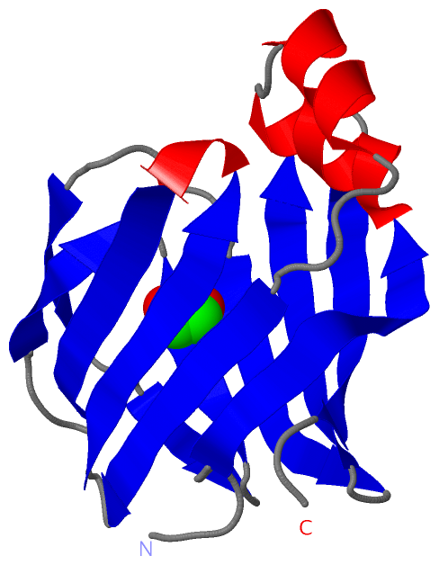
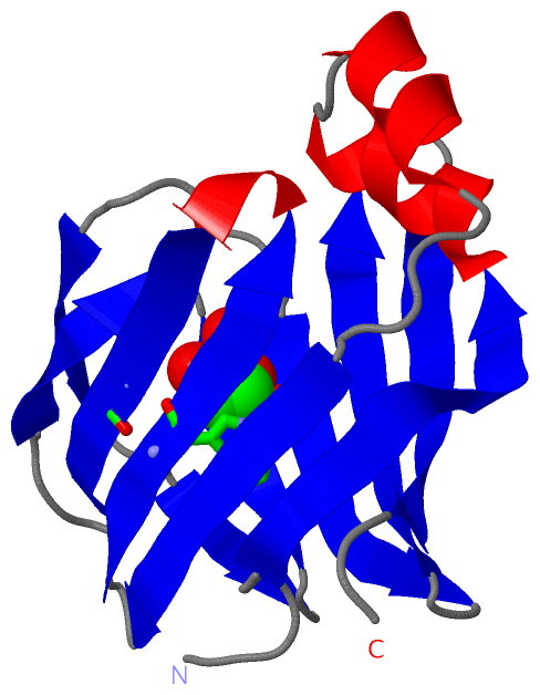
Asymmetric/Biological Unit (1, 1)
No.NameCountTypeFull Name
1EDO1Ligand/Ion1,2-ETHANEDIOL

(-) Sites  (1, 1)

Asymmetric Unit (1, 1)
No.NameEvidenceResiduesDescription
1AC1SOFTWARETRP A:49 , ASN A:61 , ILE A:92 , HOH A:208 , HOH A:255 , HOH A:299BINDING SITE FOR RESIDUE EDO A 200

(-) SS Bonds  (0, 0)

(no "SS Bond" information available for 3ELX)

(-) Cis Peptide Bonds  (1, 1)

Asymmetric/Biological Unit
No.Residues
1Gly A:-1 -Ser A:0

 Sequence-Structure Mapping

(-) SAPs(SNPs)/Variants  (0, 0)

(no "SAP(SNP)/Variant" information available for 3ELX)

(-) PROSITE Motifs  (0, 0)

(no "PROSITE Motif" information available for 3ELX)

(-) Exons   (0, 0)

(no "Exon" information available for 3ELX)

(-) Sequences/Alignments

Asymmetric/Biological Unit
   Reformat: Number of residues per line =  ('0' or empty: single-line sequence representation)
  Number of residues per labelling interval =   
  UniProt sequence: complete  aligned part    
   Show mapping: SCOP domains CATH domains Pfam domains Secondary structure (by author)
SAPs(SNPs) PROSITE motifs Exons
(details for a mapped element are shown in a popup box when the mouse pointer rests over it)
Chain A from PDB  Type:PROTEIN  Length:134
 aligned with Q6IMW5_DANRE | Q6IMW5 from UniProtKB/TrEMBL  Length:131

    Alignment length:134
                             1                                                                                                                               131  
                             |       9        19        29        39        49        59        69        79        89        99       109       119       129 |  
         Q6IMW5_DANRE     - -MAFNGKWETESQEGYEPFCKLIGIPDDVIAKGRDFKLVTEIVQNGDDFTWTQYYPNNHVVTNKFIVGKESDMETVGGKKFKGIVSMEGGKLTISFPKYQQTTEISGGKLVETSTASGAQGTAVLVRTSKKV--   -
               SCOP domains d3elxa_ A: automated matches                                                                                                           SCOP domains
               CATH domains 3elxA00 A:-1-132  [code=2.40.128.20, no name defined]                                                                                  CATH domains
               Pfam domains -------------------------------------------------------------------------------------------------------------------------------------- Pfam domains
         Sec.struct. author .....eeeeeeeeehhhhhhhhh..hhhhhhhh.....eeeeeee..eeeeeeeehhheeeeeeee....eeee.....eeee..eee..eeeee....eeeeeee..eeeeeeeee....eeeeeeeeee... Sec.struct. author
                 SAPs(SNPs) -------------------------------------------------------------------------------------------------------------------------------------- SAPs(SNPs)
                    PROSITE -------------------------------------------------------------------------------------------------------------------------------------- PROSITE
                 Transcript -------------------------------------------------------------------------------------------------------------------------------------- Transcript
                 3elx A  -1 GSAFNGKWETESQEGYEPFCKLIGIPDDVIAKGRDFKLVTEIVQNGDDFTWTQYYPNNHVVTNKFIVGKESDMETVGGKKFKGIVSMEGGKLTISFPKYQQTTEISGGKLVETSTASGAQGTAVLVRTSKKVLV 132
                                     8        18        28        38        48        58        68        78        88        98       108       118       128    

   Legend:   → Mismatch (orange background)
  - → Gap (green background, '-', border residues have a numbering label)
    → Modified Residue (blue background, lower-case, 'x' indicates undefined single-letter code, labelled with number + name)
  x → Chemical Group (purple background, 'x', labelled with number + name, e.g. ACE or NH2)
  extra numbering lines below/above indicate numbering irregularities and modified residue names etc., number ends below/above '|'

 Classification and Annotation

(-) SCOP Domains  (1, 1)

Asymmetric/Biological Unit

(-) CATH Domains  (1, 1)

Asymmetric/Biological Unit
(-)
Class: Mainly Beta (13760)

(-) Pfam Domains  (0, 0)

(no "Pfam Domain" information available for 3ELX)

(-) Gene Ontology  (6, 6)

Asymmetric/Biological Unit(hide GO term definitions)
Chain A   (Q6IMW5_DANRE | Q6IMW5)
molecular function
    GO:0032052    bile acid binding    Interacting selectively and non-covalently with bile acids, any of a group of steroid carboxylic acids occurring in bile.
    GO:0008289    lipid binding    Interacting selectively and non-covalently with a lipid.
    GO:0005215    transporter activity    Enables the directed movement of substances (such as macromolecules, small molecules, ions) into, out of or within a cell, or between cells.
biological process
    GO:0008206    bile acid metabolic process    The chemical reactions and pathways involving bile acids, any of a group of steroid carboxylic acids occurring in bile, where they are present as the sodium salts of their amides with glycine or taurine.
    GO:0006810    transport    The directed movement of substances (such as macromolecules, small molecules, ions) or cellular components (such as complexes and organelles) into, out of or within a cell, or between cells, or within a multicellular organism by means of some agent such as a transporter, pore or motor protein.
cellular component
    GO:0005737    cytoplasm    All of the contents of a cell excluding the plasma membrane and nucleus, but including other subcellular structures.

 Visualization

(-) Interactive Views

Asymmetric/Biological Unit
  Complete Structure
    Jena3D(integrated viewing of ligand, site, SAP, PROSITE, SCOP information)
    WebMol | AstexViewer[tm]@PDBe
(Java Applets, require no local installation except for Java; loading may be slow)
    STRAP
(Java WebStart application, automatic local installation, requires Java; full application with system access!)
    RasMol
(require local installation)
    Molscript (VRML)
(requires installation of a VRML viewer; select preferred view via VRML and generate a mono or stereo PDF format file)
 
  Ligands, Modified Residues, Ions
    EDO  [ RasMol | Jena3D ]  +environment [ RasMol | Jena3D ]
 
  Sites
    AC1  [ RasMol ]  +environment [ RasMol ]
 
  Cis Peptide Bonds
    Gly A:-1 - Ser A:0   [ RasMol ]  
 

(-) Still Images

Jmol
  protein: cartoon or spacefill or dots and stick; nucleic acid: cartoon and stick; ligands: spacefill; active site: stick
Molscript
  protein, nucleic acid: cartoon; ligands: spacefill; active site: ball and stick

 Databases and Analysis Tools

(-) Databases

Access by PDB/NDB ID
  3elx
    Family and Domain InformationProDom | SYSTERS
    General Structural InformationGlycoscienceDB | MMDB | NDB | OCA | PDB | PDBe | PDBj | PDBsum | PDBWiki | PQS | PROTEOPEDIA
    Orientation in MembranesOPM
    Protein SurfaceSURFACE
    Secondary StructureDSSP (structure derived) | HSSP (homology derived)
    Structural GenomicsGeneCensus
    Structural NeighboursCE | VAST
    Structure ClassificationCATH | Dali | SCOP
    Validation and Original DataBMRB Data View | BMRB Restraints Grid | EDS | PROCHECK | RECOORD | WHAT_CHECK
 
Access by UniProt ID/Accession number
  Q6IMW5_DANRE | Q6IMW5
    Comparative Protein Structure ModelsModBase
    Genomic InformationEnsembl
    Protein-protein InteractionDIP
    Sequence, Family and Domain InformationInterPro | Pfam | SMART | UniProtKB/TrEMBL
 
Access by Enzyme Classificator   (EC Number)
  (no 'Enzyme Classificator' available)
    General Enzyme InformationBRENDA | EC-PDB | Enzyme | IntEnz
    PathwayKEGG | MetaCyc
 
Access by Disease Identifier   (MIM ID)
  (no 'MIM ID' available)
    Disease InformationOMIM
 
Access by GenAge ID
  (no 'GenAge ID' available)
    Age Related InformationGenAge

(-) Analysis Tools

Access by PDB/NDB ID
    Domain InformationXDom
    Interatomic Contacts of Structural UnitsCSU
    Ligand-protein ContactsLPC
    Protein CavitiescastP
    Sequence and Secondary StructurePDBCartoon
    Structure AlignmentSTRAP(Java WebStart application, automatic local installation, requires Java; full application with system access!)
    Structure and Sequence BrowserSTING
 
Access by UniProt ID/Accession number
  Q6IMW5_DANRE | Q6IMW5
    Protein Disorder PredictionDisEMBL | FoldIndex | GLOBPLOT (for more information see DisProt)

 Related Entries

(-) Entries Sharing at Least One Protein Chain (UniProt ID)

UniProtKB/TrEMBL
        Q6IMW5_DANRE | Q6IMW53elz 3em0

(-) Related Entries Specified in the PDB File

3elz CRYSTAL STRUCTURE OF ZEBRAFISH ILEAL BILE ACID-BINDIN PROTEIN COMPLEXED WITH CHOLIC ACID (CRYSTAL FORM A).
3em0 CRYSTAL STRUCTURE OF ZEBRAFISH ILEAL BILE ACID-BINDIN PROTEIN COMPLEXED WITH CHOLIC ACID (CRYSTAL FORM B).