|
|
|
|
 Description
|
|
Compounds
|
||||||||||||||||||||||||||||||||||||||||||||||||
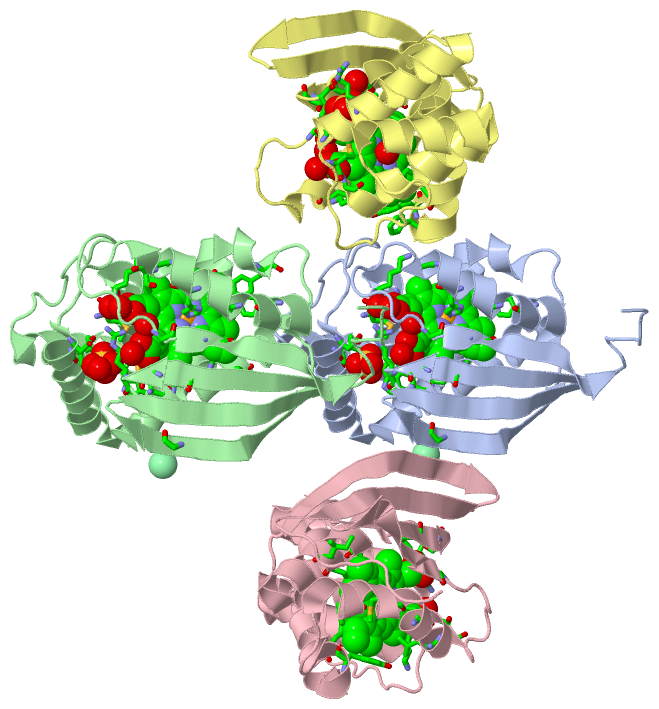
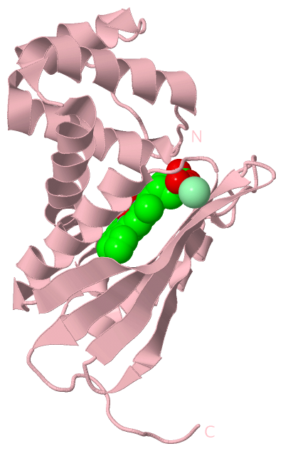
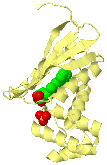
Chains, Units
Summary Information (see also Sequences/Alignments below) |
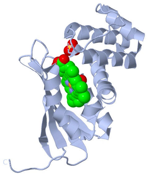
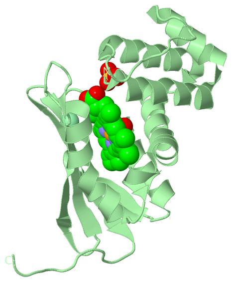
Ligands, Modified Residues, Ions (4, 13)
Asymmetric Unit (4, 13)
|
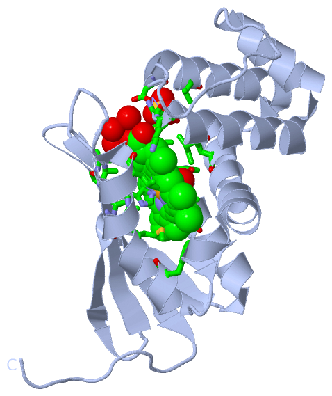
Sites (13, 13)
Asymmetric Unit (13, 13)
|
SS Bonds (0, 0)| (no "SS Bond" information available for 3EEE) |
Cis Peptide Bonds (0, 0)| (no "Cis Peptide Bond" information available for 3EEE) |
SAPs(SNPs)/Variants (0, 0)| (no "SAP(SNP)/Variant" information available for 3EEE) |
PROSITE Motifs (0, 0)| (no "PROSITE Motif" information available for 3EEE) |
Exons (0, 0)| (no "Exon" information available for 3EEE) |
Sequences/Alignments
Asymmetric UnitChain A from PDB Type:PROTEIN Length:188 aligned with Q8RBX6_CALS4 | Q8RBX6 from UniProtKB/TrEMBL Length:602 Alignment length:188 10 20 30 40 50 60 70 80 90 100 110 120 130 140 150 160 170 180 Q8RBX6_CALS4 1 MKGTIVGTWIKTLRDLYGNDVVDESLKSVGWEPDRVITPLEDIDDDEVRRIFAKVSEKTGKNVNEIWREVGRQNIKTFSEWFPSYFAGRRLVNFLMMMDEVHLQLTKMIKGATPPRLIAKPVAKDAIEMEYVSKRKMYDYFLGLIEGSSKFFKEEISVEEVERGEKDGFSRLKVRIKFKNPVFEYKKN 188 SCOP domains d3eeea_ A: Methyl-accepting chemotaxis protein SCOP domains CATH domains 3eeeA00 A:1-188 H-NOX domain CATH domains Pfam domains -------------------------------------------------------------------------------------------------------------------------------------------------------------------------------------------- Pfam domains SAPs(SNPs) -------------------------------------------------------------------------------------------------------------------------------------------------------------------------------------------- SAPs(SNPs) PROSITE -------------------------------------------------------------------------------------------------------------------------------------------------------------------------------------------- PROSITE Transcript -------------------------------------------------------------------------------------------------------------------------------------------------------------------------------------------- Transcript 3eee A 1 MKGTIVGTWIKTLRDLYGNDVVDESLKSVGWEPDRVITPLEDIDDDEVRRIFAKVSEKTGKNVNEIWREVGRQNIKTFSEWFPSYFAGRRLVNFLMMMDEVHLQLTKMIKGATPARLIAKPVAKDAIEMEYVSKRKMYDYFLGLIEGSSKFFKEEISVEEVERGEKDGFSRLKVRIKFKNPVFEYKKN 188 10 20 30 40 50 60 70 80 90 100 110 120 130 140 150 160 170 180 Chain B from PDB Type:PROTEIN Length:188 aligned with Q8RBX6_CALS4 | Q8RBX6 from UniProtKB/TrEMBL Length:602 Alignment length:188 10 20 30 40 50 60 70 80 90 100 110 120 130 140 150 160 170 180 Q8RBX6_CALS4 1 MKGTIVGTWIKTLRDLYGNDVVDESLKSVGWEPDRVITPLEDIDDDEVRRIFAKVSEKTGKNVNEIWREVGRQNIKTFSEWFPSYFAGRRLVNFLMMMDEVHLQLTKMIKGATPPRLIAKPVAKDAIEMEYVSKRKMYDYFLGLIEGSSKFFKEEISVEEVERGEKDGFSRLKVRIKFKNPVFEYKKN 188 SCOP domains d3eeeb_ B: Methyl-accepting chemotaxis protein SCOP domains CATH domains 3eeeB00 B:1-188 H-NOX domain CATH domains Pfam domains -------------------------------------------------------------------------------------------------------------------------------------------------------------------------------------------- Pfam domains SAPs(SNPs) -------------------------------------------------------------------------------------------------------------------------------------------------------------------------------------------- SAPs(SNPs) PROSITE -------------------------------------------------------------------------------------------------------------------------------------------------------------------------------------------- PROSITE Transcript -------------------------------------------------------------------------------------------------------------------------------------------------------------------------------------------- Transcript 3eee B 1 MKGTIVGTWIKTLRDLYGNDVVDESLKSVGWEPDRVITPLEDIDDDEVRRIFAKVSEKTGKNVNEIWREVGRQNIKTFSEWFPSYFAGRRLVNFLMMMDEVHLQLTKMIKGATPARLIAKPVAKDAIEMEYVSKRKMYDYFLGLIEGSSKFFKEEISVEEVERGEKDGFSRLKVRIKFKNPVFEYKKN 188 10 20 30 40 50 60 70 80 90 100 110 120 130 140 150 160 170 180 Chain C from PDB Type:PROTEIN Length:188 aligned with Q8RBX6_CALS4 | Q8RBX6 from UniProtKB/TrEMBL Length:602 Alignment length:188 10 20 30 40 50 60 70 80 90 100 110 120 130 140 150 160 170 180 Q8RBX6_CALS4 1 MKGTIVGTWIKTLRDLYGNDVVDESLKSVGWEPDRVITPLEDIDDDEVRRIFAKVSEKTGKNVNEIWREVGRQNIKTFSEWFPSYFAGRRLVNFLMMMDEVHLQLTKMIKGATPPRLIAKPVAKDAIEMEYVSKRKMYDYFLGLIEGSSKFFKEEISVEEVERGEKDGFSRLKVRIKFKNPVFEYKKN 188 SCOP domains d3eeec_ C: Methyl-accepting chemotaxis protein SCOP domains CATH domains 3eeeC00 C:1-188 H-NOX domain CATH domains Pfam domains -------------------------------------------------------------------------------------------------------------------------------------------------------------------------------------------- Pfam domains SAPs(SNPs) -------------------------------------------------------------------------------------------------------------------------------------------------------------------------------------------- SAPs(SNPs) PROSITE -------------------------------------------------------------------------------------------------------------------------------------------------------------------------------------------- PROSITE Transcript -------------------------------------------------------------------------------------------------------------------------------------------------------------------------------------------- Transcript 3eee C 1 MKGTIVGTWIKTLRDLYGNDVVDESLKSVGWEPDRVITPLEDIDDDEVRRIFAKVSEKTGKNVNEIWREVGRQNIKTFSEWFPSYFAGRRLVNFLMMMDEVHLQLTKMIKGATPARLIAKPVAKDAIEMEYVSKRKMYDYFLGLIEGSSKFFKEEISVEEVERGEKDGFSRLKVRIKFKNPVFEYKKN 188 10 20 30 40 50 60 70 80 90 100 110 120 130 140 150 160 170 180 Chain D from PDB Type:PROTEIN Length:188 aligned with Q8RBX6_CALS4 | Q8RBX6 from UniProtKB/TrEMBL Length:602 Alignment length:188 10 20 30 40 50 60 70 80 90 100 110 120 130 140 150 160 170 180 Q8RBX6_CALS4 1 MKGTIVGTWIKTLRDLYGNDVVDESLKSVGWEPDRVITPLEDIDDDEVRRIFAKVSEKTGKNVNEIWREVGRQNIKTFSEWFPSYFAGRRLVNFLMMMDEVHLQLTKMIKGATPPRLIAKPVAKDAIEMEYVSKRKMYDYFLGLIEGSSKFFKEEISVEEVERGEKDGFSRLKVRIKFKNPVFEYKKN 188 SCOP domains d3eeed_ D: Methyl-accepting chemotaxis protein SCOP domains CATH domains 3eeeD00 D:1-188 H-NOX domain CATH domains Pfam domains -------------------------------------------------------------------------------------------------------------------------------------------------------------------------------------------- Pfam domains SAPs(SNPs) -------------------------------------------------------------------------------------------------------------------------------------------------------------------------------------------- SAPs(SNPs) PROSITE -------------------------------------------------------------------------------------------------------------------------------------------------------------------------------------------- PROSITE Transcript -------------------------------------------------------------------------------------------------------------------------------------------------------------------------------------------- Transcript 3eee D 1 MKGTIVGTWIKTLRDLYGNDVVDESLKSVGWEPDRVITPLEDIDDDEVRRIFAKVSEKTGKNVNEIWREVGRQNIKTFSEWFPSYFAGRRLVNFLMMMDEVHLQLTKMIKGATPARLIAKPVAKDAIEMEYVSKRKMYDYFLGLIEGSSKFFKEEISVEEVERGEKDGFSRLKVRIKFKNPVFEYKKN 188 10 20 30 40 50 60 70 80 90 100 110 120 130 140 150 160 170 180
|
||||||||||||||||||||
SCOP Domains (1, 4)
Asymmetric Unit
 |
CATH Domains (1, 4)
Asymmetric Unit
|
Pfam Domains (0, 0)| (no "Pfam Domain" information available for 3EEE) |
Gene Ontology (6, 6)|
Asymmetric Unit(hide GO term definitions) Chain A,B,C,D (Q8RBX6_CALS4 | Q8RBX6)
|
||||||||||||||||||||||||||||||||||||||||||||||||||||||
Interactive Views
|
||||||||||||||||||||||||||||||||||||||||||||||||||||||||||||||||||||||||||||||||||||||||||||||||||||||||||||||||||||||||||||||||||||||||||||||||||||||||||||||||||||||||||||||||||||||||||||||||||||||||||||||||||||||||||||||||||||||||||||||||||||||||||||||||
Still Images
|
||||||||||||||||
Databases
|
||||||||||||||||||||||||||||||||||||||||||||||||||||||||||||||||||||||||||||||||||||||||||||||||||||||||||||||||||||||||||||||||||||||||||||||||||||||||||||||||
Analysis Tools
|
|||||||||||||||||||||||||||||||||||||||||||||||||||||||||||||
Entries Sharing at Least One Protein Chain (UniProt ID)
Related Entries Specified in the PDB File
|
|