|
|
|
|
 Description
|
|
Compounds
|
||||||||||||||||||||||||||||||||||||||||||||||||
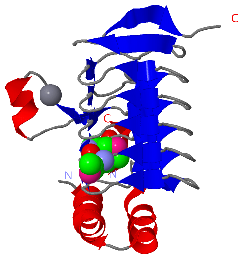
Chains, Units
Summary Information (see also Sequences/Alignments below) |
Ligands, Modified Residues, Ions (2, 5)| Asymmetric Unit (2, 5) Biological Unit 1 (1, 9) |
Sites (2, 2)
Asymmetric Unit (2, 2)
|
SS Bonds (0, 0)| (no "SS Bond" information available for 3ECT) |
Cis Peptide Bonds (2, 2)
Asymmetric Unit
|
||||||||||||
SAPs(SNPs)/Variants (0, 0)| (no "SAP(SNP)/Variant" information available for 3ECT) |
PROSITE Motifs (0, 0)| (no "PROSITE Motif" information available for 3ECT) |
Exons (0, 0)| (no "Exon" information available for 3ECT) |
Sequences/Alignments
Asymmetric UnitChain A from PDB Type:PROTEIN Length:176 aligned with Q9KLB0_VIBCH | Q9KLB0 from UniProtKB/TrEMBL Length:192 Alignment length:180 15 25 35 45 55 65 75 85 95 105 115 125 135 145 155 165 175 185 Q9KLB0_VIBCH 6 LEKMLKGEHFDGASAEIEALRSQAGRLKLEINQSLDEAERYALQRELFGHLGHKSCVQPPFHCEFGKTIRIGDHTFINMNVVMLDGAPITIGDHVLIGPSTQFYTASHSLDYRRRQAWETICKPIVIEDDVWIGGNVVINQGVTIGARSVVAANSVVNQDVPPDTLVGGTPARILRSLKD 185 SCOP domains d3ecta_ A: automated matches SCOP domains CATH domains 3ectA00 A:6-185 Hexapeptide repeat proteins CATH domains Pfam domains ------------------------------------------------------------------------------------------------------------------------------------------------------------------------------------ Pfam domains SAPs(SNPs) ------------------------------------------------------------------------------------------------------------------------------------------------------------------------------------ SAPs(SNPs) PROSITE ------------------------------------------------------------------------------------------------------------------------------------------------------------------------------------ PROSITE Transcript ------------------------------------------------------------------------------------------------------------------------------------------------------------------------------------ Transcript 3ect A 6 LEKmLKG-H---ASAEIEALRSQAGRLKLEINQSLDEAERYALQRELFGHLGHKSCVQPPFHCEFGKTIRIGDHTFINmNVVmLDGAPITIGDHVLIGPSTQFYTASHSLDYRRRQAWETICKPIVIEDDVWIGGNVVINQGVTIGARSVVAANSVVNQDVPPDTLVGGTPARILRSLKD 185 | | |- | 25 35 45 55 65 75 85 | 95 105 115 125 135 145 155 165 175 185 | 12 | 18 84-MSE 9-MSE4 88-MSE
|
||||||||||||||||||||
SCOP Domains (1, 1)
Asymmetric Unit

|
CATH Domains (1, 1)
Asymmetric Unit
|
Pfam Domains (0, 0)| (no "Pfam Domain" information available for 3ECT) |
Gene Ontology (3, 3)|
Asymmetric Unit(hide GO term definitions) Chain A (Q9KLB0_VIBCH | Q9KLB0)
|
||||||||||||||||||||||||
Interactive Views
|
||||||||||||||||||||||||||||||||||||||||||||||||||||||||||||||||||||||||||||||||||||||||||||||||||||||||||||||||||||||||||||||||||||||||||||||||||||||||||||||
Still Images
|
||||||||||||||||
Databases
|
||||||||||||||||||||||||||||||||||||||||||||||||||||||||||||||||||||||||||||||||||||||||||||||||||||||||||||||||||||||||||||||||||||||||||||||||||||||||||||||||
Analysis Tools
|
|||||||||||||||||||||||||||||||||||||||||||||||||||||||||||||
Entries Sharing at Least One Protein Chain (UniProt ID)
Related Entries Specified in the PDB File
|
|