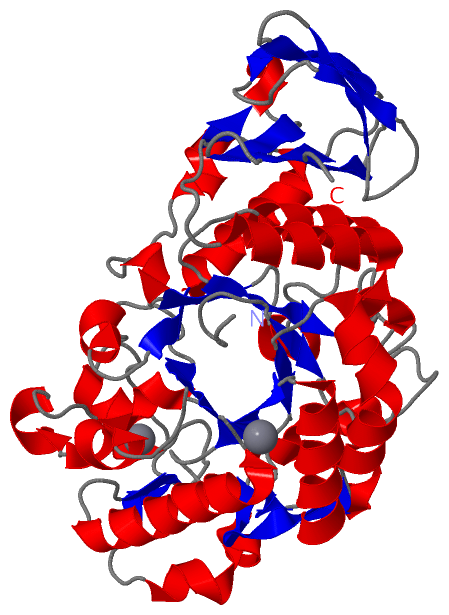
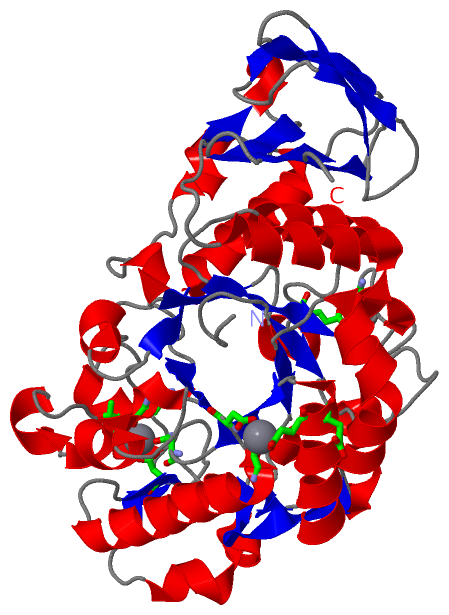
Show PDB file:   
         Plain Text   HTML   (compressed file size)
QuickSearch:   
by PDB,NDB,UniProt,PROSITE Code or Search Term(s)  
(-)Asym./Biol. Unit
(-)Asym./Biol. Unit - sites
collapse expand < >
Image Asym./Biol. Unit
Asym./Biol. Unit  (Jmol Viewer)
Image Asym./Biol. Unit - sites
Asym./Biol. Unit - sites  (Jmol Viewer)

(-) Description

Title :  CRYSTAL STRUCTURE OF NATIVE ALPHA-AMYLASE FROM BACILLUS SP. KR-8104
 
Authors :  J. Alikhajeh, K. Khajeh, B. Ranjbar, M. Naderi-Manesh, H. Naderi- Manesh, C. J. Chen
Date :  03 Jun 08  (Deposition) - 17 Jun 08  (Release) - 24 Feb 09  (Revision)
Method :  X-RAY DIFFRACTION
Resolution :  2.78
Chains :  Asym./Biol. Unit :  A
Keywords :  Crystal Structure, Native Alpha-Amylase, Hydrolase (Keyword Search: [Gene Ontology, PubMed, Web (Google))
 
Reference :  J. Alikhajeh, K. Khajeh, B. Ranjbar, M. Naderi-Manesh, H. Naderi-Manesh, C. J. Chen
Crystal Structure Of Native Alpha-Amylase From Bacillus Sp. Kr-8104
To Be Published
PubMed: search
(for further references see the PDB file header)

(-) Compounds

Molecule 1 - ALPHA-AMYLASE
    ChainsA
    EC Number3.2.1.1
    EngineeredYES
    Expression SystemESCHERICHIA COLI
    Expression System PlasmidPET28A
    Expression System StrainBL21
    Expression System Taxid511693
    Expression System Vector TypePLASMID
    Organism ScientificBACILLUS SP. KR8104
    Organism Taxid535911

 Structural Features

(-) Chains, Units

  1
Asymmetric/Biological Unit A

Summary Information (see also Sequences/Alignments below)

(-) Ligands, Modified Residues, Ions  (1, 3)

Asymmetric/Biological Unit (1, 3)
No.NameCountTypeFull Name
1CA3Ligand/IonCALCIUM ION

(-) Sites  (3, 3)

Asymmetric Unit (3, 3)
No.NameEvidenceResiduesDescription
1AC1SOFTWAREGLU A:88 , GLY A:169 , ASP A:171BINDING SITE FOR RESIDUE CA A 1001
2AC2SOFTWAREGLU A:89 , GLU A:276 , GLY A:313BINDING SITE FOR RESIDUE CA A 1002
3AC3SOFTWAREASN A:101 , THR A:137 , ASP A:146 , HIS A:180 , HOH A:1006BINDING SITE FOR RESIDUE CA A 1004

(-) SS Bonds  (0, 0)

(no "SS Bond" information available for 3DC0)

(-) Cis Peptide Bonds  (0, 0)

(no "Cis Peptide Bond" information available for 3DC0)

 Sequence-Structure Mapping

(-) SAPs(SNPs)/Variants  (0, 0)

(no "SAP(SNP)/Variant" information available for 3DC0)

(-) PROSITE Motifs  (0, 0)

(no "PROSITE Motif" information available for 3DC0)

(-) Exons   (0, 0)

(no "Exon" information available for 3DC0)

(-) Sequences/Alignments

Asymmetric/Biological Unit
   Reformat: Number of residues per line =  ('0' or empty: single-line sequence representation)
  Number of residues per labelling interval =   
  UniProt sequence: complete  aligned part    
   Show mapping: SCOP domains CATH domains Pfam domains Secondary structure (by author)
SAPs(SNPs) PROSITE motifs Exons
(details for a mapped element are shown in a popup box when the mouse pointer rests over it)
Chain A from PDB  Type:PROTEIN  Length:422
 aligned with B3GQD0_9BACI | B3GQD0 from UniProtKB/TrEMBL  Length:659

    Alignment length:422
                                    54        64        74        84        94       104       114       124       134       144       154       164       174       184       194       204       214       224       234       244       254       264       274       284       294       304       314       324       334       344       354       364       374       384       394       404       414       424       434       444       454       464  
         B3GQD0_9BACI    45 PSIKSGTILHAWNWSFNTLKNNMKDIHDAGYTAIQTSPIKQVKEGNNGDKSMGNWYWLYQPTSYQIGNRYLGSEEEFKEMCAAAEEYGVKVIVDAVINHTTSDYAAISNEIKSISNWTHGNTQIKNWSDRWDVTQNSLLGLYDWNTQNTQVQSYLKRFLERALNDGADGFRYDAAKHIELPDDGNYGSQFWPNITNTSAEFQYGEILQDSASRDAAYANYMNVTASNYGHSIRSALKNRNLSVSNISHYASDVSADKLVTWVESHDTYANDDEESTWMSDDDIRLGWAVIASRSGSTPLFFSRPDGGGNGVRFPGKTQIGDRGSALFEDQAIVAVNTFHNVMAGQPEELSNPNGNNQIFMNQRGSKGVVLANAGSSSVSINASTKLPDGSYDNKAGTGSFQVRDGKLTGTINARSVAVLYPD 466
               SCOP domains d3dc0a1 A:4-347 automated matches                                                                                                                                                                                                                                                                                                                       d3dc0a2 A:348-425 automated matches                                            SCOP domains
               CATH domains 3dc0A01 A:4-347 Glycosidases                                                                                                                                                                                                                                                                                                                            3dc0A02 A:348-425 Golgi alpha-mannosidase II                                   CATH domains
               Pfam domains -------------------------------------------------------------------------------------------------------------------------------------------------------------------------------------------------------------------------------------------------------------------------------------------------------------------------------------------------------------------------------------------------------------------------------------- Pfam domains
         Sec.struct. author .......eee....hhhhhhhhhhhhhhh...eee....eee..hhhhh.hhhhhhhhh.eeeeeeee...eehhhhhhhhhhhhhh..eeeeee.......hhhhhhhhhhh....eee........hhhhhhhh......ee...hhhhhhhhhhhhhhhhhhh..eeee.hhhhh....hhhhh.hhhhhhh.....eeee........hhhhhh...eeehhhhhhhhhhhhhhh..hhhhhh......hhh.eee...hhhhhhh........hhhhhhhhhhhhh.....eeeee......................hhhhhhhhhhhhhhhhhhh......eehhhhh..eeeeee...eeeeee.....eeeeee.....eeee......eeee...eeeeee...eeeee... Sec.struct. author
                 SAPs(SNPs) -------------------------------------------------------------------------------------------------------------------------------------------------------------------------------------------------------------------------------------------------------------------------------------------------------------------------------------------------------------------------------------------------------------------------------------- SAPs(SNPs)
                    PROSITE -------------------------------------------------------------------------------------------------------------------------------------------------------------------------------------------------------------------------------------------------------------------------------------------------------------------------------------------------------------------------------------------------------------------------------------- PROSITE
                 Transcript -------------------------------------------------------------------------------------------------------------------------------------------------------------------------------------------------------------------------------------------------------------------------------------------------------------------------------------------------------------------------------------------------------------------------------------- Transcript
                 3dc0 A   4 PSIKSGTILHAWNWSFNTLKNNMKDIHDAGYTAIQTSPINQVKEGNKGDKSMGNWYWLYQPTSYQIGNRYLGSEEEFKEMCAAAEEYGVKVIVDAVINHTTSDYAAISNEIKSISNWTHGNTQIKNWSDRWDVTQNSLLGLYDWNTQNTQVQSYLKRFLERALNDGADGFRYDAAKHIELPDDGNYGSQFWPNITNTSAEFQYGEILQDSASRDAAYANYMNVTASNYGHSIRSALKNRNLSVSNISHYASDVSADKLVTWVESHDTYANDDEESTWMSDDDIRLGWAVIASRSGSTPLFFSRPDGGGNGVRFPGKTQIGDRGSALFEDQAIVAVNTFHNVMAGQPEELSNPNGNNQIFMNQRGSKGVVLANAGSSSVSINASTKLPDGSYDNKAGTGSFQVRDGKLTGTINARSVAVLYPD 425
                                    13        23        33        43        53        63        73        83        93       103       113       123       133       143       153       163       173       183       193       203       213       223       233       243       253       263       273       283       293       303       313       323       333       343       353       363       373       383       393       403       413       423  

   Legend:   → Mismatch (orange background)
  - → Gap (green background, '-', border residues have a numbering label)
    → Modified Residue (blue background, lower-case, 'x' indicates undefined single-letter code, labelled with number + name)
  x → Chemical Group (purple background, 'x', labelled with number + name, e.g. ACE or NH2)
  extra numbering lines below/above indicate numbering irregularities and modified residue names etc., number ends below/above '|'

 Classification and Annotation

(-) SCOP Domains  (2, 2)

Asymmetric/Biological Unit

(-) CATH Domains  (2, 2)

Asymmetric/Biological Unit
(-)
Class: Alpha Beta (26913)
(-)
Class: Mainly Beta (13760)

(-) Pfam Domains  (0, 0)

(no "Pfam Domain" information available for 3DC0)

(-) Gene Ontology  (8, 8)

Asymmetric/Biological Unit(hide GO term definitions)
Chain A   (B3GQD0_9BACI | B3GQD0)
molecular function
    GO:0004556    alpha-amylase activity    Catalysis of the endohydrolysis of (1->4)-alpha-D-glucosidic linkages in polysaccharides containing three or more alpha-(1->4)-linked D-glucose units.
    GO:0003824    catalytic activity    Catalysis of a biochemical reaction at physiological temperatures. In biologically catalyzed reactions, the reactants are known as substrates, and the catalysts are naturally occurring macromolecular substances known as enzymes. Enzymes possess specific binding sites for substrates, and are usually composed wholly or largely of protein, but RNA that has catalytic activity (ribozyme) is often also regarded as enzymatic.
    GO:0043169    cation binding    Interacting selectively and non-covalently with cations, charged atoms or groups of atoms with a net positive charge.
    GO:0016787    hydrolase activity    Catalysis of the hydrolysis of various bonds, e.g. C-O, C-N, C-C, phosphoric anhydride bonds, etc. Hydrolase is the systematic name for any enzyme of EC class 3.
    GO:0016798    hydrolase activity, acting on glycosyl bonds    Catalysis of the hydrolysis of any glycosyl bond.
    GO:0046872    metal ion binding    Interacting selectively and non-covalently with any metal ion.
biological process
    GO:0005975    carbohydrate metabolic process    The chemical reactions and pathways involving carbohydrates, any of a group of organic compounds based of the general formula Cx(H2O)y. Includes the formation of carbohydrate derivatives by the addition of a carbohydrate residue to another molecule.
    GO:0008152    metabolic process    The chemical reactions and pathways, including anabolism and catabolism, by which living organisms transform chemical substances. Metabolic processes typically transform small molecules, but also include macromolecular processes such as DNA repair and replication, and protein synthesis and degradation.

 Visualization

(-) Interactive Views

Asymmetric/Biological Unit
  Complete Structure
    Jena3D(integrated viewing of ligand, site, SAP, PROSITE, SCOP information)
    WebMol | AstexViewer[tm]@PDBe
(Java Applets, require no local installation except for Java; loading may be slow)
    STRAP
(Java WebStart application, automatic local installation, requires Java; full application with system access!)
    RasMol
(require local installation)
    Molscript (VRML)
(requires installation of a VRML viewer; select preferred view via VRML and generate a mono or stereo PDF format file)
 
  Ligands, Modified Residues, Ions
    CA  [ RasMol | Jena3D ]  +environment [ RasMol | Jena3D ]
 
  Sites
    AC1  [ RasMol ]  +environment [ RasMol ]
    AC2  [ RasMol ]  +environment [ RasMol ]
    AC3  [ RasMol ]  +environment [ RasMol ]
 
  Cis Peptide Bonds
(no "Cis Peptide Bonds" information available for 3dc0)
 

(-) Still Images

Jmol
  protein: cartoon or spacefill or dots and stick; nucleic acid: cartoon and stick; ligands: spacefill; active site: stick
Molscript
  protein, nucleic acid: cartoon; ligands: spacefill; active site: ball and stick

 Databases and Analysis Tools

(-) Databases

Access by PDB/NDB ID
  3dc0
    Family and Domain InformationProDom | SYSTERS
    General Structural InformationGlycoscienceDB | MMDB | NDB | OCA | PDB | PDBe | PDBj | PDBsum | PDBWiki | PQS | PROTEOPEDIA
    Orientation in MembranesOPM
    Protein SurfaceSURFACE
    Secondary StructureDSSP (structure derived) | HSSP (homology derived)
    Structural GenomicsGeneCensus
    Structural NeighboursCE | VAST
    Structure ClassificationCATH | Dali | SCOP
    Validation and Original DataBMRB Data View | BMRB Restraints Grid | EDS | PROCHECK | RECOORD | WHAT_CHECK
 
Access by UniProt ID/Accession number
  B3GQD0_9BACI | B3GQD0
    Comparative Protein Structure ModelsModBase
    Genomic InformationEnsembl
    Protein-protein InteractionDIP
    Sequence, Family and Domain InformationInterPro | Pfam | SMART | UniProtKB/TrEMBL
 
Access by Enzyme Classificator   (EC Number)
  3.2.1.1
    General Enzyme InformationBRENDA | EC-PDB | Enzyme | IntEnz
    PathwayKEGG | MetaCyc
 
Access by Disease Identifier   (MIM ID)
  (no 'MIM ID' available)
    Disease InformationOMIM
 
Access by GenAge ID
  (no 'GenAge ID' available)
    Age Related InformationGenAge

(-) Analysis Tools

Access by PDB/NDB ID
    Domain InformationXDom
    Interatomic Contacts of Structural UnitsCSU
    Ligand-protein ContactsLPC
    Protein CavitiescastP
    Sequence and Secondary StructurePDBCartoon
    Structure AlignmentSTRAP(Java WebStart application, automatic local installation, requires Java; full application with system access!)
    Structure and Sequence BrowserSTING
 
Access by UniProt ID/Accession number
  B3GQD0_9BACI | B3GQD0
    Protein Disorder PredictionDisEMBL | FoldIndex | GLOBPLOT (for more information see DisProt)

 Related Entries

(-) Entries Sharing at Least One Protein Chain (UniProt ID)

(no "Entries Sharing at Least One Protein Chain" available for 3DC0)

(-) Related Entries Specified in the PDB File

(no "Related Entries Specified in the PDB File" available for 3DC0)