Show PDB file:   
         Plain Text   HTML   (compressed file size)
QuickSearch:   
by PDB,NDB,UniProt,PROSITE Code or Search Term(s)  
(-)Asym./Biol. Unit
(-)Asym./Biol. Unit - sites
collapse expand < >
Image Asym./Biol. Unit
Asym./Biol. Unit  (Jmol Viewer)
Image Asym./Biol. Unit - sites
Asym./Biol. Unit - sites  (Jmol Viewer)

(-) Description

Title :  CRYSTAL STRUCTURE OF ACTIBIND A T2 RNASE
 
Authors :  Gonzalez A. , Almog O.
Date :  13 May 08  (Deposition) - 02 Jun 09  (Release) - 13 Jul 11  (Revision)
Method :  X-RAY DIFFRACTION
Resolution :  1.70
Chains :  Asym./Biol. Unit :  A
Keywords :  Rnase, T2, Hydrolase (Keyword Search: [Gene Ontology, PubMed, Web (Google))
 
Reference :  O. Almog, A. Gonzalez
Crystal Structue Of Actibind A T2 Rnase
To Be Published
PubMed: search

(-) Compounds

Molecule 1 - ACTIBIND
    ChainsA
    EC Number3.1.27.1
    Organism ScientificASPERGILLUS NIGER
    Organism Taxid5061
    StrainCMI CC 324626

 Structural Features

(-) Chains, Units

  1
Asymmetric/Biological Unit A

Summary Information (see also Sequences/Alignments below)
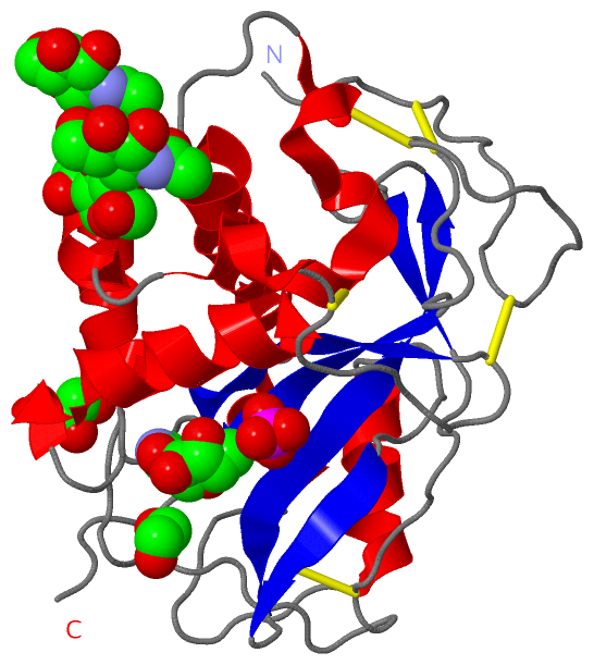
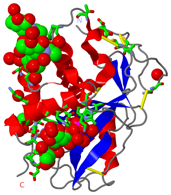
(-) Ligands, Modified Residues, Ions  (4, 7)

Asymmetric/Biological Unit (4, 7)
No.NameCountTypeFull Name
1D3Z1Ligand/Ion1-(5-DEOXY-BETA-L-XYLOFURANOSYL)PYRIMIDINE-2,4(1H,3H)-DIONE
2EDO2Ligand/Ion1,2-ETHANEDIOL
3NAG3Ligand/IonN-ACETYL-D-GLUCOSAMINE
4PO41Ligand/IonPHOSPHATE ION

(-) Sites  (7, 7)

Asymmetric Unit (7, 7)
No.NameEvidenceResiduesDescription
1AC1SOFTWAREASN A:74 , SER A:77 , ALA A:81 , NAG A:249 , HOH A:1022 , HOH A:1099 , HOH A:1106 , HOH A:1240 , HOH A:1241BINDING SITE FOR RESIDUE NAG A 248
2AC2SOFTWAREGLN A:133 , NAG A:248 , HOH A:1139 , HOH A:1280 , HOH A:1308BINDING SITE FOR RESIDUE NAG A 249
3AC3SOFTWAREGLN A:14 , ALA A:81 , GLN A:82 , ASN A:83 , TYR A:131 , HOH A:1084 , HOH A:1201 , HOH A:1341BINDING SITE FOR RESIDUE NAG A 250
4AC4SOFTWAREHIS A:51 , TRP A:54 , HIS A:110 , GLU A:111 , LYS A:114 , HIS A:115 , D3Z A:252 , HOH A:1063 , HOH A:1101 , HOH A:1191 , HOH A:1271BINDING SITE FOR RESIDUE PO4 A 251
5AC5SOFTWAREASP A:3 , PHE A:35 , ASP A:37 , HIS A:51 , PRO A:96 , ASP A:97 , TYR A:98 , PHE A:107 , HIS A:110 , THR A:128 , PO4 A:251 , HOH A:1000 , HOH A:1001 , HOH A:1071 , HOH A:1101 , HOH A:1125 , HOH A:1185 , HOH A:1189 , HOH A:1211BINDING SITE FOR RESIDUE D3Z A 252
6AC6SOFTWAREASP A:42 , GLY A:43 , LYS A:92 , GLU A:93 , HOH A:1108 , HOH A:1110BINDING SITE FOR RESIDUE EDO A 253
7AC7SOFTWARESER A:20 , CYS A:21 , CYS A:22 , ASP A:39 , PRO A:40 , HOH A:1142 , HOH A:1276BINDING SITE FOR RESIDUE EDO A 254

(-) SS Bonds  (5, 5)

Asymmetric/Biological Unit
No.Residues
1A:5 -A:22
2A:13 -A:58
3A:21 -A:126
4A:66 -A:118
5A:191 -A:225

(-) Cis Peptide Bonds  (2, 2)

Asymmetric/Biological Unit
No.Residues
1Ser A:25 -Pro A:26
2Asp A:39 -Pro A:40

 Sequence-Structure Mapping

(-) SAPs(SNPs)/Variants  (0, 0)

(no "SAP(SNP)/Variant" information available for 3D3Z)

(-) PROSITE Motifs  (0, 0)

(no "PROSITE Motif" information available for 3D3Z)

(-) Exons   (0, 0)

(no "Exon" information available for 3D3Z)

(-) Sequences/Alignments

Asymmetric/Biological Unit
   Reformat: Number of residues per line =  ('0' or empty: single-line sequence representation)
  Number of residues per labelling interval =   
  UniProt sequence: complete  aligned part    
   Show mapping: SCOP domains CATH domains Pfam domains Secondary structure (by author)
SAPs(SNPs) PROSITE motifs Exons
(details for a mapped element are shown in a popup box when the mouse pointer rests over it)
Chain A from PDB  Type:PROTEIN  Length:238
 aligned with Q45U61_ASPNG | Q45U61 from UniProtKB/TrEMBL  Length:270

    Alignment length:238
                                    33        43        53        63        73        83        93       103       113       123       133       143       153       163       173       183       193       203       213       223       233       243       253        
         Q45U61_ASPNG    24 TIDTCSSDSPLSCQTDNEASCCFNSPGGSLLQTQFWDYDPSDGPSGSWTIHGLWPDNCDGTYQEYCDESREYSNITSILEAQNRTELLSYMKEYWPDYEGADEDESFWEHEWNKHGTCINTIEPSCYTDYYAQEEVGDFFQQVVDLFKTLDSYTALSDAGITPSEDATYKLSDIEDALAAIHDGYPPYVGCEDGALSQLYYYFNVKGSAIGGTYVASERLEDSNCKDSGIKYPPKYSS 261
               SCOP domains d3d3za_ A: automated matches                                                                                                                                                                                                                   SCOP domains
               CATH domains ---------------------------------------------------------------------------------------------------------------------------------------------------------------------------------------------------------------------------------------------- CATH domains
               Pfam domains ---------------------------------------------------------------------------------------------------------------------------------------------------------------------------------------------------------------------------------------------- Pfam domains
         Sec.struct. author ............................eeeeeee.............eeeeeeee..........hhhhh..hhhhhhhhh.hhhhhhhhhhhh....hhhhhhhhhhhhhhhhhhhhhhhhhhhh.....hhhhhhhhhhhhhhhh..hhhhhhhhh..........hhhhhhhhhhh.......eeeee..eeeeeeeeeeee......eeee...................... Sec.struct. author
                 SAPs(SNPs) ---------------------------------------------------------------------------------------------------------------------------------------------------------------------------------------------------------------------------------------------- SAPs(SNPs)
                    PROSITE ---------------------------------------------------------------------------------------------------------------------------------------------------------------------------------------------------------------------------------------------- PROSITE
                 Transcript ---------------------------------------------------------------------------------------------------------------------------------------------------------------------------------------------------------------------------------------------- Transcript
                 3d3z A   1 TIDTCSSDSPLSCQTDNEASCCFNSPGGSLLQTQFWDYDPSDGPSDSWTIHGLWPDNCDGTYQEYCDESREYSNITSILEAQNRTELLSYMKEYWPDYEGADEDESFWEHEWNKHGTCINTIEPSCYTDYYAQEEVGDFFQQVVDLFKTLDSYTALSDAGITPSEDATYKLSDIEDALAAIHDGYPPYVGCEDGALSQLYYYFNVKGSAIGGTYVASERLEDSNCKDSGIKYPPKYSS 238
                                    10        20        30        40        50        60        70        80        90       100       110       120       130       140       150       160       170       180       190       200       210       220       230        

   Legend:   → Mismatch (orange background)
  - → Gap (green background, '-', border residues have a numbering label)
    → Modified Residue (blue background, lower-case, 'x' indicates undefined single-letter code, labelled with number + name)
  x → Chemical Group (purple background, 'x', labelled with number + name, e.g. ACE or NH2)
  extra numbering lines below/above indicate numbering irregularities and modified residue names etc., number ends below/above '|'

 Classification and Annotation

(-) SCOP Domains  (1, 1)

Asymmetric/Biological Unit

(-) CATH Domains  (0, 0)

(no "CATH Domain" information available for 3D3Z)

(-) Pfam Domains  (0, 0)

(no "Pfam Domain" information available for 3D3Z)

(-) Gene Ontology  (3, 3)

Asymmetric/Biological Unit(hide GO term definitions)
Chain A   (Q45U61_ASPNG | Q45U61)
molecular function
    GO:0003723    RNA binding    Interacting selectively and non-covalently with an RNA molecule or a portion thereof.
    GO:0033897    ribonuclease T2 activity    Catalysis of the two-stage endonucleolytic cleavage to nucleoside 3'-phosphates and 3'-phosphooligonucleotides with 2',3'-cyclic phosphate intermediates.
biological process
    GO:0090502    RNA phosphodiester bond hydrolysis, endonucleolytic    The chemical reactions and pathways involving the hydrolysis of internal 3',5'-phosphodiester bonds in one or two strands of ribonucleotides.

 Visualization

(-) Interactive Views

Asymmetric/Biological Unit
  Complete Structure
    Jena3D(integrated viewing of ligand, site, SAP, PROSITE, SCOP information)
    WebMol | AstexViewer[tm]@PDBe
(Java Applets, require no local installation except for Java; loading may be slow)
    STRAP
(Java WebStart application, automatic local installation, requires Java; full application with system access!)
    RasMol
(require local installation)
    Molscript (VRML)
(requires installation of a VRML viewer; select preferred view via VRML and generate a mono or stereo PDF format file)
 
  Ligands, Modified Residues, Ions
    D3Z  [ RasMol | Jena3D ]  +environment [ RasMol | Jena3D ]
    EDO  [ RasMol | Jena3D ]  +environment [ RasMol | Jena3D ]
    NAG  [ RasMol | Jena3D ]  +environment [ RasMol | Jena3D ]
    PO4  [ RasMol | Jena3D ]  +environment [ RasMol | Jena3D ]
 
  Sites
    AC1  [ RasMol ]  +environment [ RasMol ]
    AC2  [ RasMol ]  +environment [ RasMol ]
    AC3  [ RasMol ]  +environment [ RasMol ]
    AC4  [ RasMol ]  +environment [ RasMol ]
    AC5  [ RasMol ]  +environment [ RasMol ]
    AC6  [ RasMol ]  +environment [ RasMol ]
    AC7  [ RasMol ]  +environment [ RasMol ]
 
  Cis Peptide Bonds
    Asp A:39 - Pro A:40   [ RasMol ]  
    Ser A:25 - Pro A:26   [ RasMol ]  
 

(-) Still Images

Jmol
  protein: cartoon or spacefill or dots and stick; nucleic acid: cartoon and stick; ligands: spacefill; active site: stick
Molscript
  protein, nucleic acid: cartoon; ligands: spacefill; active site: ball and stick

 Databases and Analysis Tools

(-) Databases

Access by PDB/NDB ID
  3d3z
    Family and Domain InformationProDom | SYSTERS
    General Structural InformationGlycoscienceDB | MMDB | NDB | OCA | PDB | PDBe | PDBj | PDBsum | PDBWiki | PQS | PROTEOPEDIA
    Orientation in MembranesOPM
    Protein SurfaceSURFACE
    Secondary StructureDSSP (structure derived) | HSSP (homology derived)
    Structural GenomicsGeneCensus
    Structural NeighboursCE | VAST
    Structure ClassificationCATH | Dali | SCOP
    Validation and Original DataBMRB Data View | BMRB Restraints Grid | EDS | PROCHECK | RECOORD | WHAT_CHECK
 
Access by UniProt ID/Accession number
  Q45U61_ASPNG | Q45U61
    Comparative Protein Structure ModelsModBase
    Genomic InformationEnsembl
    Protein-protein InteractionDIP
    Sequence, Family and Domain InformationInterPro | Pfam | SMART | UniProtKB/TrEMBL
 
Access by Enzyme Classificator   (EC Number)
  3.1.27.1
    General Enzyme InformationBRENDA | EC-PDB | Enzyme | IntEnz
    PathwayKEGG | MetaCyc
 
Access by Disease Identifier   (MIM ID)
  (no 'MIM ID' available)
    Disease InformationOMIM
 
Access by GenAge ID
  (no 'GenAge ID' available)
    Age Related InformationGenAge

(-) Analysis Tools

Access by PDB/NDB ID
    Domain InformationXDom
    Interatomic Contacts of Structural UnitsCSU
    Ligand-protein ContactsLPC
    Protein CavitiescastP
    Sequence and Secondary StructurePDBCartoon
    Structure AlignmentSTRAP(Java WebStart application, automatic local installation, requires Java; full application with system access!)
    Structure and Sequence BrowserSTING
 
Access by UniProt ID/Accession number
  Q45U61_ASPNG | Q45U61
    Protein Disorder PredictionDisEMBL | FoldIndex | GLOBPLOT (for more information see DisProt)

 Related Entries

(-) Entries Sharing at Least One Protein Chain (UniProt ID)

UniProtKB/TrEMBL
        Q45U61_ASPNG | Q45U613tbj

(-) Related Entries Specified in the PDB File

(no "Related Entries Specified in the PDB File" available for 3D3Z)