Show PDB file:   
         Plain Text   HTML   (compressed file size)
QuickSearch:   
by PDB,NDB,UniProt,PROSITE Code or Search Term(s)  
(-)Biol.Unit 1 - manually
(-)Asymmetric Unit
(-)Asym. Unit - sites
(-)Biological Unit 1
(-)Biol. Unit 1 - sites
(-)Biological Unit 2
collapse expand < >
Image Biol.Unit 1 - manually
Biol.Unit 1 - manually  (Jmol Viewer)
Image Asymmetric Unit
Asymmetric Unit  (Jmol Viewer)
Image Asym. Unit - sites
Asym. Unit - sites  (Jmol Viewer)
Image Biological Unit 1
Biological Unit 1  (Jmol Viewer)
Image Biol. Unit 1 - sites
Biol. Unit 1 - sites  (Jmol Viewer)
Image Biological Unit 2
Biological Unit 2  (Jmol Viewer)

(-) Description

Title :  STRUCTURE OF THE EUKARYOTIC TPP-SPECIFIC RIBOSWITCH BOUND TO THE ANTIBACTERIAL COMPOUND PYRITHIAMINE PYROPHOSPHATE
 
Authors :  S. Thore, C. Frick, N. Ban
Date :  09 May 08  (Deposition) - 01 Jul 08  (Release) - 16 Nov 11  (Revision)
Method :  X-RAY DIFFRACTION
Resolution :  2.00
Chains :  Asym. Unit :  A,B
Biol. Unit 1:  A  (1x)
Biol. Unit 2:  B  (1x)
Keywords :  Rna, Riboswitch, Antibiotic, Thiamine Pyrophosphate, Pyrithiamine Pyrophosphate, Complex (Keyword Search: [Gene Ontology, PubMed, Web (Google))
 
Reference :  S. Thore, C. Frick, N. Ban
Structural Basis Of Thiamine Pyrophosphate Analogues Bindin To The Eukaryotic Riboswitch
J. Am. Chem. Soc. V. 130 8116 2008
PubMed-ID: 18533652  |  Reference-DOI: 10.1021/JA801708E

(-) Compounds

Molecule 1 - TPP-SPECIFIC RIBOSWITCH
    ChainsA, B
    EngineeredYES
    Expression SystemCELL-FREE SYNTHESIS
    Organism ScientificARABIDOPSIS THALIANA
    Other DetailsIN VITRO TRANSCRIPTION

 Structural Features

(-) Chains, Units

  12
Asymmetric Unit AB
Biological Unit 1 (1x)A 
Biological Unit 2 (1x) B

Summary Information (see also Sequences/Alignments below)

(-) Ligands, Modified Residues, Ions  (2, 12)

Asymmetric Unit (2, 12)
No.NameCountTypeFull Name
1MG10Ligand/IonMAGNESIUM ION
2PYI2Ligand/Ion1-[(4-AMINO-2-METHYLPYRIMIDIN-5-YL)METHYL]-3-(2-{[HYDROXY(PHOSPHONOOXY)PHOSPHORYL]OXY}ETHYL)-2-METHYLPYRIDINIUM
Biological Unit 1 (1, 1)
No.NameCountTypeFull Name
1MG-1Ligand/IonMAGNESIUM ION
2PYI1Ligand/Ion1-[(4-AMINO-2-METHYLPYRIMIDIN-5-YL)METHYL]-3-(2-{[HYDROXY(PHOSPHONOOXY)PHOSPHORYL]OXY}ETHYL)-2-METHYLPYRIDINIUM
Biological Unit 2 (1, 1)
No.NameCountTypeFull Name
1MG-1Ligand/IonMAGNESIUM ION
2PYI1Ligand/Ion1-[(4-AMINO-2-METHYLPYRIMIDIN-5-YL)METHYL]-3-(2-{[HYDROXY(PHOSPHONOOXY)PHOSPHORYL]OXY}ETHYL)-2-METHYLPYRIDINIUM

(-) Sites  (12, 12)

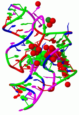
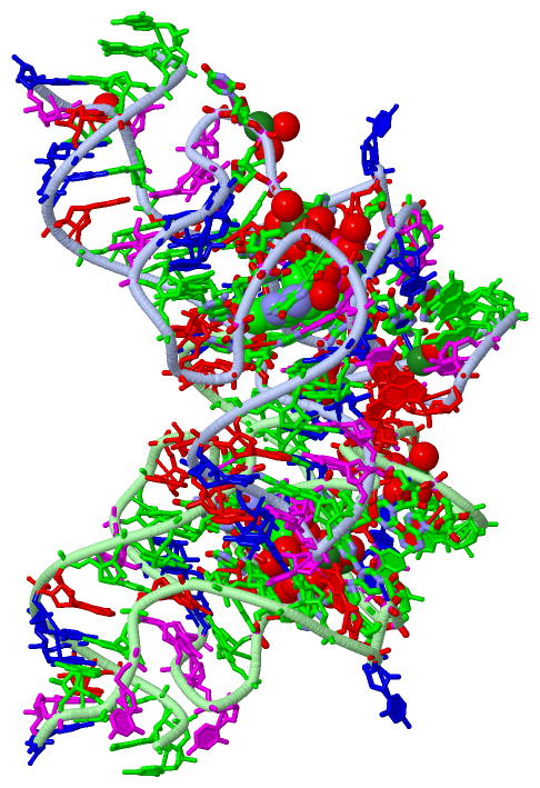
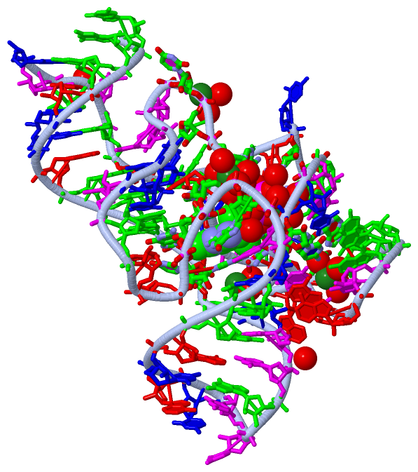
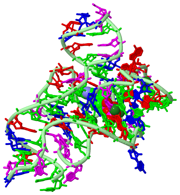
Asymmetric Unit (12, 12)
No.NameEvidenceResiduesDescription
01AC1SOFTWAREPYI A:84 , HOH A:94 , HOH A:96 , HOH A:98 , HOH A:341BINDING SITE FOR RESIDUE MG A 78
02AC2SOFTWAREC A:51 , A A:52 , HOH A:88 , HOH A:108 , HOH A:345 , HOH B:98BINDING SITE FOR RESIDUE MG A 79
03AC3SOFTWAREU A:41 , G A:42 , C A:65 , HOH A:167 , HOH A:332BINDING SITE FOR RESIDUE MG A 80
04AC4SOFTWAREG A:48 , G A:66 , PYI A:84 , HOH A:85 , HOH A:89 , HOH A:339BINDING SITE FOR RESIDUE MG A 81
05AC5SOFTWAREA A:29 , HOH A:114 , HOH A:151 , HOH A:208 , HOH A:319 , HOH A:321BINDING SITE FOR RESIDUE MG A 82
06AC6SOFTWAREG A:48 , HOH A:107 , HOH A:258 , HOH B:118 , HOH B:296BINDING SITE FOR RESIDUE MG A 83
07AC7SOFTWAREHOH A:102 , C B:51 , A B:52 , HOH B:88 , HOH B:111 , HOH B:300BINDING SITE FOR RESIDUE MG B 78
08AC8SOFTWAREPYI B:82 , HOH B:87 , HOH B:180 , HOH B:315 , HOH B:316BINDING SITE FOR RESIDUE MG B 79
09AC9SOFTWAREG B:48 , G B:66 , PYI B:82 , HOH B:83 , HOH B:89 , HOH B:314BINDING SITE FOR RESIDUE MG B 80
10BC1SOFTWAREHOH A:236 , A B:29 , HOH B:90 , HOH B:127 , HOH B:172 , HOH B:182BINDING SITE FOR RESIDUE MG B 81
11BC2SOFTWAREG A:11 , U A:12 , G A:28 , G A:30 , A A:31 , A A:43 , C A:45 , G A:60 , C A:61 , C A:65 , G A:66 , MG A:78 , MG A:81 , HOH A:93 , HOH A:94 , HOH A:101 , HOH A:142 , HOH A:144 , HOH A:339 , HOH A:341BINDING SITE FOR RESIDUE PYI A 84
12BC3SOFTWAREG B:11 , U B:12 , G B:28 , G B:30 , A B:31 , A B:43 , C B:45 , G B:60 , C B:61 , C B:65 , G B:66 , MG B:79 , MG B:80 , HOH B:83 , HOH B:87 , HOH B:96 , HOH B:104 , HOH B:120 , HOH B:135 , HOH B:153 , HOH B:162 , HOH B:314 , HOH B:315 , HOH B:331BINDING SITE FOR RESIDUE PYI B 82

(-) SS Bonds  (0, 0)

(no "SS Bond" information available for 3D2V)

(-) Cis Peptide Bonds  (0, 0)

(no "Cis Peptide Bond" information available for 3D2V)

 Sequence-Structure Mapping

(-) SAPs(SNPs)/Variants  (0, 0)

(no "SAP(SNP)/Variant" information available for 3D2V)

(-) PROSITE Motifs  (0, 0)

(no "PROSITE Motif" information available for 3D2V)

(-) Exons   (0, 0)

(no "Exon" information available for 3D2V)

(-) Sequences/Alignments

Asymmetric Unit
   Reformat: Number of residues per line =  ('0' or empty: single-line sequence representation)
  Number of residues per labelling interval =   
  UniProt sequence: complete  aligned part    
   Show mapping: SCOP domains CATH domains Pfam domains Secondary structure (by author)
SAPs(SNPs) PROSITE motifs Exons
(details for a mapped element are shown in a popup box when the mouse pointer rests over it)
Chain A from PDB  Type:RNA  Length:77
                                                                                                            
                  3d2v A  1 GGGACCAGGGGUGCUUGUUCACAGGCUGAGAAAGUCCCUUUGAACCUGAACAGGGUAAUGCCUGCGCAGGGAGUGUC 77
                                    10        20        30        40        50        60        70       

Chain B from PDB  Type:RNA  Length:77
                                                                                                            
                  3d2v B  1 GGGACCAGGGGUGCUUGUUCACAGGCUGAGAAAGUCCCUUUGAACCUGAACAGGGUAAUGCCUGCGCAGGGAGUGUC 77
                                    10        20        30        40        50        60        70       

   Legend:   → Mismatch (orange background)
  - → Gap (green background, '-', border residues have a numbering label)
    → Modified Residue (blue background, lower-case, 'x' indicates undefined single-letter code, labelled with number + name)
  x → Chemical Group (purple background, 'x', labelled with number + name, e.g. ACE or NH2)
  extra numbering lines below/above indicate numbering irregularities and modified residue names etc., number ends below/above '|'

 Classification and Annotation

(-) SCOP Domains  (0, 0)

(no "SCOP Domain" information available for 3D2V)

(-) CATH Domains  (0, 0)

(no "CATH Domain" information available for 3D2V)

(-) Pfam Domains  (0, 0)

(no "Pfam Domain" information available for 3D2V)

(-) Gene Ontology  (0, 0)

Asymmetric Unit(hide GO term definitions)
    (no "Gene Ontology" information available for 3D2V)

 Visualization

(-) Interactive Views

Asymmetric Unit
  Complete Structure
    Jena3D(integrated viewing of ligand, site, SAP, PROSITE, SCOP information)
    WebMol | AstexViewer[tm]@PDBe
(Java Applets, require no local installation except for Java; loading may be slow)
    STRAP
(Java WebStart application, automatic local installation, requires Java; full application with system access!)
    RasMol
(require local installation)
    Molscript (VRML)
(requires installation of a VRML viewer; select preferred view via VRML and generate a mono or stereo PDF format file)
 
  Ligands, Modified Residues, Ions
    MG  [ RasMol | Jena3D ]  +environment [ RasMol | Jena3D ]
    PYI  [ RasMol | Jena3D ]  +environment [ RasMol | Jena3D ]
 
  Sites
    AC1  [ RasMol ]  +environment [ RasMol ]
    AC2  [ RasMol ]  +environment [ RasMol ]
    AC3  [ RasMol ]  +environment [ RasMol ]
    AC4  [ RasMol ]  +environment [ RasMol ]
    AC5  [ RasMol ]  +environment [ RasMol ]
    AC6  [ RasMol ]  +environment [ RasMol ]
    AC7  [ RasMol ]  +environment [ RasMol ]
    AC8  [ RasMol ]  +environment [ RasMol ]
    AC9  [ RasMol ]  +environment [ RasMol ]
    BC1  [ RasMol ]  +environment [ RasMol ]
    BC2  [ RasMol ]  +environment [ RasMol ]
    BC3  [ RasMol ]  +environment [ RasMol ]
 
  Cis Peptide Bonds
(no "Cis Peptide Bonds" information available for 3d2v)
 
Biological Units
  Complete Structure
    Biological Unit 1  [ Jena3D ]
    Biological Unit 2  [ Jena3D ]

(-) Still Images

Jmol
  protein: cartoon or spacefill or dots and stick; nucleic acid: cartoon and stick; ligands: spacefill; active site: stick
Molscript
  protein, nucleic acid: cartoon; ligands: spacefill; active site: ball and stick

 Databases and Analysis Tools

(-) Databases

Access by PDB/NDB ID
  3d2v
    Family and Domain InformationProDom | SYSTERS
    General Structural InformationGlycoscienceDB | MMDB | NDB | OCA | PDB | PDBe | PDBj | PDBsum | PDBWiki | PQS | PROTEOPEDIA
    Orientation in MembranesOPM
    Protein SurfaceSURFACE
    Secondary StructureDSSP | HSSP
    Structural GenomicsGeneCensus
    Structural NeighboursCE | VAST
    Structure ClassificationCATH | Dali | SCOP
    Validation and Original DataBMRB Data View | BMRB Restraints Grid | EDS | PROCHECK | RECOORD | WHAT_CHECK
 
Access by UniProt ID/Accession number
  (no 'UniProt ID/Accession number' available) |
    Comparative Protein Structure ModelsModBase
    Genomic InformationEnsembl
    Protein-protein InteractionDIP
    Sequence, Family and Domain InformationInterPro | Pfam | SMART | UniProt
 
Access by Enzyme Classificator   (EC Number)
  (no 'Enzyme Classificator' available)
    General Enzyme InformationBRENDA | EC-PDB | Enzyme | IntEnz
    PathwayKEGG | MetaCyc
 
Access by Disease Identifier   (MIM ID)
  (no 'MIM ID' available)
    Disease InformationOMIM
 
Access by GenAge ID
  (no 'GenAge ID' available)
    Age Related InformationGenAge

(-) Analysis Tools

Access by PDB/NDB ID
    Domain InformationXDom
    Interatomic Contacts of Structural UnitsCSU
    Ligand-protein ContactsLPC
    Protein CavitiescastP
    Sequence and Secondary StructurePDBCartoon
    Structure AlignmentSTRAP(Java WebStart application, automatic local installation, requires Java; full application with system access!)
    Structure and Sequence BrowserSTING
 
Access by UniProt ID/Accession number
  (no 'UniProt ID/Accession number' available) |
    Protein Disorder PredictionDisEMBL | FoldIndex | GLOBPLOT

 Related Entries

(-) Entries Sharing at Least One Protein Chain (UniProt ID)

(no "Entries Sharing at Least One Protein Chain" available for 3D2V)

(-) Related Entries Specified in the PDB File

2cky 3d2g 3d2x