Show PDB file:   
         Plain Text   HTML   (compressed file size)
QuickSearch:   
by PDB,NDB,UniProt,PROSITE Code or Search Term(s)  
(-)Asym./Biol. Unit
(-)Asym./Biol. Unit - sites
collapse expand < >
Image Asym./Biol. Unit
Asym./Biol. Unit  (Jmol Viewer)
Image Asym./Biol. Unit - sites
Asym./Biol. Unit - sites  (Jmol Viewer)

(-) Description

Title :  STRUCTURE OF BTHTX-I COMPLEXED WITH ALPHA-TOCOPHEROL
 
Authors :  J. I. Dos Santos, M. R. M. Fontes
Date :  24 Apr 08  (Deposition) - 05 May 09  (Release) - 28 Jul 09  (Revision)
Method :  X-RAY DIFFRACTION
Resolution :  1.83
Chains :  Asym./Biol. Unit :  A,B
Keywords :  Myotoxin, Bothrops Jaracussu, Lys49-Pla2, Snake Venom, Phospholipase A2, X-Ray Crystallography, Secreted (Keyword Search: [Gene Ontology, PubMed, Web (Google))
 
Reference :  J. I. Dos Santos, A. M. Soares, M. R. Fontes
Comparative Structural Studies On Lys49-Phospholipases A(2) From Bothrops Genus Reveal Their Myotoxic Site.
J. Struct. Biol. V. 167 106 2009
PubMed-ID: 19401234  |  Reference-DOI: 10.1016/J.JSB.2009.04.003
(for further references see the PDB file header)

(-) Compounds

Molecule 1 - MYOTOXIC PHOSPHOLIPASE A2-LIKE
    ChainsA, B
    FragmentUNP RESIDUES 17-137
    Organism CommonJARARACUSSU
    Organism ScientificBOTHROPS JARARACUSSU
    SynonymMYOTOXIC A2-LIKE PHOSPHOLIPASE

 Structural Features

(-) Chains, Units

  12
Asymmetric/Biological Unit AB

Summary Information (see also Sequences/Alignments below)

(-) Ligands, Modified Residues, Ions  (3, 7)

Asymmetric/Biological Unit (3, 7)
No.NameCountTypeFull Name
1PE41Ligand/Ion2-{2-[2-(2-{2-[2-(2-ETHOXY-ETHOXY)-ETHOXY]-ETHOXY}-ETHOXY)-ETHOXY]-ETHOXY}-ETHANOL
2SO44Ligand/IonSULFATE ION
3VIT2Ligand/IonVITAMIN E

(-) Sites  (14, 14)

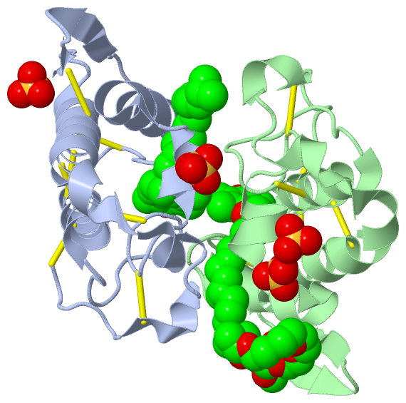
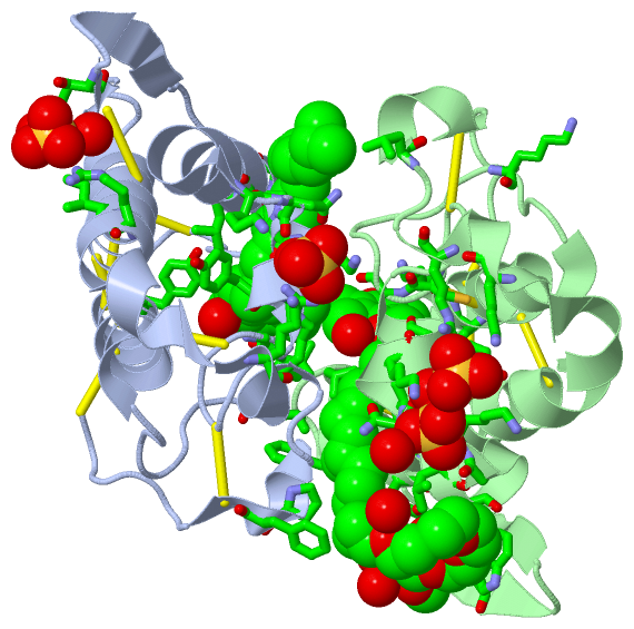
Asymmetric Unit (14, 14)
No.NameEvidenceResiduesDescription
01AC1unknownLYS A:16 , LYS A:20 , LYS A:115 , ARG A:118BINDING SITE FOR RESIDUE SO4 A 303
02AC2unknownTHR A:81 , ILE A:104 , ARG A:107 , LYS B:36BINDING SITE FOR RESIDUE SO4 A 305
03AC3unknownLYS B:20 , THR B:59 , LYS B:115 , ARG B:118BINDING SITE FOR RESIDUE SO4 B 308
04AC4unknownGLY B:15 , LYS B:16 , ASN B:17 , LYS B:20 , PRO B:90BINDING SITE FOR RESIDUE SO4 B 309
05AC5unknownPHE A:125 , LYS B:7 , LEU B:10 , GLN B:11 , TYR B:75 , TRP B:77BINDING SITE FOR RESIDUE PE4 B 310
06AC6unknownLEU A:32 , PRO A:123 , LEU B:2 , PHE B:3 , LEU B:5 , GLY B:6 , LYS B:7 , ILE B:9 , PRO B:18 , TYR B:22 , CYS B:29 , GLY B:30 , HIS B:48 , LYS B:49 , TYR B:52BINDING SITE FOR RESIDUE VIT B 311
07AC7unknownLEU A:2 , PHE A:3 , LEU A:5 , GLY A:6 , ASN A:17 , PRO A:18 , ALA A:19 , TYR A:22 , GLY A:23 , GLY A:30 , TYR A:52 , LEU B:121BINDING SITE FOR RESIDUE VIT A 306
08AC8SOFTWARELYS A:16 , LYS A:20 , LYS A:115 , ARG A:118BINDING SITE FOR RESIDUE SO4 A 303
09AC9SOFTWARETHR A:81 , ILE A:104 , ARG A:107 , HOH A:313 , HOH A:316 , LYS B:36BINDING SITE FOR RESIDUE SO4 A 305
10AD1SOFTWARELYS B:20 , THR B:59 , LYS B:115 , ARG B:118 , SO4 B:309BINDING SITE FOR RESIDUE SO4 B 308
11AD2SOFTWAREGLY B:15 , LYS B:16 , ASN B:17 , LYS B:20 , PRO B:90 , SO4 B:308 , HOH B:430 , HOH B:469BINDING SITE FOR RESIDUE SO4 B 309
12AD3SOFTWARELEU A:2 , PHE A:3 , LEU A:5 , GLY A:6 , ASN A:17 , PRO A:18 , ALA A:19 , TYR A:22 , GLY A:23 , GLY A:30 , VAL A:31 , TYR A:52 , HOH A:336 , LEU B:121BINDING SITE FOR RESIDUE VIT A 306
13AD4SOFTWAREPHE A:125 , LYS B:7 , LEU B:10 , GLN B:11 , TYR B:75 , TRP B:77 , VIT B:311 , HOH B:495 , HOH B:499BINDING SITE FOR RESIDUE PE4 B 310
14AD5SOFTWAREVAL A:31 , LEU A:32 , PRO A:123 , LEU B:2 , PHE B:3 , LEU B:5 , GLY B:6 , LYS B:7 , ILE B:9 , PRO B:18 , TYR B:22 , CYS B:29 , GLY B:30 , HIS B:48 , LYS B:49 , TYR B:52 , PE4 B:310 , HOH B:484BINDING SITE FOR RESIDUE VIT B 311

(-) SS Bonds  (14, 14)

Asymmetric/Biological Unit
No.Residues
1A:27 -A:126
2A:29 -A:45
3A:44 -A:105
4A:50 -A:133
5A:51 -A:98
6A:61 -A:91
7A:84 -A:96
8B:27 -B:126
9B:29 -B:45
10B:44 -B:105
11B:50 -B:133
12B:51 -B:98
13B:61 -B:91
14B:84 -B:96

(-) Cis Peptide Bonds  (0, 0)

(no "Cis Peptide Bond" information available for 3CXI)

 Sequence-Structure Mapping

(-) SAPs(SNPs)/Variants  (3, 6)

Asymmetric/Biological Unit (3, 6)
  dbSNPPDB
No.SourceVariant IDVariantUniProt IDStatusIDChainVariant
1UniProtVAR_PA2B1_BOTJR_001 *F19LPA2B1_BOTJR  ---  ---A/BF3L
2UniProtVAR_PA2B1_BOTJR_002 *Y37HPA2B1_BOTJR  ---  ---A/BY22H
3UniProtVAR_PA2B1_BOTJR_003 *P136APA2B1_BOTJR  ---  ---A/BP132A
   * ID not provided by source

  SNP/SAP Summary Statistics (UniProtKB/Swiss-Prot)

(-) PROSITE Motifs  (2, 4)

Asymmetric/Biological Unit (2, 4)
 PROSITEUniProtKBPDB
No.IDACDescriptionIDLocationCountLocation
1PA2_HISPS00118 Phospholipase A2 histidine active site.PA2B1_BOTJR59-66
 
  2A:44-51
B:44-51
2PA2_ASPPS00119 Phospholipase A2 aspartic acid active site.PA2B1_BOTJR101-111
 
  2A:95-105
B:95-105

(-) Exons   (0, 0)

(no "Exon" information available for 3CXI)

(-) Sequences/Alignments

Asymmetric/Biological Unit
   Reformat: Number of residues per line =  ('0' or empty: single-line sequence representation)
  Number of residues per labelling interval =   
  UniProt sequence: complete  aligned part    
   Show mapping: SCOP domains CATH domains Pfam domains Secondary structure (by author)
SAPs(SNPs) PROSITE motifs Exons
(details for a mapped element are shown in a popup box when the mouse pointer rests over it)
Chain A from PDB  Type:PROTEIN  Length:121
 aligned with PA2B1_BOTJR | Q90249 from UniProtKB/Swiss-Prot  Length:137

    Alignment length:121
                                    26        36        46        56        66        76        86        96       106       116       126       136 
          PA2B1_BOTJR    17 SLFELGKMILQETGKNPAKSYGAYGCNCGVLGRGKPKDATDRCCYVHKCCYKKLTGCDPKKDRYSYSWKDKTIVCGENNPCLKELCECDKAVAICLRENLGTYNKKYRYHLKPFCKKADPC 137
               SCOP domains d3cxia_ A: Snake phospholipase A2                                                                                         SCOP domains
               CATH domains 3cxiA00 A:1-133 Phospholipase A2                                                                                          CATH domains
               Pfam domains ------------------------------------------------------------------------------------------------------------------------- Pfam domains
         Sec.struct. author hhhhhhhhhhhhhh.hhhhhhh...............hhhhhhhhhhhhhhhhh...........eeee..eeee...hhhhhhhhhhhhhhhhhhhhhhhhhhhhhh..hhhhh...... Sec.struct. author
                 SAPs(SNPs) --L-----------------H--------------------------------------------------------------------------------------------------A- SAPs(SNPs)
                    PROSITE ------------------------------------------PA2_HIS ----------------------------------PA2_ASP    -------------------------- PROSITE
                 Transcript ------------------------------------------------------------------------------------------------------------------------- Transcript
                 3cxi A   1 SLFELGKMILQETGKNPAKSYGAYGCNCGVLGRGKPKDATDRCCYVHKCCYKKLTGCDPKKDRYSYSWKDKTIVCGENNPCLKELCECDKAVAICLRENLGTYNKKYRYHLKPFCKKADPC 133
                                    10  ||    21        31        41        51 ||   ||69        79        90       100       110       120  || ||132 
                                       13|                                    53|  61|                   88|                              123| ||    
                                        15                                     57   67                    90                               125 ||    
                                                                                                                                             127|    
                                                                                                                                              129    

Chain B from PDB  Type:PROTEIN  Length:121
 aligned with PA2B1_BOTJR | Q90249 from UniProtKB/Swiss-Prot  Length:137

    Alignment length:121
                                    26        36        46        56        66        76        86        96       106       116       126       136 
          PA2B1_BOTJR    17 SLFELGKMILQETGKNPAKSYGAYGCNCGVLGRGKPKDATDRCCYVHKCCYKKLTGCDPKKDRYSYSWKDKTIVCGENNPCLKELCECDKAVAICLRENLGTYNKKYRYHLKPFCKKADPC 137
               SCOP domains d3cxib_ B: Snake phospholipase A2                                                                                         SCOP domains
               CATH domains 3cxiB00 B:1-133 Phospholipase A2                                                                                          CATH domains
               Pfam domains ------------------------------------------------------------------------------------------------------------------------- Pfam domains
         Sec.struct. author hhhhhhhhhhhhhh.hhhhhhh...............hhhhhhhhhhhhhhhh............eeee..eeee...hhhhhhhhhhhhhhhhhhhhhhhhhhhhhh..hhhhh...... Sec.struct. author
                 SAPs(SNPs) --L-----------------H--------------------------------------------------------------------------------------------------A- SAPs(SNPs)
                    PROSITE ------------------------------------------PA2_HIS ----------------------------------PA2_ASP    -------------------------- PROSITE
                 Transcript ------------------------------------------------------------------------------------------------------------------------- Transcript
                 3cxi B   1 SLFELGKMILQETGKNPAKSYGAYGCNCGVLGRGKPKDATDRCCYVHKCCYKKLTGCDPKKDRYSYSWKDKTIVCGENNPCLKELCECDKAVAICLRENLGTYNKKYRYHLKPFCKKADPC 133
                                    10  ||    21        31        41        51 ||   ||69        79        90       100       110       120  || ||132 
                                       13|                                    53|  61|                   88|                              123| ||    
                                        15                                     57   67                    90                               125 ||    
                                                                                                                                             127|    
                                                                                                                                              129    

   Legend:   → Mismatch (orange background)
  - → Gap (green background, '-', border residues have a numbering label)
    → Modified Residue (blue background, lower-case, 'x' indicates undefined single-letter code, labelled with number + name)
  x → Chemical Group (purple background, 'x', labelled with number + name, e.g. ACE or NH2)
  extra numbering lines below/above indicate numbering irregularities and modified residue names etc., number ends below/above '|'

 Classification and Annotation

(-) SCOP Domains  (1, 2)

Asymmetric/Biological Unit

(-) CATH Domains  (1, 2)

Asymmetric/Biological Unit

(-) Pfam Domains  (0, 0)

(no "Pfam Domain" information available for 3CXI)

(-) Gene Ontology  (5, 5)

Asymmetric/Biological Unit(hide GO term definitions)
Chain A,B   (PA2B1_BOTJR | Q90249)
molecular function
    GO:0005509    calcium ion binding    Interacting selectively and non-covalently with calcium ions (Ca2+).
    GO:0004623    phospholipase A2 activity    Catalysis of the reaction: phosphatidylcholine + H2O = 1-acylglycerophosphocholine + a carboxylate.
biological process
    GO:0042742    defense response to bacterium    Reactions triggered in response to the presence of a bacterium that act to protect the cell or organism.
    GO:0016042    lipid catabolic process    The chemical reactions and pathways resulting in the breakdown of lipids, compounds soluble in an organic solvent but not, or sparingly, in an aqueous solvent.
cellular component
    GO:0005576    extracellular region    The space external to the outermost structure of a cell. For cells without external protective or external encapsulating structures this refers to space outside of the plasma membrane. This term covers the host cell environment outside an intracellular parasite.

 Visualization

(-) Interactive Views

Asymmetric/Biological Unit
  Complete Structure
    Jena3D(integrated viewing of ligand, site, SAP, PROSITE, SCOP information)
    WebMol | AstexViewer[tm]@PDBe
(Java Applets, require no local installation except for Java; loading may be slow)
    STRAP
(Java WebStart application, automatic local installation, requires Java; full application with system access!)
    RasMol
(require local installation)
    Molscript (VRML)
(requires installation of a VRML viewer; select preferred view via VRML and generate a mono or stereo PDF format file)
 
  Ligands, Modified Residues, Ions
    PE4  [ RasMol | Jena3D ]  +environment [ RasMol | Jena3D ]
    SO4  [ RasMol | Jena3D ]  +environment [ RasMol | Jena3D ]
    VIT  [ RasMol | Jena3D ]  +environment [ RasMol | Jena3D ]
 
  Sites
    AC1  [ RasMol ]  +environment [ RasMol ]
    AC2  [ RasMol ]  +environment [ RasMol ]
    AC3  [ RasMol ]  +environment [ RasMol ]
    AC4  [ RasMol ]  +environment [ RasMol ]
    AC5  [ RasMol ]  +environment [ RasMol ]
    AC6  [ RasMol ]  +environment [ RasMol ]
    AC7  [ RasMol ]  +environment [ RasMol ]
    AC8  [ RasMol ]  +environment [ RasMol ]
    AC9  [ RasMol ]  +environment [ RasMol ]
    AD1  [ RasMol ]  +environment [ RasMol ]
    AD2  [ RasMol ]  +environment [ RasMol ]
    AD3  [ RasMol ]  +environment [ RasMol ]
    AD4  [ RasMol ]  +environment [ RasMol ]
    AD5  [ RasMol ]  +environment [ RasMol ]
 
  Cis Peptide Bonds
(no "Cis Peptide Bonds" information available for 3cxi)
 

(-) Still Images

Jmol
  protein: cartoon or spacefill or dots and stick; nucleic acid: cartoon and stick; ligands: spacefill; active site: stick
Molscript
  protein, nucleic acid: cartoon; ligands: spacefill; active site: ball and stick

 Databases and Analysis Tools

(-) Databases

Access by PDB/NDB ID
  3cxi
    Family and Domain InformationProDom | SYSTERS
    General Structural InformationGlycoscienceDB | MMDB | NDB | OCA | PDB | PDBe | PDBj | PDBsum | PDBWiki | PQS | PROTEOPEDIA
    Orientation in MembranesOPM
    Protein SurfaceSURFACE
    Secondary StructureDSSP (structure derived) | HSSP (homology derived)
    Structural GenomicsGeneCensus
    Structural NeighboursCE | VAST
    Structure ClassificationCATH | Dali | SCOP
    Validation and Original DataBMRB Data View | BMRB Restraints Grid | EDS | PROCHECK | RECOORD | WHAT_CHECK
 
Access by UniProt ID/Accession number
  PA2B1_BOTJR | Q90249
    Comparative Protein Structure ModelsModBase
    Genomic InformationEnsembl
    Protein-protein InteractionDIP
    Sequence, Family and Domain InformationInterPro | Pfam | SMART | UniProtKB/SwissProt
 
Access by Enzyme Classificator   (EC Number)
  (no 'Enzyme Classificator' available)
    General Enzyme InformationBRENDA | EC-PDB | Enzyme | IntEnz
    PathwayKEGG | MetaCyc
 
Access by Disease Identifier   (MIM ID)
  (no 'MIM ID' available)
    Disease InformationOMIM
 
Access by GenAge ID
  (no 'GenAge ID' available)
    Age Related InformationGenAge

(-) Analysis Tools

Access by PDB/NDB ID
    Domain InformationXDom
    Interatomic Contacts of Structural UnitsCSU
    Ligand-protein ContactsLPC
    Protein CavitiescastP
    Sequence and Secondary StructurePDBCartoon
    Structure AlignmentSTRAP(Java WebStart application, automatic local installation, requires Java; full application with system access!)
    Structure and Sequence BrowserSTING
 
Access by UniProt ID/Accession number
  PA2B1_BOTJR | Q90249
    Protein Disorder PredictionDisEMBL | FoldIndex | GLOBPLOT (for more information see DisProt)

 Related Entries

(-) Entries Sharing at Least One Protein Chain (UniProt ID)

UniProtKB/Swiss-Prot
        PA2B1_BOTJR | Q902492h8i 3hzd 3hzw 3i03 3i3h 3i3i 3iq3 4k06 4k09 4wtb

(-) Related Entries Specified in the PDB File

1clp 1pa0 1pc9 1y4l 2q2j 3cyl