Show PDB file:   
         Plain Text   HTML   (compressed file size)
QuickSearch:   
by PDB,NDB,UniProt,PROSITE Code or Search Term(s)  
(-)Biol.Unit 1 - manually
(-)Asymmetric Unit
(-)Asym. Unit - sites
(-)Biological Unit 1
(-)Biol. Unit 1 - sites
collapse expand < >
Image Biol.Unit 1 - manually
Biol.Unit 1 - manually  (Jmol Viewer)
Image Asymmetric Unit
Asymmetric Unit  (Jmol Viewer)
Image Asym. Unit - sites
Asym. Unit - sites  (Jmol Viewer)
Image Biological Unit 1
Biological Unit 1  (Jmol Viewer)
Image Biol. Unit 1 - sites
Biol. Unit 1 - sites  (Jmol Viewer)

(-) Description

Title :  CRYSTAL STRUCTURE OF CENTRAL DOMAIN OF 3-HYDROXYACYL-COA DEHYDROGENASE FROM ARCHAEOGLOBUS FULGIDUS
 
Authors :  J. Osipiuk, E. Evdokimova, M. Kudritska, A. Savchenko, A. M. Edwards, A. Joachimiak, Midwest Center For Structural Genomics (Mcsg)
Date :  14 Apr 08  (Deposition) - 29 Apr 08  (Release) - 24 Feb 09  (Revision)
Method :  X-RAY DIFFRACTION
Resolution :  2.46
Chains :  Asym. Unit :  A
Biol. Unit 1:  A  (2x)
Keywords :  Structural Genomics, Apc7539, 3-Hydroxyacyl-Coa Dehydrogenase, Psi-2, Protein Structure Initiative, Midwest Center For Structural Genomics, Mcsg, Fatty Acid Metabolism, Lipid Metabolism, Lyase, Multifunctional Enzyme, Nad, Oxidoreductase (Keyword Search: [Gene Ontology, PubMed, Web (Google))
 
Reference :  J. Osipiuk, E. Evdokimova, M. Kudritska, A. Savchenko, A. M. Edwards, A. Joachimiak
X-Ray Crystal Structure Of Central Domain Of 3-Hydroxyacyl-Coa Dehydrogenase From Archaeoglobus Fulgidus.
To Be Published
PubMed: search
(for further references see the PDB file header)

(-) Compounds

Molecule 1 - 3-HYDROXYACYL-COA DEHYDROGENASE
    Atcc49558
    ChainsA
    EngineeredYES
    Expression SystemESCHERICHIA COLI BL21(DE3)
    Expression System PlasmidMODIFIED PET15B
    Expression System StrainBL21(DE3)
    Expression System Taxid469008
    Expression System Vector TypePLASMID
    GeneAF_2273
    Organism ScientificARCHAEOGLOBUS FULGIDUS DSM 4304
    Organism Taxid224325
    StrainDSM 4304 / VC-16 / JCM 9628 / NBRC 100126
    SynonymHBD-10

 Structural Features

(-) Chains, Units

  1
Asymmetric Unit A
Biological Unit 1 (2x)A

Summary Information (see also Sequences/Alignments below)
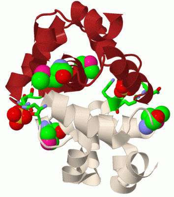
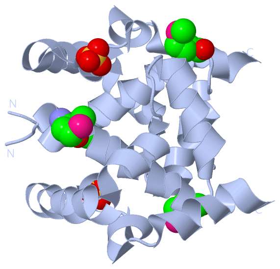
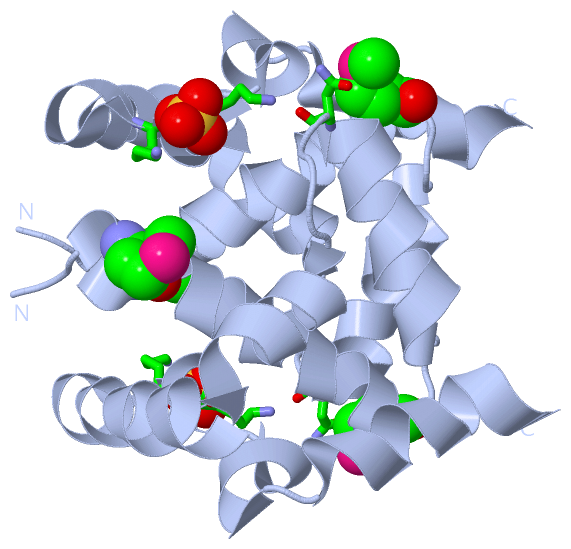
(-) Ligands, Modified Residues, Ions  (2, 3)

Asymmetric Unit (2, 3)
No.NameCountTypeFull Name
1MSE2Mod. Amino AcidSELENOMETHIONINE
2SO41Ligand/IonSULFATE ION
Biological Unit 1 (2, 6)
No.NameCountTypeFull Name
1MSE4Mod. Amino AcidSELENOMETHIONINE
2SO42Ligand/IonSULFATE ION

(-) Sites  (1, 1)

Asymmetric Unit (1, 1)
No.NameEvidenceResiduesDescription
1AC1SOFTWAREASN A:52 , LYS A:82 , LYS A:83BINDING SITE FOR RESIDUE SO4 A 201

(-) SS Bonds  (0, 0)

(no "SS Bond" information available for 3CTV)

(-) Cis Peptide Bonds  (0, 0)

(no "Cis Peptide Bond" information available for 3CTV)

 Sequence-Structure Mapping

(-) SAPs(SNPs)/Variants  (0, 0)

(no "SAP(SNP)/Variant" information available for 3CTV)

(-) PROSITE Motifs  (0, 0)

(no "PROSITE Motif" information available for 3CTV)

(-) Exons   (0, 0)

(no "Exon" information available for 3CTV)

(-) Sequences/Alignments

Asymmetric Unit
   Reformat: Number of residues per line =  ('0' or empty: single-line sequence representation)
  Number of residues per labelling interval =   
  UniProt sequence: complete  aligned part    
   Show mapping: SCOP domains CATH domains Pfam domains Secondary structure (by author)
SAPs(SNPs) PROSITE motifs Exons
(details for a mapped element are shown in a popup box when the mouse pointer rests over it)
Chain A from PDB  Type:PROTEIN  Length:92
 aligned with O28011_ARCFU | O28011 from UniProtKB/TrEMBL  Length:668

    Alignment length:92
                                   318       328       338       348       358       368       378       388       398  
         O28011_ARCFU   309 KINPMDFTFVEINEAVKLVEMGVATPQDIDTAIKLGLNRPFGPFELAKQFGAEQIAKRLEELAKQFGKKIFEPAKTLKEGKLEELLKAGKAE 400
               SCOP domains -------------------------------------------------------------------------------------------- SCOP domains
               CATH domains 3ctvA00 A:15-106 N-(1-d-carboxylethyl)-l-norvaline Dehydrogenase; domain 2                   CATH domains
               Pfam domains -------------------------------------------------------------------------------------------- Pfam domains
         Sec.struct. author ..hhhhhhhhhhhhhhhhhhh...hhhhhhhhhhhhh....hhhhhhhhhhhhhhhhhhhhhhhhhhhhhhh.hhhhhh.hhhhhhhhhhhh Sec.struct. author
                 SAPs(SNPs) -------------------------------------------------------------------------------------------- SAPs(SNPs)
                    PROSITE -------------------------------------------------------------------------------------------- PROSITE
                 Transcript -------------------------------------------------------------------------------------------- Transcript
                 3ctv A  15 KINPmDFTFVEINEAVKLVEmGVATPQDIDTAIKLGLNRPFGPFELAKQFGAEQIAKRLEELAKQFGKKIFEPAKTLKEGKLEELLKAGKAE 106
                                |   24        34|       44        54        64        74        84        94       104  
                                |              35-MSE                                                                   
                               19-MSE                                                                                   

   Legend:   → Mismatch (orange background)
  - → Gap (green background, '-', border residues have a numbering label)
    → Modified Residue (blue background, lower-case, 'x' indicates undefined single-letter code, labelled with number + name)
  x → Chemical Group (purple background, 'x', labelled with number + name, e.g. ACE or NH2)
  extra numbering lines below/above indicate numbering irregularities and modified residue names etc., number ends below/above '|'

 Classification and Annotation

(-) SCOP Domains  (0, 0)

(no "SCOP Domain" information available for 3CTV)

(-) CATH Domains  (1, 1)

Asymmetric Unit

(-) Pfam Domains  (0, 0)

(no "Pfam Domain" information available for 3CTV)

(-) Gene Ontology  (8, 8)

Asymmetric Unit(hide GO term definitions)
Chain A   (O28011_ARCFU | O28011)
molecular function
    GO:0003857    3-hydroxyacyl-CoA dehydrogenase activity    Catalysis of the reaction: (S)-3-hydroxyacyl-CoA + NAD+ = 3-oxoacyl-CoA + NADH + H(+).
    GO:0003824    catalytic activity    Catalysis of a biochemical reaction at physiological temperatures. In biologically catalyzed reactions, the reactants are known as substrates, and the catalysts are naturally occurring macromolecular substances known as enzymes. Enzymes possess specific binding sites for substrates, and are usually composed wholly or largely of protein, but RNA that has catalytic activity (ribozyme) is often also regarded as enzymatic.
    GO:0016491    oxidoreductase activity    Catalysis of an oxidation-reduction (redox) reaction, a reversible chemical reaction in which the oxidation state of an atom or atoms within a molecule is altered. One substrate acts as a hydrogen or electron donor and becomes oxidized, while the other acts as hydrogen or electron acceptor and becomes reduced.
biological process
    GO:0006631    fatty acid metabolic process    The chemical reactions and pathways involving fatty acids, aliphatic monocarboxylic acids liberated from naturally occurring fats and oils by hydrolysis.
    GO:0008152    metabolic process    The chemical reactions and pathways, including anabolism and catabolism, by which living organisms transform chemical substances. Metabolic processes typically transform small molecules, but also include macromolecular processes such as DNA repair and replication, and protein synthesis and degradation.
    GO:0055114    oxidation-reduction process    A metabolic process that results in the removal or addition of one or more electrons to or from a substance, with or without the concomitant removal or addition of a proton or protons.
cellular component
    GO:0016021    integral component of membrane    The component of a membrane consisting of the gene products and protein complexes having at least some part of their peptide sequence embedded in the hydrophobic region of the membrane.
    GO:0016020    membrane    A lipid bilayer along with all the proteins and protein complexes embedded in it an attached to it.

 Visualization

(-) Interactive Views

Asymmetric Unit
  Complete Structure
    Jena3D(integrated viewing of ligand, site, SAP, PROSITE, SCOP information)
    WebMol | AstexViewer[tm]@PDBe
(Java Applets, require no local installation except for Java; loading may be slow)
    STRAP
(Java WebStart application, automatic local installation, requires Java; full application with system access!)
    RasMol
(require local installation)
    Molscript (VRML)
(requires installation of a VRML viewer; select preferred view via VRML and generate a mono or stereo PDF format file)
 
  Ligands, Modified Residues, Ions
    MSE  [ RasMol | Jena3D ]  +environment [ RasMol | Jena3D ]
    SO4  [ RasMol | Jena3D ]  +environment [ RasMol | Jena3D ]
 
  Sites
    AC1  [ RasMol ]  +environment [ RasMol ]
 
  Cis Peptide Bonds
(no "Cis Peptide Bonds" information available for 3ctv)
 
Biological Unit
  Complete Structure
    Biological Unit 1  [ Jena3D ]

(-) Still Images

Jmol
  protein: cartoon or spacefill or dots and stick; nucleic acid: cartoon and stick; ligands: spacefill; active site: stick
Molscript
  protein, nucleic acid: cartoon; ligands: spacefill; active site: ball and stick

 Databases and Analysis Tools

(-) Databases

Access by PDB/NDB ID
  3ctv
    Family and Domain InformationProDom | SYSTERS
    General Structural InformationGlycoscienceDB | MMDB | NDB | OCA | PDB | PDBe | PDBj | PDBsum | PDBWiki | PQS | PROTEOPEDIA
    Orientation in MembranesOPM
    Protein SurfaceSURFACE
    Secondary StructureDSSP (structure derived) | HSSP (homology derived)
    Structural GenomicsGeneCensus
    Structural NeighboursCE | VAST
    Structure ClassificationCATH | Dali | SCOP
    Validation and Original DataBMRB Data View | BMRB Restraints Grid | EDS | PROCHECK | RECOORD | WHAT_CHECK
 
Access by UniProt ID/Accession number
  O28011_ARCFU | O28011
    Comparative Protein Structure ModelsModBase
    Genomic InformationEnsembl
    Protein-protein InteractionDIP
    Sequence, Family and Domain InformationInterPro | Pfam | SMART | UniProtKB/TrEMBL
 
Access by Enzyme Classificator   (EC Number)
  (no 'Enzyme Classificator' available)
    General Enzyme InformationBRENDA | EC-PDB | Enzyme | IntEnz
    PathwayKEGG | MetaCyc
 
Access by Disease Identifier   (MIM ID)
  (no 'MIM ID' available)
    Disease InformationOMIM
 
Access by GenAge ID
  (no 'GenAge ID' available)
    Age Related InformationGenAge

(-) Analysis Tools

Access by PDB/NDB ID
    Domain InformationXDom
    Interatomic Contacts of Structural UnitsCSU
    Ligand-protein ContactsLPC
    Protein CavitiescastP
    Sequence and Secondary StructurePDBCartoon
    Structure AlignmentSTRAP(Java WebStart application, automatic local installation, requires Java; full application with system access!)
    Structure and Sequence BrowserSTING
 
Access by UniProt ID/Accession number
  O28011_ARCFU | O28011
    Protein Disorder PredictionDisEMBL | FoldIndex | GLOBPLOT (for more information see DisProt)

 Related Entries

(-) Entries Sharing at Least One Protein Chain (UniProt ID)

(no "Entries Sharing at Least One Protein Chain" available for 3CTV)

(-) Related Entries Specified in the PDB File

3hdh PIG HEART SHORT CHAIN L-3-HYDROXYACYL COA DEHYDROGENASE