Show PDB file:   
         Plain Text   HTML   (compressed file size)
QuickSearch:   
by PDB,NDB,UniProt,PROSITE Code or Search Term(s)  
(-)Biol.Unit 1 - manually
(-)Asym.Unit - manually
(-)Asymmetric Unit
(-)Asym. Unit - sites
(-)Biological Unit 1
(-)Biol. Unit 1 - sites
collapse expand < >
Image Biol.Unit 1 - manually
Biol.Unit 1 - manually  (Jmol Viewer)
Image Asym.Unit - manually
Asym.Unit - manually  (Jmol Viewer)
Image Asymmetric Unit
Asymmetric Unit  (Jmol Viewer)
Image Asym. Unit - sites
Asym. Unit - sites  (Jmol Viewer)
Image Biological Unit 1
Biological Unit 1  (Jmol Viewer)
Image Biol. Unit 1 - sites
Biol. Unit 1 - sites  (Jmol Viewer)

(-) Description

Title :  CRYSTAL STRUCTURE OF A MYO-INOSITOL-1-PHOSPHATE SYNTHASE-RELATED PROTEIN (TM_1419) FROM THERMOTOGA MARITIMA MSB8 AT 1.70 A RESOLUTION
 
Authors :  Joint Center For Structural Genomics (Jcsg)
Date :  11 Mar 08  (Deposition) - 25 Mar 08  (Release) - 13 Jul 11  (Revision)
Method :  X-RAY DIFFRACTION
Resolution :  1.70
Chains :  Asym. Unit :  A
Biol. Unit 1:  A  (4x)
Keywords :  Myo-Inositol-1-Phosphate Synthase-Related Protein, Structural Genomics, Joint Center For Structural Genomics, Jcsg, Protein Structure Initiative, Psi-2, Isomerase (Keyword Search: [Gene Ontology, PubMed, Web (Google))
 
Reference :  Joint Center For Structural Genomics (Jcsg)
Crystal Structure Of Myo-Inositol-1-Phosphate Synthase-Related Protein (Tm1419) From Thermotoga Maritima At 1. 70 A Resolution
To Be Published
PubMed: search

(-) Compounds

Molecule 1 - MYO-INOSITOL-1-PHOSPHATE SYNTHASE-RELATED PROTEIN
    Atcc43589
    ChainsA
    EC Number5.5.1.4
    EngineeredYES
    Expression SystemESCHERICHIA COLI
    Expression System PlasmidMH1
    Expression System StrainDL41
    Expression System Taxid562
    Expression System Vector TypePLASMID
    GeneTM1419, TM_1419
    Organism ScientificTHERMOTOGA MARITIMA MSB8
    Organism Taxid243274
    StrainMSB8 / DSM 3109 / JCM 10099

 Structural Features

(-) Chains, Units

  1
Asymmetric Unit A
Biological Unit 1 (4x)A

Summary Information (see also Sequences/Alignments below)
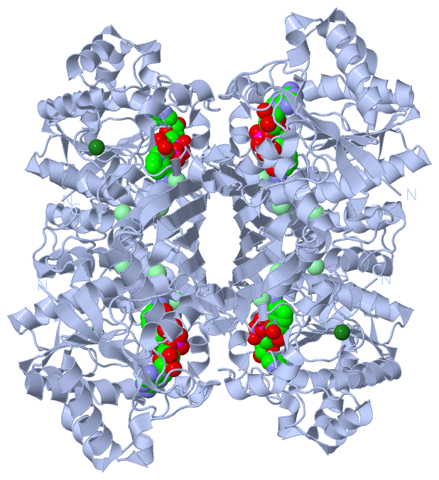
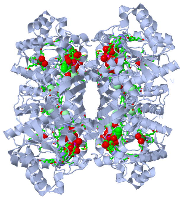
(-) Ligands, Modified Residues, Ions  (3, 5)

Asymmetric Unit (3, 5)
No.NameCountTypeFull Name
1CL3Ligand/IonCHLORIDE ION
2MG1Ligand/IonMAGNESIUM ION
3NAD1Ligand/IonNICOTINAMIDE-ADENINE-DINUCLEOTIDE
Biological Unit 1 (1, 4)
No.NameCountTypeFull Name
1CL-1Ligand/IonCHLORIDE ION
2MG-1Ligand/IonMAGNESIUM ION
3NAD4Ligand/IonNICOTINAMIDE-ADENINE-DINUCLEOTIDE

(-) Sites  (5, 5)

Asymmetric Unit (5, 5)
No.NameEvidenceResiduesDescription
1AC1SOFTWAREGLY A:212 , ARG A:229 , HOH A:506BINDING SITE FOR RESIDUE CL A 383
2AC2SOFTWAREALA A:218 , ASP A:219 , SER A:222 , PRO A:363 , ARG A:364BINDING SITE FOR RESIDUE CL A 384
3AC3SOFTWAREALA A:213 , ASN A:238 , LYS A:290 , HOH A:433BINDING SITE FOR RESIDUE CL A 385
4AC4SOFTWAREASN A:189 , PRO A:354 , PRO A:356 , GLU A:359 , HOH A:401 , HOH A:402BINDING SITE FOR RESIDUE MG A 386
5AC5SOFTWAREGLY A:8 , GLN A:9 , GLY A:10 , TYR A:11 , ASP A:53 , LYS A:58 , SER A:91 , THR A:130 , CYS A:131 , THR A:132 , THR A:133 , ALA A:159 , TYR A:163 , ILE A:183 , ASP A:208 , THR A:211 , MET A:243 , ASP A:244 , ASP A:316 , LEU A:320 , HOH A:411 , HOH A:416 , HOH A:421 , HOH A:426 , HOH A:474 , HOH A:563BINDING SITE FOR RESIDUE NAD A 400

(-) SS Bonds  (0, 0)

(no "SS Bond" information available for 3CIN)

(-) Cis Peptide Bonds  (2, 2)

Asymmetric Unit
No.Residues
1Gly A:206 -Asp A:207
2Gly A:355 -Pro A:356

 Sequence-Structure Mapping

(-) SAPs(SNPs)/Variants  (0, 0)

(no "SAP(SNP)/Variant" information available for 3CIN)

(-) PROSITE Motifs  (0, 0)

(no "PROSITE Motif" information available for 3CIN)

(-) Exons   (0, 0)

(no "Exon" information available for 3CIN)

(-) Sequences/Alignments

Asymmetric Unit
   Reformat: Number of residues per line =  ('0' or empty: single-line sequence representation)
  Number of residues per labelling interval =   
  UniProt sequence: complete  aligned part    
   Show mapping: SCOP domains CATH domains Pfam domains Secondary structure (by author)
SAPs(SNPs) PROSITE motifs Exons
(details for a mapped element are shown in a popup box when the mouse pointer rests over it)
Chain A from PDB  Type:PROTEIN  Length:382
 aligned with Q9X1D6_THEMA | Q9X1D6 from UniProtKB/TrEMBL  Length:382

    Alignment length:382
                             1                                                                                                                                                                                                                                                                                                                                                                                            
                             |       9        19        29        39        49        59        69        79        89        99       109       119       129       139       149       159       169       179       189       199       209       219       229       239       249       259       269       279       289       299       309       319       329       339       349       359       369       379  
         Q9X1D6_THEMA     - -MVKVLILGQGYVASTFVAGLEKLRKGEIEPYGVPLARELPIGFEDIKIVGSYDVDRAKIGKKLSEVVKQYWNDVDSLTSDPEIRKGVHLGSVRNLPIEAEGLEDSMTLKEAVDTLVKEWTELDPDVIVNTCTTEAFVPFGNKEDLLKAIENNDKERLTATQVYAYAAALYANKRGGAAFVNVIPTFIANDPAFVELAKENNLVVFGDDGATGATPFTADVLSHLAQRNRYVKDVAQFNIGGNMDFLALTDDGKNKSKEFTKSSIVKDILGYDAPHYIKPTGYLEPLGDKKFIAIHIEYVSFNGATDELMINGRINDSPALGGLLVDLVRLGKIALDRKEFGTVYPVNAFYMKNPGPAEEKNIPRIIAYEKMRIWAGLKPKW 381
               SCOP domains d3cina1 A:0-209,A:317-381 Hypothetical protein TM1419                                                                                                                                                             d3cina2 A:210-316 Hypothetical protein TM1419                                                              d3cina1 A:0-209,A:317-381 Hypothetical protein TM1419             SCOP domains
               CATH domains 3cinA01 A:0-211,A:316-381 NAD(P)-binding Rossmann-like Domain                                                                                                                                                       3cinA02 A:212-315 Dihydrodipicolinate Reductase; domain 2                                               3cinA01 A:0-211,A:316-381 NAD(P)-binding Rossmann-like Domain      CATH domains
               Pfam domains ---------------------------------------------------------------------------------------------------------------------------------------------------------------------------------------------------------------------------------------------------------------------------------------------------------------------------------------------------------------------------------------------- Pfam domains
         Sec.struct. author ..eeeeee..hhhhhhhhhhhhhhhh................hhh.eeeeeeee........hhhhhhhhhh............ee...............hhhhh.hhhhhhhhhhhhhhhhh..eeee...........hhhhhhhhhhh......hhhhhhhhhhhhhhhhhh.eeeee........hhhhhhhhhhh.eeee......hhhhhhhhhhhhhhhh..eeeeeeeeeee.hhhhhh.hhhhhhhhhhhhh.hhhhhhhh....ee....ee.hhhh.eeeeeeeeeee.hhh.eeeeeeeeeeehhhhhhhhhhhhhhhhhhhhhh...eehhhhhhhhh...........hhhhhhhhhhhhh...... Sec.struct. author
                 SAPs(SNPs) ---------------------------------------------------------------------------------------------------------------------------------------------------------------------------------------------------------------------------------------------------------------------------------------------------------------------------------------------------------------------------------------------- SAPs(SNPs)
                    PROSITE ---------------------------------------------------------------------------------------------------------------------------------------------------------------------------------------------------------------------------------------------------------------------------------------------------------------------------------------------------------------------------------------------- PROSITE
                 Transcript ---------------------------------------------------------------------------------------------------------------------------------------------------------------------------------------------------------------------------------------------------------------------------------------------------------------------------------------------------------------------------------------------- Transcript
                 3cin A   0 HMVKVLILGQGYVASTFVAGLEKLRKGEIEPYGVPLARELPIGFEDIKIVGSYDVDRAKIGKKLSEVVKQYWNDVDSLTSDPEIRKGVHLGSVRNLPIEAEGLEDSMTLKEAVDTLVKEWTELDPDVIVNTCTTEAFVPFGNKEDLLKAIENNDKERLTATQVYAYAAALYANKRGGAAFVNVIPTFIANDPAFVELAKENNLVVFGDDGATGATPFTADVLSHLAQRNRYVKDVAQFNIGGNMDFLALTDDGKNKSKEFTKSSIVKDILGYDAPHYIKPTGYLEPLGDKKFIAIHIEYVSFNGATDELMINGRINDSPALGGLLVDLVRLGKIALDRKEFGTVYPVNAFYMKNPGPAEEKNIPRIIAYEKMRIWAGLKPKW 381
                                     9        19        29        39        49        59        69        79        89        99       109       119       129       139       149       159       169       179       189       199       209       219       229       239       249       259       269       279       289       299       309       319       329       339       349       359       369       379  

   Legend:   → Mismatch (orange background)
  - → Gap (green background, '-', border residues have a numbering label)
    → Modified Residue (blue background, lower-case, 'x' indicates undefined single-letter code, labelled with number + name)
  x → Chemical Group (purple background, 'x', labelled with number + name, e.g. ACE or NH2)
  extra numbering lines below/above indicate numbering irregularities and modified residue names etc., number ends below/above '|'

 Classification and Annotation

(-) SCOP Domains  (2, 2)

Asymmetric Unit

(-) CATH Domains  (2, 2)

Asymmetric Unit
(-)
Class: Alpha Beta (26913)

(-) Pfam Domains  (0, 0)

(no "Pfam Domain" information available for 3CIN)

(-) Gene Ontology  (6, 6)

Asymmetric Unit(hide GO term definitions)
Chain A   (Q9X1D6_THEMA | Q9X1D6)
molecular function
    GO:0004512    inositol-3-phosphate synthase activity    Catalysis of the reaction: D-glucose 6-phosphate = 1D-myo-inositol 3-phosphate. This reaction requires NAD, which dehydrogenates the CHOH group to CO at C-5 of the glucose 6-phosphate, making C-6 into an active methylene, able to condense with the aldehyde at C-1. Finally, the enzyme-bound NADH reconverts C-5 into the CHOH form.
    GO:0016853    isomerase activity    Catalysis of the geometric or structural changes within one molecule. Isomerase is the systematic name for any enzyme of EC class 5.
    GO:0046872    metal ion binding    Interacting selectively and non-covalently with any metal ion.
    GO:0000166    nucleotide binding    Interacting selectively and non-covalently with a nucleotide, any compound consisting of a nucleoside that is esterified with (ortho)phosphate or an oligophosphate at any hydroxyl group on the ribose or deoxyribose.
biological process
    GO:0006021    inositol biosynthetic process    The chemical reactions and pathways resulting in the formation of inositol, 1,2,3,4,5,6-cyclohexanehexol, a growth factor for animals and microorganisms.
    GO:0008654    phospholipid biosynthetic process    The chemical reactions and pathways resulting in the formation of phospholipids, any lipid containing phosphoric acid as a mono- or diester.

 Visualization

(-) Interactive Views

Asymmetric Unit
  Complete Structure
    Jena3D(integrated viewing of ligand, site, SAP, PROSITE, SCOP information)
    WebMol | AstexViewer[tm]@PDBe
(Java Applets, require no local installation except for Java; loading may be slow)
    STRAP
(Java WebStart application, automatic local installation, requires Java; full application with system access!)
    RasMol
(require local installation)
    Molscript (VRML)
(requires installation of a VRML viewer; select preferred view via VRML and generate a mono or stereo PDF format file)
 
  Ligands, Modified Residues, Ions
    CL  [ RasMol | Jena3D ]  +environment [ RasMol | Jena3D ]
    MG  [ RasMol | Jena3D ]  +environment [ RasMol | Jena3D ]
    NAD  [ RasMol | Jena3D ]  +environment [ RasMol | Jena3D ]
 
  Sites
    AC1  [ RasMol ]  +environment [ RasMol ]
    AC2  [ RasMol ]  +environment [ RasMol ]
    AC3  [ RasMol ]  +environment [ RasMol ]
    AC4  [ RasMol ]  +environment [ RasMol ]
    AC5  [ RasMol ]  +environment [ RasMol ]
 
  Cis Peptide Bonds
    Gly A:206 - Asp A:207   [ RasMol ]  
    Gly A:355 - Pro A:356   [ RasMol ]  
 
Biological Unit
  Complete Structure
    Biological Unit 1  [ Jena3D ]

(-) Still Images

Jmol
  protein: cartoon or spacefill or dots and stick; nucleic acid: cartoon and stick; ligands: spacefill; active site: stick
Molscript
  protein, nucleic acid: cartoon; ligands: spacefill; active site: ball and stick

 Databases and Analysis Tools

(-) Databases

Access by PDB/NDB ID
  3cin
    Family and Domain InformationProDom | SYSTERS
    General Structural InformationGlycoscienceDB | MMDB | NDB | OCA | PDB | PDBe | PDBj | PDBsum | PDBWiki | PQS | PROTEOPEDIA
    Orientation in MembranesOPM
    Protein SurfaceSURFACE
    Secondary StructureDSSP (structure derived) | HSSP (homology derived)
    Structural GenomicsGeneCensus
    Structural NeighboursCE | VAST
    Structure ClassificationCATH | Dali | SCOP
    Validation and Original DataBMRB Data View | BMRB Restraints Grid | EDS | PROCHECK | RECOORD | WHAT_CHECK
 
Access by UniProt ID/Accession number
  Q9X1D6_THEMA | Q9X1D6
    Comparative Protein Structure ModelsModBase
    Genomic InformationEnsembl
    Protein-protein InteractionDIP
    Sequence, Family and Domain InformationInterPro | Pfam | SMART | UniProtKB/TrEMBL
 
Access by Enzyme Classificator   (EC Number)
  5.5.1.4
    General Enzyme InformationBRENDA | EC-PDB | Enzyme | IntEnz
    PathwayKEGG | MetaCyc
 
Access by Disease Identifier   (MIM ID)
  (no 'MIM ID' available)
    Disease InformationOMIM
 
Access by GenAge ID
  (no 'GenAge ID' available)
    Age Related InformationGenAge

(-) Analysis Tools

Access by PDB/NDB ID
    Domain InformationXDom
    Interatomic Contacts of Structural UnitsCSU
    Ligand-protein ContactsLPC
    Protein CavitiescastP
    Sequence and Secondary StructurePDBCartoon
    Structure AlignmentSTRAP(Java WebStart application, automatic local installation, requires Java; full application with system access!)
    Structure and Sequence BrowserSTING
 
Access by UniProt ID/Accession number
  Q9X1D6_THEMA | Q9X1D6
    Protein Disorder PredictionDisEMBL | FoldIndex | GLOBPLOT (for more information see DisProt)

 Related Entries

(-) Entries Sharing at Least One Protein Chain (UniProt ID)

(no "Entries Sharing at Least One Protein Chain" available for 3CIN)

(-) Related Entries Specified in the PDB File

(no "Related Entries Specified in the PDB File" available for 3CIN)