Show PDB file:   
         Plain Text   HTML   (compressed file size)
QuickSearch:   
by PDB,NDB,UniProt,PROSITE Code or Search Term(s)  
(-)Asym./Biol. Unit
(-)Asym./Biol. Unit - sites
collapse expand < >
Image Asym./Biol. Unit
Asym./Biol. Unit  (Jmol Viewer)
Image Asym./Biol. Unit - sites
Asym./Biol. Unit - sites  (Jmol Viewer)

(-) Description

Title :  ACTIVE-SITE MUTANT E212Q DETERMINED AT PH 6.0 WITH CELLOBIOSE BOUND IN THE ACTIVE SITE
 
Authors :  C. Divne, J. Stahlberg, T. A. Jones
Date :  24 Aug 96  (Deposition) - 12 Mar 97  (Release) - 13 Jul 11  (Revision)
Method :  X-RAY DIFFRACTION
Resolution :  2.00
Chains :  Asym./Biol. Unit :  A
Keywords :  Cellulose Degradation, Hydrolase, Glycosidase, Glycoprotein (Keyword Search: [Gene Ontology, PubMed, Web (Google))
 
Reference :  J. Stahlberg, C. Divne, A. Koivula, K. Piens, M. Claeyssens, T. T. Teeri, T. A. Jones
Activity Studies And Crystal Structures Of Catalytically Deficient Mutants Of Cellobiohydrolase I From Trichoderma Reesei.
J. Mol. Biol. V. 264 337 1996
PubMed-ID: 8951380  |  Reference-DOI: 10.1006/JMBI.1996.0644
(for further references see the PDB file header)

(-) Compounds

Molecule 1 - 1,4-BETA-D-GLUCAN CELLOBIOHYDROLASE I
    ChainsA
    EC Number3.2.1.91
    EngineeredYES
    Expression SystemHYPOCREA JECORINA
    Expression System GeneCBH1
    Expression System PlasmidPEM-F5
    Expression System Taxid51453
    FragmentCATALYTIC DOMAIN, RESIDUES 1 - 434
    GeneCBH1
    MutationYES
    Organism ScientificHYPOCREA JECORINA
    Organism Taxid51453
    StrainQM 9414
    SynonymEXOGLUCANASE, EXOCELLULASE
    VariantVTT-D-93201

 Structural Features

(-) Chains, Units

  1
Asymmetric/Biological Unit A

Summary Information (see also Sequences/Alignments below)
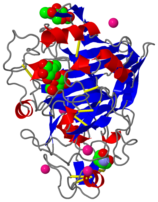
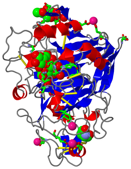
(-) Ligands, Modified Residues, Ions  (4, 9)

Asymmetric/Biological Unit (4, 9)
No.NameCountTypeFull Name
1BGC2Ligand/IonBETA-D-GLUCOSE
2CD5Ligand/IonCADMIUM ION
3NAG1Ligand/IonN-ACETYL-D-GLUCOSAMINE
4PCA1Mod. Amino AcidPYROGLUTAMIC ACID

(-) Sites  (10, 10)

Asymmetric Unit (10, 10)
No.NameEvidenceResiduesDescription
01AC1SOFTWARESER A:21 , GLY A:22 , ASN A:270 , PRO A:314 , HOH A:577 , HOH A:578 , HOH A:650BINDING SITE FOR RESIDUE NAG A 435
02AC2SOFTWAREARG A:251 , ARG A:394 , BGC A:437 , HOH A:544BINDING SITE FOR RESIDUE BGC A 436
03AC3SOFTWARETHR A:246 , ARG A:251 , PRO A:258 , ASP A:259 , TRP A:376 , HOH A:545 , HOH A:553 , HOH A:702 , HOH A:703 , HOH A:710 , BGC A:436BINDING SITE FOR RESIDUE BGC A 437
04AC4SOFTWAREGLU A:65 , GLU A:190BINDING SITE FOR RESIDUE CD A 438
05AC5SOFTWAREHIS A:206 , GLU A:239BINDING SITE FOR RESIDUE CD A 439
06AC6SOFTWAREASP A:52 , GLU A:317BINDING SITE FOR RESIDUE CD A 440
07AC7SOFTWAREASP A:94 , HOH A:730BINDING SITE FOR RESIDUE CD A 441
08AC8SOFTWAREGLU A:295 , GLU A:325BINDING SITE FOR RESIDUE CD A 1000
09CADUNKNOWNCD A:1000 , GLU A:295 , GLU A:325CALCIUM-BINDING SITE ON CRYSTALLOGRAPHIC DYAD. NOTE THAT THE CADMIUM ION IS BOUND BY GLU 295 AND GLU 325 FROM TWO CRYSTALLOGRAPHICALLY RELATED MOLECULES.
10CTAUNKNOWNGLN A:212 , ASP A:214 , GLU A:217 , HIS A:228CATALYTIC SITE INCLUDING MUTATION E212Q.

(-) SS Bonds  (10, 10)

Asymmetric/Biological Unit
No.Residues
1A:4 -A:72
2A:19 -A:25
3A:50 -A:71
4A:61 -A:67
5A:138 -A:397
6A:172 -A:210
7A:176 -A:209
8A:230 -A:256
9A:238 -A:243
10A:261 -A:331

(-) Cis Peptide Bonds  (1, 1)

Asymmetric/Biological Unit
No.Residues
1Tyr A:381 -Pro A:382

 Sequence-Structure Mapping

(-) SAPs(SNPs)/Variants  (0, 0)

(no "SAP(SNP)/Variant" information available for 3CEL)

(-) PROSITE Motifs  (0, 0)

(no "PROSITE Motif" information available for 3CEL)

(-) Exons   (0, 0)

(no "Exon" information available for 3CEL)

(-) Sequences/Alignments

Asymmetric/Biological Unit
   Reformat: Number of residues per line =  ('0' or empty: single-line sequence representation)
  Number of residues per labelling interval =   
  UniProt sequence: complete  aligned part    
   Show mapping: SCOP domains CATH domains Pfam domains Secondary structure (by author)
SAPs(SNPs) PROSITE motifs Exons
(details for a mapped element are shown in a popup box when the mouse pointer rests over it)
Chain A from PDB  Type:PROTEIN  Length:434
 aligned with GUX1_HYPJE | P62694 from UniProtKB/Swiss-Prot  Length:513

    Alignment length:434
                                    27        37        47        57        67        77        87        97       107       117       127       137       147       157       167       177       187       197       207       217       227       237       247       257       267       277       287       297       307       317       327       337       347       357       367       377       387       397       407       417       427       437       447    
           GUX1_HYPJE    18 QSACTLQSETHPPLTWQKCSSGGTCTQQTGSVVIDANWRWTHATNSSTNCYDGNTWSSTLCPDNETCAKNCCLDGAAYASTYGVTTSGNSLSIGFVTQSAQKNVGARLYLMASDTTYQEFTLLGNEFSFDVDVSQLPCGLNGALYFVSMDADGGVSKYPTNTAGAKYGTGYCDSQCPRDLKFINGQANVEGWEPSSNNANTGIGGHGSCCSEMDIWEANSISEALTPHPCTTVGQEICEGDGCGGTYSDNRYGGTCDPDGCDWNPYRLGNTSFYGPGSSFTLDTTKKLTVVTQFETSGAINRYYVQNGVTFQQPNAELGSYSGNELNDDYCTAEEAEFGGSSFSDKGGLTQFKKATSGGMVLVMSLWDDYYANMLWLDSTYPTNETSSTPGAVRGSCSTSSGVPAQVESQSPNAKVTFSNIKFGPIGSTGNPSG 451
               SCOP domains d3cela_ A: Cellobiohydrolase I (cellulase, Endoglucanase I, CBH1)                                                                                                                                                                                                                                                                                                                                                                                  SCOP domains
               CATH domains -3celA00 A:2-434 1,4-Beta-D-Glucan Cellobiohydrolase I, subunit A                                                                                                                                                                                                                                                                                                                                                                                  CATH domains
               Pfam domains -------------------------------------------------------------------------------------------------------------------------------------------------------------------------------------------------------------------------------------------------------------------------------------------------------------------------------------------------------------------------------------------------------------------------------------------------- Pfam domains
         Sec.struct. author ............eeeeeee.....eeeeeeeeee.hhh..eee....................hhhhhhheee....hhhhh.eeee..eeeee.eee...eee.eeeeee.......eee...eeeeeeee.......eeeeeeee.................hhh......................................eeee..eee.eee......eeee.......eeeehhhh.................eee.hhhh...................eeeeeee.....eeeeeee..eeee...eee..eee....hhhhhhhhhhh...hhhh..hhhhhhhhh..eeeeeeeeee......hhhh.........................hhhhhhh....eeeee..eeee......... Sec.struct. author
                 SAPs(SNPs) -------------------------------------------------------------------------------------------------------------------------------------------------------------------------------------------------------------------------------------------------------------------------------------------------------------------------------------------------------------------------------------------------------------------------------------------------- SAPs(SNPs)
                    PROSITE -------------------------------------------------------------------------------------------------------------------------------------------------------------------------------------------------------------------------------------------------------------------------------------------------------------------------------------------------------------------------------------------------------------------------------------------------- PROSITE
                 Transcript -------------------------------------------------------------------------------------------------------------------------------------------------------------------------------------------------------------------------------------------------------------------------------------------------------------------------------------------------------------------------------------------------------------------------------------------------- Transcript
                 3cel A   1 xSACTLQSETHPPLTWQKCSSGGTCTQQTGSVVIDANWRWTHATNSSTNCYDGNTWSSTLCPDNETCAKNCCLDGAAYASTYGVTTSGNSLSIDFVTQSAQKNVGARLYLMASDTTYQEFTLLGNEFSFDVDVSQLPCGLNGALYFVSMDADGGVSKYPTNTAGAKYGTGYCDSQCPRDLKFINGQANVEGWEPSSNNANTGIGGHGSCCSQMDIWEANSISEALTPHPCTTVGQEICEGDGCGGTYSDNRYGGTCDPDGCDWNPYRLGNTSFYGPGSSFTLDTTKKLTVVTQFETSGAINRYYVQNGVTFQQPNAELGSYSGNELNDDYCTAEEAEFGGSSFSDKGGLTQFKKATSGGMVLVMSLWDDYYANMLWLDSTYPTNETSSTPGAVRGSCSTSSGVPAQVESQSPNAKVTFSNIKFGPIGSTGNPSG 434
                            |       10        20        30        40        50        60        70        80        90       100       110       120       130       140       150       160       170       180       190       200       210       220       230       240       250       260       270       280       290       300       310       320       330       340       350       360       370       380       390       400       410       420       430    
                            |                                                                                                                                                                                                                                                                                                                                                                                                                                                 
                            1-PCA                                                                                                                                                                                                                                                                                                                                                                                                                                             

Chain A from PDB  Type:PROTEIN  Length:434
 aligned with GUX1_TRIKO | P62695 from UniProtKB/Swiss-Prot  Length:513

    Alignment length:434
                                    27        37        47        57        67        77        87        97       107       117       127       137       147       157       167       177       187       197       207       217       227       237       247       257       267       277       287       297       307       317       327       337       347       357       367       377       387       397       407       417       427       437       447    
           GUX1_TRIKO    18 QSACTLQSETHPPLTWQKCSSGGTCTQQTGSVVIDANWRWTHATNSSTNCYDGNTWSSTLCPDNETCAKNCCLDGAAYASTYGVTTSGNSLSIGFVTQSAQKNVGARLYLMASDTTYQEFTLLGNEFSFDVDVSQLPCGLNGALYFVSMDADGGVSKYPTNTAGAKYGTGYCDSQCPRDLKFINGQANVEGWEPSSNNANTGIGGHGSCCSEMDIWEANSISEALTPHPCTTVGQEICEGDGCGGTYSDNRYGGTCDPDGCDWNPYRLGNTSFYGPGSSFTLDTTKKLTVVTQFETSGAINRYYVQNGVTFQQPNAELGSYSGNELNDDYCTAEEAEFGGSSFSDKGGLTQFKKATSGGMVLVMSLWDDYYANMLWLDSTYPTNETSSTPGAVRGSCSTSSGVPAQVESQSPNAKVTFSNIKFGPIGSTGNPSG 451
               SCOP domains d3cela_ A: Cellobiohydrolase I (cellulase, Endoglucanase I, CBH1)                                                                                                                                                                                                                                                                                                                                                                                  SCOP domains
               CATH domains -3celA00 A:2-434 1,4-Beta-D-Glucan Cellobiohydrolase I, subunit A                                                                                                                                                                                                                                                                                                                                                                                  CATH domains
               Pfam domains -------------------------------------------------------------------------------------------------------------------------------------------------------------------------------------------------------------------------------------------------------------------------------------------------------------------------------------------------------------------------------------------------------------------------------------------------- Pfam domains
         Sec.struct. author ............eeeeeee.....eeeeeeeeee.hhh..eee....................hhhhhhheee....hhhhh.eeee..eeeee.eee...eee.eeeeee.......eee...eeeeeeee.......eeeeeeee.................hhh......................................eeee..eee.eee......eeee.......eeeehhhh.................eee.hhhh...................eeeeeee.....eeeeeee..eeee...eee..eee....hhhhhhhhhhh...hhhh..hhhhhhhhh..eeeeeeeeee......hhhh.........................hhhhhhh....eeeee..eeee......... Sec.struct. author
                 SAPs(SNPs) -------------------------------------------------------------------------------------------------------------------------------------------------------------------------------------------------------------------------------------------------------------------------------------------------------------------------------------------------------------------------------------------------------------------------------------------------- SAPs(SNPs)
                    PROSITE -------------------------------------------------------------------------------------------------------------------------------------------------------------------------------------------------------------------------------------------------------------------------------------------------------------------------------------------------------------------------------------------------------------------------------------------------- PROSITE
                 Transcript -------------------------------------------------------------------------------------------------------------------------------------------------------------------------------------------------------------------------------------------------------------------------------------------------------------------------------------------------------------------------------------------------------------------------------------------------- Transcript
                 3cel A   1 xSACTLQSETHPPLTWQKCSSGGTCTQQTGSVVIDANWRWTHATNSSTNCYDGNTWSSTLCPDNETCAKNCCLDGAAYASTYGVTTSGNSLSIDFVTQSAQKNVGARLYLMASDTTYQEFTLLGNEFSFDVDVSQLPCGLNGALYFVSMDADGGVSKYPTNTAGAKYGTGYCDSQCPRDLKFINGQANVEGWEPSSNNANTGIGGHGSCCSQMDIWEANSISEALTPHPCTTVGQEICEGDGCGGTYSDNRYGGTCDPDGCDWNPYRLGNTSFYGPGSSFTLDTTKKLTVVTQFETSGAINRYYVQNGVTFQQPNAELGSYSGNELNDDYCTAEEAEFGGSSFSDKGGLTQFKKATSGGMVLVMSLWDDYYANMLWLDSTYPTNETSSTPGAVRGSCSTSSGVPAQVESQSPNAKVTFSNIKFGPIGSTGNPSG 434
                            |       10        20        30        40        50        60        70        80        90       100       110       120       130       140       150       160       170       180       190       200       210       220       230       240       250       260       270       280       290       300       310       320       330       340       350       360       370       380       390       400       410       420       430    
                            1-PCA                                                                                                                                                                                                                                                                                                                                                                                                                                             

   Legend:   → Mismatch (orange background)
  - → Gap (green background, '-', border residues have a numbering label)
    → Modified Residue (blue background, lower-case, 'x' indicates undefined single-letter code, labelled with number + name)
  x → Chemical Group (purple background, 'x', labelled with number + name, e.g. ACE or NH2)
  extra numbering lines below/above indicate numbering irregularities and modified residue names etc., number ends below/above '|'

 Classification and Annotation

(-) SCOP Domains  (1, 1)

Asymmetric/Biological Unit

(-) CATH Domains  (1, 1)

Asymmetric/Biological Unit
(-)
Class: Mainly Beta (13760)

(-) Pfam Domains  (0, 0)

(no "Pfam Domain" information available for 3CEL)

(-) Gene Ontology  (10, 20)

Asymmetric/Biological Unit(hide GO term definitions)
Chain A   (GUX1_TRIKO | P62695)
molecular function
    GO:0016162    cellulose 1,4-beta-cellobiosidase activity    Catalysis of the hydrolysis of (1->4)-beta-D-glucosidic linkages in cellulose and cellotetraose, releasing cellobiose from the non-reducing ends of the chains.
    GO:0030248    cellulose binding    Interacting selectively and non-covalently with cellulose.
    GO:0016787    hydrolase activity    Catalysis of the hydrolysis of various bonds, e.g. C-O, C-N, C-C, phosphoric anhydride bonds, etc. Hydrolase is the systematic name for any enzyme of EC class 3.
    GO:0016798    hydrolase activity, acting on glycosyl bonds    Catalysis of the hydrolysis of any glycosyl bond.
    GO:0004553    hydrolase activity, hydrolyzing O-glycosyl compounds    Catalysis of the hydrolysis of any O-glycosyl bond.
biological process
    GO:0005975    carbohydrate metabolic process    The chemical reactions and pathways involving carbohydrates, any of a group of organic compounds based of the general formula Cx(H2O)y. Includes the formation of carbohydrate derivatives by the addition of a carbohydrate residue to another molecule.
    GO:0030245    cellulose catabolic process    The chemical reactions and pathways resulting in the breakdown of cellulose, a linear beta1-4 glucan of molecular mass 50-400 kDa with the pyranose units in the -4C1 conformation.
    GO:0008152    metabolic process    The chemical reactions and pathways, including anabolism and catabolism, by which living organisms transform chemical substances. Metabolic processes typically transform small molecules, but also include macromolecular processes such as DNA repair and replication, and protein synthesis and degradation.
    GO:0000272    polysaccharide catabolic process    The chemical reactions and pathways resulting in the breakdown of a polysaccharide, a polymer of many (typically more than 10) monosaccharide residues linked glycosidically.
cellular component
    GO:0005576    extracellular region    The space external to the outermost structure of a cell. For cells without external protective or external encapsulating structures this refers to space outside of the plasma membrane. This term covers the host cell environment outside an intracellular parasite.

Chain A   (GUX1_HYPJE | P62694)
molecular function
    GO:0016162    cellulose 1,4-beta-cellobiosidase activity    Catalysis of the hydrolysis of (1->4)-beta-D-glucosidic linkages in cellulose and cellotetraose, releasing cellobiose from the non-reducing ends of the chains.
    GO:0030248    cellulose binding    Interacting selectively and non-covalently with cellulose.
    GO:0016787    hydrolase activity    Catalysis of the hydrolysis of various bonds, e.g. C-O, C-N, C-C, phosphoric anhydride bonds, etc. Hydrolase is the systematic name for any enzyme of EC class 3.
    GO:0016798    hydrolase activity, acting on glycosyl bonds    Catalysis of the hydrolysis of any glycosyl bond.
    GO:0004553    hydrolase activity, hydrolyzing O-glycosyl compounds    Catalysis of the hydrolysis of any O-glycosyl bond.
biological process
    GO:0005975    carbohydrate metabolic process    The chemical reactions and pathways involving carbohydrates, any of a group of organic compounds based of the general formula Cx(H2O)y. Includes the formation of carbohydrate derivatives by the addition of a carbohydrate residue to another molecule.
    GO:0030245    cellulose catabolic process    The chemical reactions and pathways resulting in the breakdown of cellulose, a linear beta1-4 glucan of molecular mass 50-400 kDa with the pyranose units in the -4C1 conformation.
    GO:0008152    metabolic process    The chemical reactions and pathways, including anabolism and catabolism, by which living organisms transform chemical substances. Metabolic processes typically transform small molecules, but also include macromolecular processes such as DNA repair and replication, and protein synthesis and degradation.
    GO:0000272    polysaccharide catabolic process    The chemical reactions and pathways resulting in the breakdown of a polysaccharide, a polymer of many (typically more than 10) monosaccharide residues linked glycosidically.
cellular component
    GO:0005576    extracellular region    The space external to the outermost structure of a cell. For cells without external protective or external encapsulating structures this refers to space outside of the plasma membrane. This term covers the host cell environment outside an intracellular parasite.

 Visualization

(-) Interactive Views

Asymmetric/Biological Unit
  Complete Structure
    Jena3D(integrated viewing of ligand, site, SAP, PROSITE, SCOP information)
    WebMol | AstexViewer[tm]@PDBe
(Java Applets, require no local installation except for Java; loading may be slow)
    STRAP
(Java WebStart application, automatic local installation, requires Java; full application with system access!)
    RasMol
(require local installation)
    Molscript (VRML)
(requires installation of a VRML viewer; select preferred view via VRML and generate a mono or stereo PDF format file)
 
  Ligands, Modified Residues, Ions
    BGC  [ RasMol | Jena3D ]  +environment [ RasMol | Jena3D ]
    CD  [ RasMol | Jena3D ]  +environment [ RasMol | Jena3D ]
    NAG  [ RasMol | Jena3D ]  +environment [ RasMol | Jena3D ]
    PCA  [ RasMol | Jena3D ]  +environment [ RasMol | Jena3D ]
 
  Sites
    AC1  [ RasMol ]  +environment [ RasMol ]
    AC2  [ RasMol ]  +environment [ RasMol ]
    AC3  [ RasMol ]  +environment [ RasMol ]
    AC4  [ RasMol ]  +environment [ RasMol ]
    AC5  [ RasMol ]  +environment [ RasMol ]
    AC6  [ RasMol ]  +environment [ RasMol ]
    AC7  [ RasMol ]  +environment [ RasMol ]
    AC8  [ RasMol ]  +environment [ RasMol ]
    CAD  [ RasMol ]  +environment [ RasMol ]
    CTA  [ RasMol ]  +environment [ RasMol ]
 
  Cis Peptide Bonds
    Tyr A:381 - Pro A:382   [ RasMol ]  
 

(-) Still Images

Jmol
  protein: cartoon or spacefill or dots and stick; nucleic acid: cartoon and stick; ligands: spacefill; active site: stick
Molscript
  protein, nucleic acid: cartoon; ligands: spacefill; active site: ball and stick

 Databases and Analysis Tools

(-) Databases

Access by PDB/NDB ID
  3cel
    Family and Domain InformationProDom | SYSTERS
    General Structural InformationGlycoscienceDB | MMDB | NDB | OCA | PDB | PDBe | PDBj | PDBsum | PDBWiki | PQS | PROTEOPEDIA
    Orientation in MembranesOPM
    Protein SurfaceSURFACE
    Secondary StructureDSSP (structure derived) | HSSP (homology derived)
    Structural GenomicsGeneCensus
    Structural NeighboursCE | VAST
    Structure ClassificationCATH | Dali | SCOP
    Validation and Original DataBMRB Data View | BMRB Restraints Grid | EDS | PROCHECK | RECOORD | WHAT_CHECK
 
Access by UniProt ID/Accession number
  GUX1_HYPJE | P62694
    Comparative Protein Structure ModelsModBase
    Genomic InformationEnsembl
    Protein-protein InteractionDIP
    Sequence, Family and Domain InformationInterPro | Pfam | SMART | UniProtKB/SwissProt
  GUX1_TRIKO | P62695
    Comparative Protein Structure ModelsModBase
    Genomic InformationEnsembl
    Protein-protein InteractionDIP
    Sequence, Family and Domain InformationInterPro | Pfam | SMART | UniProtKB/SwissProt
 
Access by Enzyme Classificator   (EC Number)
  3.2.1.91
    General Enzyme InformationBRENDA | EC-PDB | Enzyme | IntEnz
    PathwayKEGG | MetaCyc
 
Access by Disease Identifier   (MIM ID)
  (no 'MIM ID' available)
    Disease InformationOMIM
 
Access by GenAge ID
  (no 'GenAge ID' available)
    Age Related InformationGenAge

(-) Analysis Tools

Access by PDB/NDB ID
    Domain InformationXDom
    Interatomic Contacts of Structural UnitsCSU
    Ligand-protein ContactsLPC
    Protein CavitiescastP
    Sequence and Secondary StructurePDBCartoon
    Structure AlignmentSTRAP(Java WebStart application, automatic local installation, requires Java; full application with system access!)
    Structure and Sequence BrowserSTING
 
Access by UniProt ID/Accession number
  GUX1_HYPJE | P62694
    Protein Disorder PredictionDisEMBL | FoldIndex | GLOBPLOT (for more information see DisProt)
  GUX1_TRIKO | P62695
    Protein Disorder PredictionDisEMBL | FoldIndex | GLOBPLOT (for more information see DisProt)

 Related Entries

(-) Entries Sharing at Least One Protein Chain (UniProt ID)

UniProtKB/Swiss-Prot
        GUX1_HYPJE | P626941az6 1azh 1azj 1azk 1cbh 1cel 1dy4 1egn 1q2b 1q2e 2cbh 2cel 2mwj 2mwk 2v3i 2v3r 4c4c 4c4d 4cel 4d5i 4d5j 4d5o 4d5p 4d5q 4d5v 4p1h 4p1j 4uwt 4v0z 5cel 5x34 5x35 5x36 5x37 5x38 5x39 5x3c 6cel 7cel 8cel
        GUX1_TRIKO | P626951cel 1q2e 5cel 6cel 7cel

(-) Related Entries Specified in the PDB File

(no "Related Entries Specified in the PDB File" available for 3CEL)