Show PDB file:   
         Plain Text   HTML   (compressed file size)
QuickSearch:   
by PDB,NDB,UniProt,PROSITE Code or Search Term(s)  
(-)Asymmetric Unit
(-)Asym. Unit - sites
(-)Biological Unit 1
(-)Biol. Unit 1 - sites
(-)Biological Unit 2
(-)Biological Unit 3
collapse expand < >
Image Asymmetric Unit
Asymmetric Unit  (Jmol Viewer)
Image Asym. Unit - sites
Asym. Unit - sites  (Jmol Viewer)
Image Biological Unit 1
Biological Unit 1  (Jmol Viewer)
Image Biol. Unit 1 - sites
Biol. Unit 1 - sites  (Jmol Viewer)
Image Biological Unit 2
Biological Unit 2  (Jmol Viewer)
Image Biological Unit 3
Biological Unit 3  (Jmol Viewer)

(-) Description

Title :  MONOMERIC FAR-RED FLUORESCENT PROTEIN MKATE CRYSTALLIZED AT PH 4.2
 
Authors :  S. Pletnev, N. Pletneva, V. Pletnev
Date :  12 Jan 08  (Deposition) - 22 Jul 08  (Release) - 13 Jul 11  (Revision)
Method :  X-RAY DIFFRACTION
Resolution :  1.75
Chains :  Asym. Unit :  A,B
Biol. Unit 1:  A  (1x)
Biol. Unit 2:  B  (1x)
Biol. Unit 3:  A,B  (1x)
Keywords :  Far-Red Fluorescent Protein, E. Quadricolor, Chromophore Structure, Ph-Induced Cis-Trans Izomerization, Fluorescent Protein (Keyword Search: [Gene Ontology, PubMed, Web (Google))
 
Reference :  S. Pletnev, D. Shcherbo, D. M. Chudakov, N. Pletneva, E. M. Merzlyak, A. Wlodawer, Z. Dauter, V. Pletnev
A Crystallographic Study Of Bright Far-Red Fluorescent Protein Mkate Reveals Ph-Induced Cis-Trans Isomerization Of The Chromophore.
J. Biol. Chem. V. 283 28980 2008
PubMed-ID: 18682399  |  Reference-DOI: 10.1074/JBC.M800599200

(-) Compounds

Molecule 1 - FAR-RED FLUORESCENT PROTEIN MKATE
    ChainsA, B
    EngineeredYES
    Expression SystemESCHERICHIA COLI
    Expression System PlasmidPQE30
    Expression System StrainXL1 BLUE
    Expression System Taxid562
    Expression System Vector TypePLASMID
    Organism ScientificENTACMAEA QUADRICOLOR
    Organism Taxid6118

 Structural Features

(-) Chains, Units

  12
Asymmetric Unit AB
Biological Unit 1 (1x)A 
Biological Unit 2 (1x) B
Biological Unit 3 (1x)AB

Summary Information (see also Sequences/Alignments below)

(-) Ligands, Modified Residues, Ions  (2, 3)

Asymmetric Unit (2, 3)
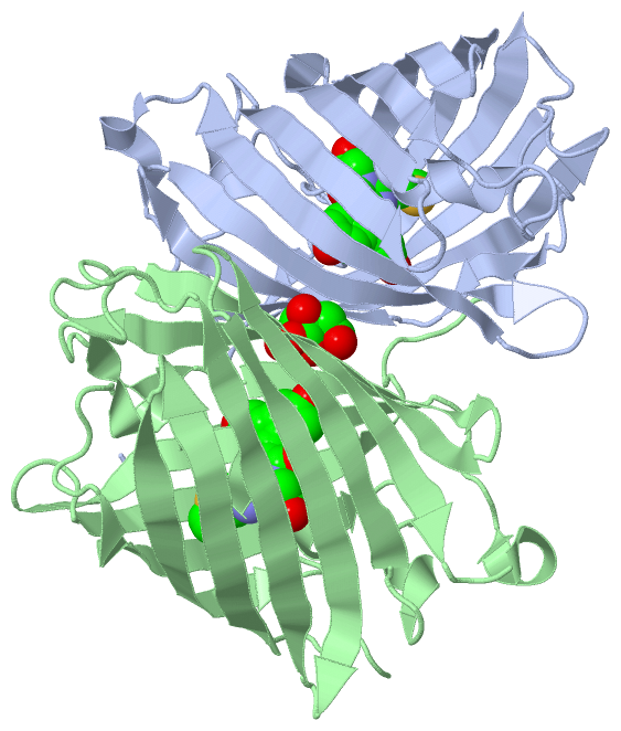
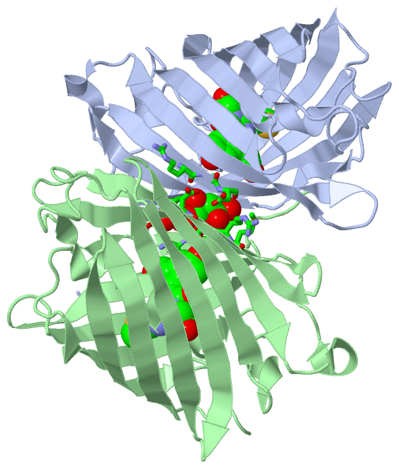
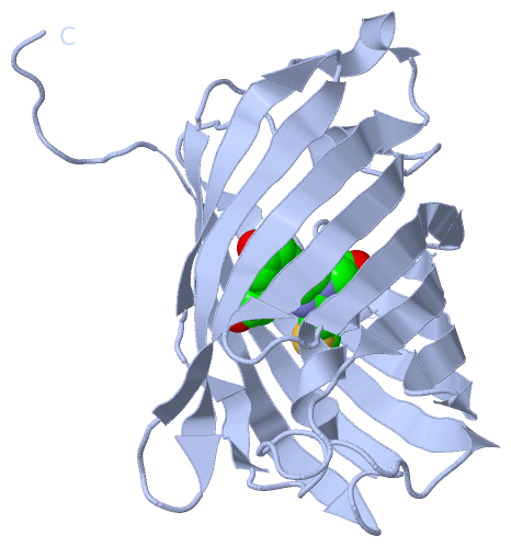
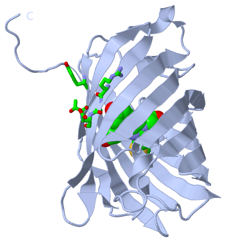
No.NameCountTypeFull Name
1CIT1Ligand/IonCITRIC ACID
2NRQ2Mod. Amino Acid{(4Z)-4-(4-HYDROXYBENZYLIDENE)-2-[3-(METHYLTHIO)PROPANIMIDOYL]-5-OXO-4,5-DIHYDRO-1H-IMIDAZOL-1-YL}ACETIC ACID
Biological Unit 1 (1, 1)
No.NameCountTypeFull Name
1CIT-1Ligand/IonCITRIC ACID
2NRQ1Mod. Amino Acid{(4Z)-4-(4-HYDROXYBENZYLIDENE)-2-[3-(METHYLTHIO)PROPANIMIDOYL]-5-OXO-4,5-DIHYDRO-1H-IMIDAZOL-1-YL}ACETIC ACID
Biological Unit 2 (2, 2)
No.NameCountTypeFull Name
1CIT1Ligand/IonCITRIC ACID
2NRQ1Mod. Amino Acid{(4Z)-4-(4-HYDROXYBENZYLIDENE)-2-[3-(METHYLTHIO)PROPANIMIDOYL]-5-OXO-4,5-DIHYDRO-1H-IMIDAZOL-1-YL}ACETIC ACID
Biological Unit 3 (2, 3)
No.NameCountTypeFull Name
1CIT1Ligand/IonCITRIC ACID
2NRQ2Mod. Amino Acid{(4Z)-4-(4-HYDROXYBENZYLIDENE)-2-[3-(METHYLTHIO)PROPANIMIDOYL]-5-OXO-4,5-DIHYDRO-1H-IMIDAZOL-1-YL}ACETIC ACID

(-) Sites  (1, 1)

Asymmetric Unit (1, 1)
No.NameEvidenceResiduesDescription
1AC1SOFTWARETHR A:144 , ARG A:157 , SER A:158 , ASP A:159 , TYR A:194 , SER B:143 , THR B:144 , MET B:146 , ARG B:157 , SER B:158 , ASP B:159 , HOH B:274 , HOH B:314BINDING SITE FOR RESIDUE CIT B 234

(-) SS Bonds  (0, 0)

(no "SS Bond" information available for 3BXA)

(-) Cis Peptide Bonds  (4, 4)

Asymmetric Unit
No.Residues
1Gly A:49 -Pro A:50
2Phe A:84 -Pro A:85
3Gly B:49 -Pro B:50
4Phe B:84 -Pro B:85

 Sequence-Structure Mapping

(-) SAPs(SNPs)/Variants  (0, 0)

(no "SAP(SNP)/Variant" information available for 3BXA)

(-) PROSITE Motifs  (0, 0)

(no "PROSITE Motif" information available for 3BXA)

(-) Exons   (0, 0)

(no "Exon" information available for 3BXA)

(-) Sequences/Alignments

Asymmetric Unit
   Reformat: Number of residues per line =  ('0' or empty: single-line sequence representation)
  Number of residues per labelling interval =   
  UniProt sequence: complete  aligned part    
   Show mapping: SCOP domains CATH domains Pfam domains Secondary structure (by author)
SAPs(SNPs) PROSITE motifs Exons
(details for a mapped element are shown in a popup box when the mouse pointer rests over it)
Chain A from PDB  Type:PROTEIN  Length:224
                                                                                                                                                                                                                                                                
               SCOP domains d3bxaa_ A: automated matches                                                                                                                                                                                                     SCOP domains
               CATH domains 3bxaA00 A:3-228 Green Fluorescent Protein                                                                                                                                                                                        CATH domains
               Pfam domains -------------------------------------------------------------------------------------------------------------------------------------------------------------------------------------------------------------------------------- Pfam domains
         Sec.struct. author ......eeeeeeeeeee..eeeeeeeeeeee....eeeeeeeeee......hhhhhhhhh......ee.....hhhhhh....eeeeeeeee....eeeeeeeeeee..eeeeeeeeeee............eeee..eeeeeeee..eeeeeeeeeeee....eeeeeeeeeeee..hhhhh.....eeeeeeeeeeeee....eeeeeeeeeee........ Sec.struct. author
                 SAPs(SNPs) -------------------------------------------------------------------------------------------------------------------------------------------------------------------------------------------------------------------------------- SAPs(SNPs)
                    PROSITE -------------------------------------------------------------------------------------------------------------------------------------------------------------------------------------------------------------------------------- PROSITE
                 Transcript -------------------------------------------------------------------------------------------------------------------------------------------------------------------------------------------------------------------------------- Transcript
                 3bxa A   3 ELITENMHMKLYMEGTVNNHHFKCTSEGEGKPYEGTQTMRIKVVEGGPLPFAFDILATSFmSKTFINHTQGIPDFFKQSFPEGFTWERVTTYEDGGVLTATQDTSLQDGCLIYNVKIRGVNFPSNGPVMQKKTLGWEASTEMLYPADGGLEGRSDMALKLVGGGHLICNLKTTYRSKKPAKNLKMPGVYYVDRRLERIKEADKETYVEQHEVAVARYCDLPSKL 228
                                    12        22        32        42        52        62||      74        84        94       104       114       124       134       144       154       164       174       184       194       204       214       224    
                                                                                       63-NRQ                                                                                                                                                               
                                                                                        66                                                                                                                                                                  

Chain B from PDB  Type:PROTEIN  Length:224
                                                                                                                                                                                                                                                                
               SCOP domains d3bxab_ B: automated matches                                                                                                                                                                                                     SCOP domains
               CATH domains 3bxaB00 B:3-228 Green Fluorescent Protein                                                                                                                                                                                        CATH domains
               Pfam domains -------------------------------------------------------------------------------------------------------------------------------------------------------------------------------------------------------------------------------- Pfam domains
         Sec.struct. author ......eeeeeeeeeee..eeeeeeeeeeee....eeeeeeeeee......hhhhhhhhh.hhhhhee.......hhhhh...eeeeeeeee....eeeeeeeeeee..eeeeeeeeeee............eeee..eeeeeeee..eeeeeeeeeeee....eeeeeeeeeeee..hhhhh.....eeeeeeeeeeeee....eeeeeeeeeee........ Sec.struct. author
                 SAPs(SNPs) -------------------------------------------------------------------------------------------------------------------------------------------------------------------------------------------------------------------------------- SAPs(SNPs)
                    PROSITE -------------------------------------------------------------------------------------------------------------------------------------------------------------------------------------------------------------------------------- PROSITE
                 Transcript -------------------------------------------------------------------------------------------------------------------------------------------------------------------------------------------------------------------------------- Transcript
                 3bxa B   3 ELITENMHMKLYMEGTVNNHHFKCTSEGEGKPYEGTQTMRIKVVEGGPLPFAFDILATSFmSKTFINHTQGIPDFFKQSFPEGFTWERVTTYEDGGVLTATQDTSLQDGCLIYNVKIRGVNFPSNGPVMQKKTLGWEASTEMLYPADGGLEGRSDMALKLVGGGHLICNLKTTYRSKKPAKNLKMPGVYYVDRRLERIKEADKETYVEQHEVAVARYCDLPSKL 228
                                    12        22        32        42        52        62||      74        84        94       104       114       124       134       144       154       164       174       184       194       204       214       224    
                                                                                       63-NRQ                                                                                                                                                               
                                                                                        66                                                                                                                                                                  

   Legend:   → Mismatch (orange background)
  - → Gap (green background, '-', border residues have a numbering label)
    → Modified Residue (blue background, lower-case, 'x' indicates undefined single-letter code, labelled with number + name)
  x → Chemical Group (purple background, 'x', labelled with number + name, e.g. ACE or NH2)
  extra numbering lines below/above indicate numbering irregularities and modified residue names etc., number ends below/above '|'

 Classification and Annotation

(-) SCOP Domains  (1, 2)

Asymmetric Unit

(-) CATH Domains  (1, 2)

Asymmetric Unit
(-)
Class: Mainly Beta (13760)

(-) Pfam Domains  (0, 0)

(no "Pfam Domain" information available for 3BXA)

(-) Gene Ontology  (0, 0)

Asymmetric Unit(hide GO term definitions)
    (no "Gene Ontology" information available for 3BXA)

 Visualization

(-) Interactive Views

Asymmetric Unit
  Complete Structure
    Jena3D(integrated viewing of ligand, site, SAP, PROSITE, SCOP information)
    WebMol | AstexViewer[tm]@PDBe
(Java Applets, require no local installation except for Java; loading may be slow)
    STRAP
(Java WebStart application, automatic local installation, requires Java; full application with system access!)
    RasMol
(require local installation)
    Molscript (VRML)
(requires installation of a VRML viewer; select preferred view via VRML and generate a mono or stereo PDF format file)
 
  Ligands, Modified Residues, Ions
    CIT  [ RasMol | Jena3D ]  +environment [ RasMol | Jena3D ]
    NRQ  [ RasMol | Jena3D ]  +environment [ RasMol | Jena3D ]
 
  Sites
    AC1  [ RasMol ]  +environment [ RasMol ]
 
  Cis Peptide Bonds
    Gly A:49 - Pro A:50   [ RasMol ]  
    Gly B:49 - Pro B:50   [ RasMol ]  
    Phe A:84 - Pro A:85   [ RasMol ]  
    Phe B:84 - Pro B:85   [ RasMol ]  
 
Biological Units
  Complete Structure
    Biological Unit 1  [ Jena3D ]
    Biological Unit 2  [ Jena3D ]
    Biological Unit 3  [ Jena3D ]

(-) Still Images

Jmol
  protein: cartoon or spacefill or dots and stick; nucleic acid: cartoon and stick; ligands: spacefill; active site: stick
Molscript
  protein, nucleic acid: cartoon; ligands: spacefill; active site: ball and stick

 Databases and Analysis Tools

(-) Databases

Access by PDB/NDB ID
  3bxa
    Family and Domain InformationProDom | SYSTERS
    General Structural InformationGlycoscienceDB | MMDB | NDB | OCA | PDB | PDBe | PDBj | PDBsum | PDBWiki | PQS | PROTEOPEDIA
    Orientation in MembranesOPM
    Protein SurfaceSURFACE
    Secondary StructureDSSP (structure derived) | HSSP (homology derived)
    Structural GenomicsGeneCensus
    Structural NeighboursCE | VAST
    Structure ClassificationCATH | Dali | SCOP
    Validation and Original DataBMRB Data View | BMRB Restraints Grid | EDS | PROCHECK | RECOORD | WHAT_CHECK
 
Access by UniProt ID/Accession number
  (no 'UniProt ID/Accession number' available) |
    Comparative Protein Structure ModelsModBase
    Genomic InformationEnsembl
    Protein-protein InteractionDIP
    Sequence, Family and Domain InformationInterPro | Pfam | SMART | UniProt
 
Access by Enzyme Classificator   (EC Number)
  (no 'Enzyme Classificator' available)
    General Enzyme InformationBRENDA | EC-PDB | Enzyme | IntEnz
    PathwayKEGG | MetaCyc
 
Access by Disease Identifier   (MIM ID)
  (no 'MIM ID' available)
    Disease InformationOMIM
 
Access by GenAge ID
  (no 'GenAge ID' available)
    Age Related InformationGenAge

(-) Analysis Tools

Access by PDB/NDB ID
    Domain InformationXDom
    Interatomic Contacts of Structural UnitsCSU
    Ligand-protein ContactsLPC
    Protein CavitiescastP
    Sequence and Secondary StructurePDBCartoon
    Structure AlignmentSTRAP(Java WebStart application, automatic local installation, requires Java; full application with system access!)
    Structure and Sequence BrowserSTING
 
Access by UniProt ID/Accession number
  (no 'UniProt ID/Accession number' available) |
    Protein Disorder PredictionDisEMBL | FoldIndex | GLOBPLOT

 Related Entries

(-) Entries Sharing at Least One Protein Chain (UniProt ID)

(no "Entries Sharing at Least One Protein Chain" available for 3BXA)

(-) Related Entries Specified in the PDB File

1uis RED FLUORESCENT PROTEIN EQFP611
3bx9 MONOMERIC FAR-RED FLUORESCENT PROTEIN MKATE CRYSTALLIZED AT PH 2.0
3bxb MONOMERIC FAR-RED FLUORESCENT PROTEIN MKATE CRYSTALLIZED AT PH 7.0
3bxc MONOMERIC FAR-RED FLUORESCENT PROTEIN MKATE CRYSTALLIZED AT PH 9.0