|
|
|
|
 Description
|
|
Compounds
|
||||||||||||||||||||||||||||||||||||||||||
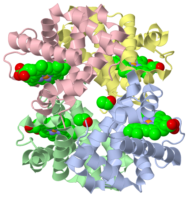
Chains, Units
Summary Information (see also Sequences/Alignments below) |
Ligands, Modified Residues, Ions (2, 6)| Asymmetric/Biological Unit (2, 6) |
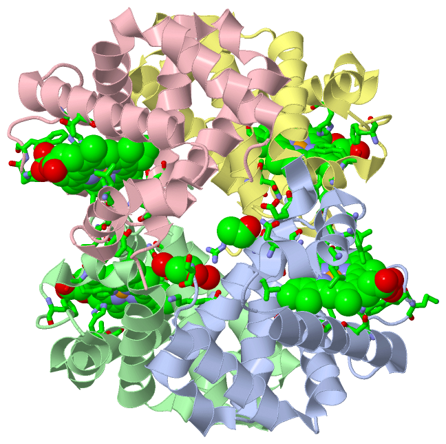
Sites (6, 6)
Asymmetric Unit (6, 6)
|
SS Bonds (0, 0)| (no "SS Bond" information available for 3BJ3) |
Cis Peptide Bonds (0, 0)| (no "Cis Peptide Bond" information available for 3BJ3) |
SAPs(SNPs)/Variants (0, 0)| (no "SAP(SNP)/Variant" information available for 3BJ3) |
PROSITE Motifs (0, 0)| (no "PROSITE Motif" information available for 3BJ3) |
Exons (0, 0)| (no "Exon" information available for 3BJ3) |
Sequences/Alignments
Asymmetric/Biological UnitChain A from PDB Type:PROTEIN Length:142 aligned with D0VWS3_PERFV | D0VWS3 from UniProtKB/TrEMBL Length:142 Alignment length:142 10 20 30 40 50 60 70 80 90 100 110 120 130 140 D0VWS3_PERFV 1 SLSSKDKDAVKALWGKIADKAEEIGADALGRMLAVYPQTKTYFSHWKDLSPGSAPVNKHGKTIMGGLVDAVASIDDLNAGLLALSELHAFTLRVDPANFKILSHCILVQLAVKFPKDFTPEVHLSYDKFFSAVARALAEKYR 142 SCOP domains d3bj3a_ A: automated matches SCOP domains CATH domains 3bj3A00 A:1-142 Globins CATH domains Pfam domains ---------------------------------------------------------------------------------------------------------------------------------------------- Pfam domains SAPs(SNPs) ---------------------------------------------------------------------------------------------------------------------------------------------- SAPs(SNPs) PROSITE ---------------------------------------------------------------------------------------------------------------------------------------------- PROSITE Transcript ---------------------------------------------------------------------------------------------------------------------------------------------- Transcript 3bj3 A 1 SLSSKDKDAVKALWGKIADKAEEIGADALGRMLAVYPQTKTYFSHWKDLSPGSAPVNKHGKTIMGGLVDAVASIDDLNAGLLALSELHAFTLRVDPANFKILSHCILVQLAVKFPKDFTPEVHLSYDKFFSAVARALAEKYR 142 10 20 30 40 50 60 70 80 90 100 110 120 130 140 Chain B from PDB Type:PROTEIN Length:146 aligned with D0VWV3_PERFV | D0VWV3 from UniProtKB/TrEMBL Length:146 Alignment length:146 10 20 30 40 50 60 70 80 90 100 110 120 130 140 D0VWV3_PERFV 1 VVWTDFERATIADIFSKLDYEAVGGATLARCLIVYPWTQRYFGNFGNLYNAAAIMGNPMIAKHGTTILHGLDRAVKNMDNIKATYAELSVLHSEKLHVDPDNFKLLSDCLTIVVAAQLGKAFSGEVQAAFQKFLSVVVSALGKQYH 146 SCOP domains d3bj3b_ B: Hemoglobin, beta-chain SCOP domains CATH domains 3bj3B00 B:1-146 Globins CATH domains Pfam domains -------------------------------------------------------------------------------------------------------------------------------------------------- Pfam domains SAPs(SNPs) -------------------------------------------------------------------------------------------------------------------------------------------------- SAPs(SNPs) PROSITE -------------------------------------------------------------------------------------------------------------------------------------------------- PROSITE Transcript -------------------------------------------------------------------------------------------------------------------------------------------------- Transcript 3bj3 B 1 VVWTDFERATIADIFSKLDYEAVGGATLARCLIVYPWTQRYFGNFGNLYNAAAIMGNPMIAKHGTTILHGLDRAVKNMDNIKATYAELSVLHSEKLHVDPDNFKLLSDCLTIVVAAQLGKAFSGEVQAAFQKFLSVVVSALGKQYH 146 10 20 30 40 50 60 70 80 90 100 110 120 130 140 Chain C from PDB Type:PROTEIN Length:143 aligned with D0VWS3_PERFV | D0VWS3 from UniProtKB/TrEMBL Length:142 Alignment length:143 142 10 20 30 40 50 60 70 80 90 100 110 120 130 140 | D0VWS3_PERFV 1 SLSSKDKDAVKALWGKIADKAEEIGADALGRMLAVYPQTKTYFSHWKDLSPGSAPVNKHGKTIMGGLVDAVASIDDLNAGLLALSELHAFTLRVDPANFKILSHCILVQLAVKFPKDFTPEVHLSYDKFFSAVARALAEKYR- - SCOP domains d3bj3c_ C: automated matches SCOP domains CATH domains 3bj3C00 C:1-142 Globins - CATH domains Pfam domains ----------------------------------------------------------------------------------------------------------------------------------------------- Pfam domains SAPs(SNPs) ----------------------------------------------------------------------------------------------------------------------------------------------- SAPs(SNPs) PROSITE ----------------------------------------------------------------------------------------------------------------------------------------------- PROSITE Transcript ----------------------------------------------------------------------------------------------------------------------------------------------- Transcript 3bj3 C 1 SLSSKDKDAVKALWGKIADKAEEIGADALGRMLAVYPQTKTYFSHWKDLSPGSAPVNKHGKTIMGGLVDAVASIDDLNAGLLALSELHAFTLRVDPANFKILSHCILVQLAVKFPKDFTPEVHLSYDKFFSAVARALAEKYRx 0 10 20 30 40 50 60 70 80 90 100 110 120 130 140 || 142| 0-ACE Chain D from PDB Type:PROTEIN Length:146 aligned with D0VWV3_PERFV | D0VWV3 from UniProtKB/TrEMBL Length:146 Alignment length:146 10 20 30 40 50 60 70 80 90 100 110 120 130 140 D0VWV3_PERFV 1 VVWTDFERATIADIFSKLDYEAVGGATLARCLIVYPWTQRYFGNFGNLYNAAAIMGNPMIAKHGTTILHGLDRAVKNMDNIKATYAELSVLHSEKLHVDPDNFKLLSDCLTIVVAAQLGKAFSGEVQAAFQKFLSVVVSALGKQYH 146 SCOP domains d3bj3d_ D: Hemoglobin, beta-chain SCOP domains CATH domains 3bj3D00 D:1-146 Globins CATH domains Pfam domains -------------------------------------------------------------------------------------------------------------------------------------------------- Pfam domains SAPs(SNPs) -------------------------------------------------------------------------------------------------------------------------------------------------- SAPs(SNPs) PROSITE -------------------------------------------------------------------------------------------------------------------------------------------------- PROSITE Transcript -------------------------------------------------------------------------------------------------------------------------------------------------- Transcript 3bj3 D 1 VVWTDFERATIADIFSKLDYEAVGGATLARCLIVYPWTQRYFGNFGNLYNAAAIMGNPMIAKHGTTILHGLDRAVKNMDNIKATYAELSVLHSEKLHVDPDNFKLLSDCLTIVVAAQLGKAFSGEVQAAFQKFLSVVVSALGKQYH 146 10 20 30 40 50 60 70 80 90 100 110 120 130 140
|
||||||||||||||||||||
SCOP Domains (2, 4)
Asymmetric/Biological Unit
 |
CATH Domains (1, 4)
Asymmetric/Biological Unit
|
Pfam Domains (0, 0)| (no "Pfam Domain" information available for 3BJ3) |
Gene Ontology (8, 16)|
Asymmetric/Biological Unit(hide GO term definitions) Chain A,C (D0VWS3_PERFV | D0VWS3)
Chain B,D (D0VWV3_PERFV | D0VWV3)
|
||||||||||||||||||||||||||||||||||||||||||||||||||||||||||||||||||||||||||||||||||||||||||||||||||||||||||||||||||||||||||||||||||||
Interactive Views
|
||||||||||||||||||||||||||||||||||||||||||||||||||||||||||||||||||||||||||||||||||||||||||||||||||||||||||||||||||||||||||||||||||||||||||||||||||||||||||||||||
Still Images
|
||||||||||||||||
Databases
|
||||||||||||||||||||||||||||||||||||||||||||||||||||||||||||||||||||||||||||||||||||||||||||||||||||||||||||||||||||||||||||||||||||||||||||||||||||||||||||||||||||||||||||||||||||||||||
Analysis Tools
|
||||||||||||||||||||||||||||||||||||||||||||||||||||||||||||||||||||||||
Entries Sharing at Least One Protein Chain (UniProt ID)
Related Entries Specified in the PDB File
|
|