| molecular function |
|---|
| | GO:0008800 | | beta-lactamase activity | | Catalysis of the reaction: a beta-lactam + H2O = a substituted beta-amino acid. |
| | GO:0004180 | | carboxypeptidase activity | | Catalysis of the hydrolysis of the terminal or penultimate peptide bond at the C-terminal end of a peptide or polypeptide. |
| | GO:0004175 | | endopeptidase activity | | Catalysis of the hydrolysis of internal, alpha-peptide bonds in a polypeptide chain. |
| | GO:0016787 | | hydrolase activity | | Catalysis of the hydrolysis of various bonds, e.g. C-O, C-N, C-C, phosphoric anhydride bonds, etc. Hydrolase is the systematic name for any enzyme of EC class 3. |
| | GO:0008658 | | penicillin binding | | Interacting selectively and non-covalently with penicillin, any antibiotic that contains the condensed beta-lactamthiazolidine ring system. |
| | GO:0008233 | | peptidase activity | | Catalysis of the hydrolysis of a peptide bond. A peptide bond is a covalent bond formed when the carbon atom from the carboxyl group of one amino acid shares electrons with the nitrogen atom from the amino group of a second amino acid. |
| | GO:0009002 | | serine-type D-Ala-D-Ala carboxypeptidase activity | | Catalysis of the reaction: (Ac)2-L-Lys-D-alanyl-D-alanine + H2O = (Ac)2-L-Lys-D-alanine + D-alanine. |
| biological process |
|---|
| | GO:0044036 | | cell wall macromolecule metabolic process | | The chemical reactions and pathways involving macromolecules forming, or destined to form, part of the cell wall. A cell wall is a rigid or semi-rigid envelope lying outside the cell membrane of plant, fungal and most prokaryotic cells, maintaining their shape and protecting them from osmotic lysis. |
| | GO:0071555 | | cell wall organization | | A process that results in the assembly, arrangement of constituent parts, or disassembly of the cell wall, the rigid or semi-rigid envelope lying outside the cell membrane of plant, fungal and most prokaryotic cells, maintaining their shape and protecting them from osmotic lysis. |
| | GO:0009252 | | peptidoglycan biosynthetic process | | The chemical reactions and pathways resulting in the formation of peptidoglycans, any of a class of glycoconjugates found in bacterial cell walls. |
| | GO:0000270 | | peptidoglycan metabolic process | | The chemical reactions and pathways involving peptidoglycans, any of a class of glycoconjugates found only in bacterial cell walls and consisting of strands of glycosaminoglycan cross-linked by oligopeptides to form a huge and rigid network. |
| | GO:0006508 | | proteolysis | | The hydrolysis of proteins into smaller polypeptides and/or amino acids by cleavage of their peptide bonds. |
| | GO:0008360 | | regulation of cell shape | | Any process that modulates the surface configuration of a cell. |
| cellular component |
|---|
| | GO:0005887 | | integral component of plasma membrane | | The component of the plasma membrane consisting of the gene products and protein complexes having at least some part of their peptide sequence embedded in the hydrophobic region of the membrane. |
| | GO:0016020 | | membrane | | A lipid bilayer along with all the proteins and protein complexes embedded in it an attached to it. |
| | GO:0005886 | | plasma membrane | | The membrane surrounding a cell that separates the cell from its external environment. It consists of a phospholipid bilayer and associated proteins. |
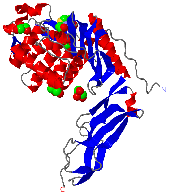
 Description
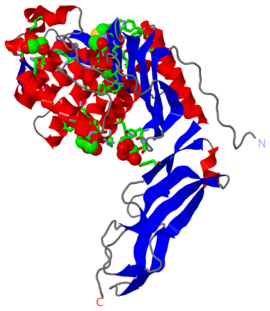