|
|
|
|
 Description
|
|
Compounds
|
||||||||||||||||||||||||||||||||||||||||||||||||||||||||
Chains, Units
Summary Information (see also Sequences/Alignments below) |
Ligands, Modified Residues, Ions (0, 0)| (no "Ligand,Modified Residues,Ions" information available for 3A4C) |
Sites (0, 0)| (no "Site" information available for 3A4C) |
SS Bonds (0, 0)| (no "SS Bond" information available for 3A4C) |
Cis Peptide Bonds (0, 0)| (no "Cis Peptide Bond" information available for 3A4C) |
SAPs(SNPs)/Variants (0, 0)| (no "SAP(SNP)/Variant" information available for 3A4C) |
PROSITE Motifs (0, 0)| (no "PROSITE Motif" information available for 3A4C) |
Exons (0, 0)| (no "Exon" information available for 3A4C) |
Sequences/Alignments
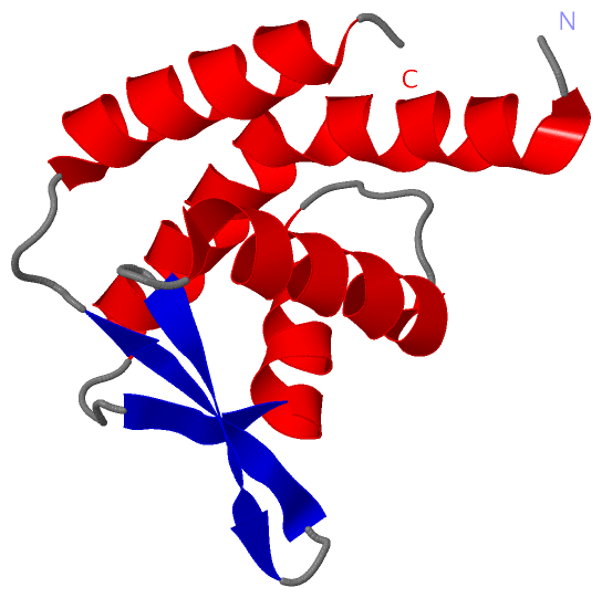
Asymmetric/Biological UnitChain A from PDB Type:PROTEIN Length:106 aligned with CDT1_MOUSE | Q8R4E9 from UniProtKB/Swiss-Prot Length:557 Alignment length:106 461 471 481 491 501 511 521 531 541 551 CDT1_MOUSE 452 RCPEQELRLQRLERLPELARVLRNVFVSERKPALTMEVVCARMVDSCQTALSPGEMEKHLVLLAELLPDWLSLHRIRTDTYVKLDKAVDLAGLTARLAHHVHAEGL 557 SCOP domains ---------------------------------------------------------------------------------------------------------- SCOP domains CATH domains ---------------------------------------------------------------------------------------------------------- CATH domains Pfam domains ---------------------------------------------------------------------------------------------------------- Pfam domains SAPs(SNPs) ---------------------------------------------------------------------------------------------------------- SAPs(SNPs) PROSITE ---------------------------------------------------------------------------------------------------------- PROSITE Transcript ---------------------------------------------------------------------------------------------------------- Transcript 3a4c A 452 RCPEQELRLQRLERLPELARVLRNVFVSERKPALTMEVVCARMVDSCQTALSPGEMEKHLVLLAELLPDWLSLHRIRTDTYVKLDKAVDLAGLTARLAHHVHAEGL 557 461 471 481 491 501 511 521 531 541 551
|
||||||||||||||||||||
SCOP Domains (0, 0)| (no "SCOP Domain" information available for 3A4C) |
CATH Domains (0, 0)| (no "CATH Domain" information available for 3A4C) |
Pfam Domains (0, 0)| (no "Pfam Domain" information available for 3A4C) |
Gene Ontology (8, 8)|
Asymmetric/Biological Unit(hide GO term definitions) Chain A (CDT1_MOUSE | Q8R4E9)
|
||||||||||||||||||||||||||||||||||||||||||||||||||||||||||||||||||
Interactive Views
|
||||||||||||||||||||||||||||||||||||||||||||||||||||||||||||||||||||||||||||||||||||||||||||||||||||||||||||||||||||
Still Images
|
||||||||||||||||
Databases
|
||||||||||||||||||||||||||||||||||||||||||||||||||||||||||||||||||||||||||||||||||||||||||||||||||||||||||||||||||||||||||||||||||||||||||||||||||||||||||||||||
Analysis Tools
|
|||||||||||||||||||||||||||||||||||||||||||||||||||||||||||||
Entries Sharing at Least One Protein Chain (UniProt ID)
Related Entries Specified in the PDB File
|
|