Show PDB file:   
         Plain Text   HTML   (compressed file size)
QuickSearch:   
by PDB,NDB,UniProt,PROSITE Code or Search Term(s)  
(-)Asymmetric Unit
(-)Asym. Unit - sites
(-)Biological Unit 1
(-)Biol. Unit 1 - sites
collapse expand < >
Image Asymmetric Unit
Asymmetric Unit  (Jmol Viewer)
Image Asym. Unit - sites
Asym. Unit - sites  (Jmol Viewer)
Image Biological Unit 1
Biological Unit 1  (Jmol Viewer)
Image Biol. Unit 1 - sites
Biol. Unit 1 - sites  (Jmol Viewer)

(-) Description

Title :  CRYSTAL STRUCTURE OF MESORHIZOBIUM LOTI 3-HYDROXY-2-METHYLPYRIDINE-4, 5-DICARBOXYLATE DECARBOXYLASE
 
Authors :  K. M. Mcculloch, T. Mukherjee, S. E. Ealick, T. P. Begley
Date :  17 Aug 07  (Deposition) - 13 Nov 07  (Release) - 13 Jul 11  (Revision)
Method :  X-RAY DIFFRACTION
Resolution :  1.90
Chains :  Asym. Unit :  A
Biol. Unit 1:  A  (4x)
Keywords :  Class Ii Aldolase Superfamily, Lyase (Keyword Search: [Gene Ontology, PubMed, Web (Google))
 
Reference :  T. Mukherjee, K. M. Mcculloch, S. E. Ealick, T. P. Begley
Gene Identification And Structural Characterization Of The Pyridoxal 5'-Phosphate Degradative Protein 3-Hydroxy-2-Methylpyridine-4, 5-Dicarboxylate Decarboxylase From Mesorhizobium Loti Maff303099
Biochemistry V. 46 13606 2007
PubMed-ID: 17973403  |  Reference-DOI: 10.1021/BI701439J

(-) Compounds

Molecule 1 - MLR6791 PROTEIN
    ChainsA
    EC Number4.1.1.51
    EngineeredYES
    Expression SystemESCHERICHIA COLI
    Expression System PlasmidPML5335.XF1
    Expression System StrainB834(DE3)
    Expression System Taxid562
    Expression System Vector TypePLASMID
    Gene5335
    Organism ScientificMESORHIZOBIUM LOTI
    Organism Taxid266835
    StrainMAFF303099
    Synonym3-HYDROXY-2-METHYLPYRIDINE-4,5-DICARBOXYLATE DECARBOXYLASE

 Structural Features

(-) Chains, Units

  1
Asymmetric Unit A
Biological Unit 1 (4x)A

Summary Information (see also Sequences/Alignments below)
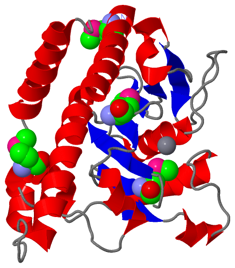
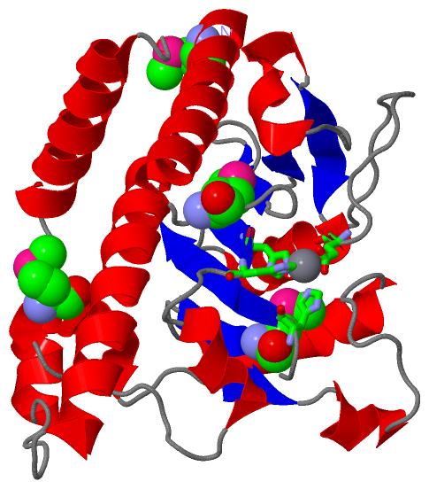
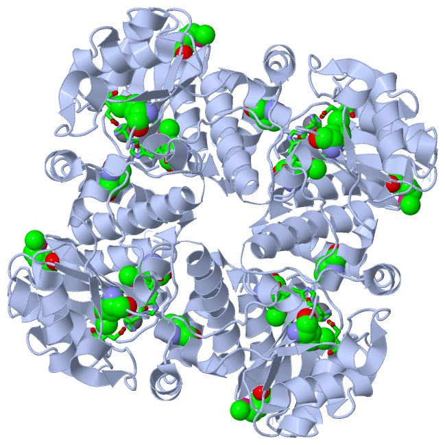
(-) Ligands, Modified Residues, Ions  (2, 5)

Asymmetric Unit (2, 5)
No.NameCountTypeFull Name
1MN1Ligand/IonMANGANESE (II) ION
2MSE4Mod. Amino AcidSELENOMETHIONINE
Biological Unit 1 (1, 16)
No.NameCountTypeFull Name
1MN-1Ligand/IonMANGANESE (II) ION
2MSE16Mod. Amino AcidSELENOMETHIONINE

(-) Sites  (1, 1)

Asymmetric Unit (1, 1)
No.NameEvidenceResiduesDescription
1AC1SOFTWAREGLU A:73 , HIS A:92 , HIS A:94 , HIS A:163BINDING SITE FOR RESIDUE MN A 235

(-) SS Bonds  (0, 0)

(no "SS Bond" information available for 2Z7B)

(-) Cis Peptide Bonds  (0, 0)

(no "Cis Peptide Bond" information available for 2Z7B)

 Sequence-Structure Mapping

(-) SAPs(SNPs)/Variants  (0, 0)

(no "SAP(SNP)/Variant" information available for 2Z7B)

(-) PROSITE Motifs  (0, 0)

(no "PROSITE Motif" information available for 2Z7B)

(-) Exons   (0, 0)

(no "Exon" information available for 2Z7B)

(-) Sequences/Alignments

Asymmetric Unit
   Reformat: Number of residues per line =  ('0' or empty: single-line sequence representation)
  Number of residues per labelling interval =   
  UniProt sequence: complete  aligned part    
   Show mapping: SCOP domains CATH domains Pfam domains Secondary structure (by author)
SAPs(SNPs) PROSITE motifs Exons
(details for a mapped element are shown in a popup box when the mouse pointer rests over it)
Chain A from PDB  Type:PROTEIN  Length:237
 aligned with Q988D0_RHILO | Q988D0 from UniProtKB/TrEMBL  Length:234

    Alignment length:237
                               1                                                                                                                                                                                                                                         
                               |     7        17        27        37        47        57        67        77        87        97       107       117       127       137       147       157       167       177       187       197       207       217       227       
         Q988D0_RHILO     - ---MRRKVFEELVTATKILLNEGIMDTFGHISARDPEDPASFFLAQKLAPSLITVDDIQRFNLDGETSDNRPSYLERYIHSEIYKTRPDVQCVLHTHSPAVLPYCFVDTPLRPVTHMGAFIGESVPVYEIRDKHGDETDLFGGSPDVCADIAESLGSQTVVLMARHGVVNVGKSVREVVFRAFYLEQEAAALTAGLKIGNVKYLSPGEIKTAGKLVGAQIDRGWNHWSQRLRQAGLA 234
               SCOP domains --------------------------------------------------------------------------------------------------------------------------------------------------------------------------------------------------------------------------------------------- SCOP domains
               CATH domains --------------------------------------------------------------------------------------------------------------------------------------------------------------------------------------------------------------------------------------------- CATH domains
               Pfam domains ---------Aldolase_II-2z7bA01 A:7-190                                                                                                                                                             -------------------------------------------- Pfam domains
         Sec.struct. author hhhhhhhhhhhhhhhhhhhhhh.......eeeee......eeeee...hhhhhhhh.eeeee.............hhhhhhhhhhhh....eeeee....hhhhhh........hhhhhhhh....ee.hhhhhh........hhhhhhhhhhhhh...eeee...eeeeee.hhhhhhhhhhhhhhhhhhhhhhhh.......hhhhhhhhh....hhhhhhhhhhhhhhhhh... Sec.struct. author
                 SAPs(SNPs) --------------------------------------------------------------------------------------------------------------------------------------------------------------------------------------------------------------------------------------------- SAPs(SNPs)
                    PROSITE --------------------------------------------------------------------------------------------------------------------------------------------------------------------------------------------------------------------------------------------- PROSITE
                 Transcript --------------------------------------------------------------------------------------------------------------------------------------------------------------------------------------------------------------------------------------------- Transcript
                 2z7b A  -2 SFTmRRKVFEELVTATKILLNEGImDTFGHISARDPEDPASFFLAQKLAPSLITVDDIQRFNLDGETSDNRPSYLERYIHSEIYKTRPDVQCVLHTHSPAVLPYCFVDTPLRPVTHmGAFIGESVPVYEIRDKHGDETDLFGGSPDVCADIAESLGSQTVVLmARHGVVNVGKSVREVVFRAFYLEQEAAALTAGLKIGNVKYLSPGEIKTAGKLVGAQIDRGWNHWSQRLRQAGLA 234
                               |     7        17    |   27        37        47        57        67        77        87        97       107      |117       127       137       147       157  |    167       177       187       197       207       217       227       
                               |                   22-MSE                                                                                     114-MSE                                       160-MSE                                                                      
                               1-MSE                                                                                                                                                                                                                                     

   Legend:   → Mismatch (orange background)
  - → Gap (green background, '-', border residues have a numbering label)
    → Modified Residue (blue background, lower-case, 'x' indicates undefined single-letter code, labelled with number + name)
  x → Chemical Group (purple background, 'x', labelled with number + name, e.g. ACE or NH2)
  extra numbering lines below/above indicate numbering irregularities and modified residue names etc., number ends below/above '|'

 Classification and Annotation

(-) SCOP Domains  (0, 0)

(no "SCOP Domain" information available for 2Z7B)

(-) CATH Domains  (0, 0)

(no "CATH Domain" information available for 2Z7B)

(-) Pfam Domains  (1, 1)

Asymmetric Unit

(-) Gene Ontology  (1, 1)

Asymmetric Unit(hide GO term definitions)
Chain A   (Q988D0_RHILO | Q988D0)
molecular function
    GO:0046872    metal ion binding    Interacting selectively and non-covalently with any metal ion.

 Visualization

(-) Interactive Views

Asymmetric Unit
  Complete Structure
    Jena3D(integrated viewing of ligand, site, SAP, PROSITE, SCOP information)
    WebMol | AstexViewer[tm]@PDBe
(Java Applets, require no local installation except for Java; loading may be slow)
    STRAP
(Java WebStart application, automatic local installation, requires Java; full application with system access!)
    RasMol
(require local installation)
    Molscript (VRML)
(requires installation of a VRML viewer; select preferred view via VRML and generate a mono or stereo PDF format file)
 
  Ligands, Modified Residues, Ions
    MN  [ RasMol | Jena3D ]  +environment [ RasMol | Jena3D ]
    MSE  [ RasMol | Jena3D ]  +environment [ RasMol | Jena3D ]
 
  Sites
    AC1  [ RasMol ]  +environment [ RasMol ]
 
  Cis Peptide Bonds
(no "Cis Peptide Bonds" information available for 2z7b)
 
Biological Unit
  Complete Structure
    Biological Unit 1  [ Jena3D ]

(-) Still Images

Jmol
  protein: cartoon or spacefill or dots and stick; nucleic acid: cartoon and stick; ligands: spacefill; active site: stick
Molscript
  protein, nucleic acid: cartoon; ligands: spacefill; active site: ball and stick

 Databases and Analysis Tools

(-) Databases

Access by PDB/NDB ID
  2z7b
    Family and Domain InformationProDom | SYSTERS
    General Structural InformationGlycoscienceDB | MMDB | NDB | OCA | PDB | PDBe | PDBj | PDBsum | PDBWiki | PQS | PROTEOPEDIA
    Orientation in MembranesOPM
    Protein SurfaceSURFACE
    Secondary StructureDSSP (structure derived) | HSSP (homology derived)
    Structural GenomicsGeneCensus
    Structural NeighboursCE | VAST
    Structure ClassificationCATH | Dali | SCOP
    Validation and Original DataBMRB Data View | BMRB Restraints Grid | EDS | PROCHECK | RECOORD | WHAT_CHECK
 
Access by UniProt ID/Accession number
  Q988D0_RHILO | Q988D0
    Comparative Protein Structure ModelsModBase
    Genomic InformationEnsembl
    Protein-protein InteractionDIP
    Sequence, Family and Domain InformationInterPro | Pfam | SMART | UniProtKB/TrEMBL
 
Access by Enzyme Classificator   (EC Number)
  4.1.1.51
    General Enzyme InformationBRENDA | EC-PDB | Enzyme | IntEnz
    PathwayKEGG | MetaCyc
 
Access by Disease Identifier   (MIM ID)
  (no 'MIM ID' available)
    Disease InformationOMIM
 
Access by GenAge ID
  (no 'GenAge ID' available)
    Age Related InformationGenAge

(-) Analysis Tools

Access by PDB/NDB ID
    Domain InformationXDom
    Interatomic Contacts of Structural UnitsCSU
    Ligand-protein ContactsLPC
    Protein CavitiescastP
    Sequence and Secondary StructurePDBCartoon
    Structure AlignmentSTRAP(Java WebStart application, automatic local installation, requires Java; full application with system access!)
    Structure and Sequence BrowserSTING
 
Access by UniProt ID/Accession number
  Q988D0_RHILO | Q988D0
    Protein Disorder PredictionDisEMBL | FoldIndex | GLOBPLOT (for more information see DisProt)

 Related Entries

(-) Entries Sharing at Least One Protein Chain (UniProt ID)

(no "Entries Sharing at Least One Protein Chain" available for 2Z7B)

(-) Related Entries Specified in the PDB File

(no "Related Entries Specified in the PDB File" available for 2Z7B)