Show PDB file:   
         Plain Text   HTML   (compressed file size)
QuickSearch:   
by PDB,NDB,UniProt,PROSITE Code or Search Term(s)  
(-)Asymmetric Unit
(-)Asym. Unit - sites
(-)Biological Unit 1
(-)Biol. Unit 1 - sites
(-)Biological Unit 2
collapse expand < >
Image Asymmetric Unit
Asymmetric Unit  (Jmol Viewer)
Image Asym. Unit - sites
Asym. Unit - sites  (Jmol Viewer)
Image Biological Unit 1
Biological Unit 1  (Jmol Viewer)
Image Biol. Unit 1 - sites
Biol. Unit 1 - sites  (Jmol Viewer)
Image Biological Unit 2
Biological Unit 2  (Jmol Viewer)

(-) Description

Title :  CRYSTALLOGRAPHIC INVESTIGATION OF INHIBITION MODE OF THE VIM-2 METALLO-BETA-LACTAMASE FROM PSEUDOMONAS AERUGINOSA WITH MERCAPTOCARBOXYLATE INHIBITOR
 
Authors :  Y. Yamaguchi, Y. Yamagata, Y. Arakawa, H. Kurosaki
Date :  02 May 07  (Deposition) - 11 Mar 08  (Release) - 24 Feb 09  (Revision)
Method :  X-RAY DIFFRACTION
Resolution :  2.30
Chains :  Asym. Unit :  A,B
Biol. Unit 1:  A  (1x)
Biol. Unit 2:  B  (1x)
Keywords :  Metallo-Beta-Lactamase, Enzyme-Inhibitor Complex, Hydrolase (Keyword Search: [Gene Ontology, PubMed, Web (Google))
 
Reference :  Y. Yamaguchi, W. Jin, K. Matsunaga, S. Ikemizu, Y. Yamagata, J. Wachino, N. Shibata, Y. Arakawa, H. Kurosaki
Crystallographic Investigation Of The Inhibition Mode Of A Vim-2 Metallo-Beta-Lactamase From Pseudomonas Aeruginosa By A Mercaptocarboxylate Inhibitor.
J. Med. Chem. V. 50 6647 2007
PubMed-ID: 18052313  |  Reference-DOI: 10.1021/JM701031N
(for further references see the PDB file header)

(-) Compounds

Molecule 1 - METALLO-BETA-LACTAMASE
    ChainsA, B
    EC Number3.5.2.6
    EngineeredYES
    Expression SystemESCHERICHIA COLI
    Expression System PlasmidPBC, PVM4K/VIM-2
    Expression System Taxid562
    Expression System Vector TypePLASMID
    GeneVIM-2, BLA VIM-2, BLAVIM-2
    Organism ScientificPSEUDOMONAS PUTIDA
    Organism Taxid303
    SynonymVIM-2, METALLO-BETA-LACTAMASE VIM-2, VIM-2 BETA- LACTAMASE

 Structural Features

(-) Chains, Units

  12
Asymmetric Unit AB
Biological Unit 1 (1x)A 
Biological Unit 2 (1x) B

Summary Information (see also Sequences/Alignments below)

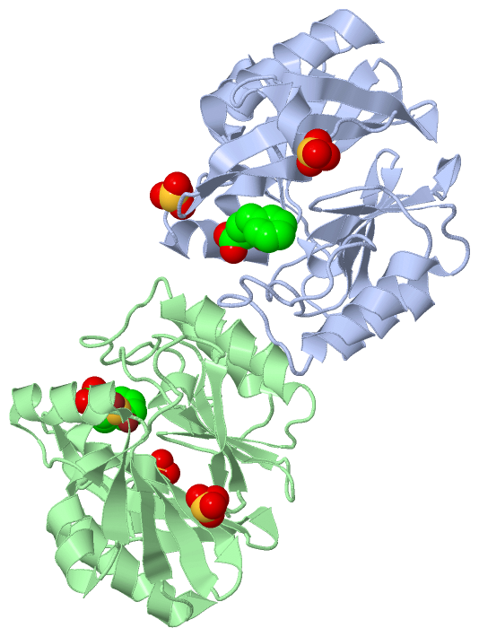
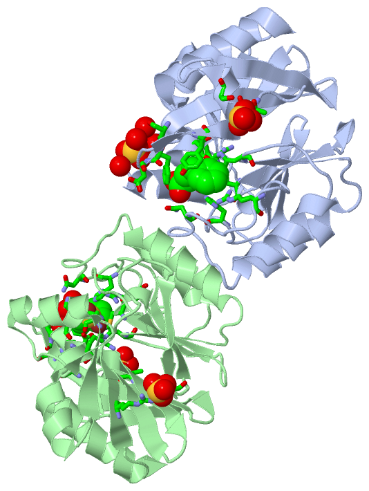
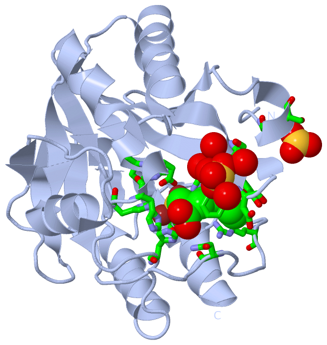
(-) Ligands, Modified Residues, Ions  (3, 10)

Asymmetric Unit (3, 10)
No.NameCountTypeFull Name
1M5P2Ligand/Ion(S)-2-(MERCAPTOMETHYL)-5-PHENYLPENTANOIC ACID
2SO44Ligand/IonSULFATE ION
3ZN4Ligand/IonZINC ION
Biological Unit 1 (2, 3)
No.NameCountTypeFull Name
1M5P1Ligand/Ion(S)-2-(MERCAPTOMETHYL)-5-PHENYLPENTANOIC ACID
2SO42Ligand/IonSULFATE ION
3ZN-1Ligand/IonZINC ION
Biological Unit 2 (2, 3)
No.NameCountTypeFull Name
1M5P1Ligand/Ion(S)-2-(MERCAPTOMETHYL)-5-PHENYLPENTANOIC ACID
2SO42Ligand/IonSULFATE ION
3ZN-1Ligand/IonZINC ION

(-) Sites  (10, 10)

Asymmetric Unit (10, 10)
No.NameEvidenceResiduesDescription
01AC1SOFTWAREHIS A:94 , HIS A:96 , HIS A:159BINDING SITE FOR RESIDUE ZN A 301
02AC2SOFTWAREASP A:98 , ARG A:99 , CYS A:178 , HIS A:220BINDING SITE FOR RESIDUE ZN A 302
03AC3SOFTWAREHIS B:94 , HIS B:96 , HIS B:159BINDING SITE FOR RESIDUE ZN B 303
04AC4SOFTWAREASP B:98 , ARG B:99 , CYS B:178 , HIS B:220BINDING SITE FOR RESIDUE ZN B 304
05AC5SOFTWARESER A:41 , HOH A:541 , THR B:15 , SER B:17BINDING SITE FOR RESIDUE SO4 B 501
06AC6SOFTWARETHR A:15 , SER A:17 , SER B:41BINDING SITE FOR RESIDUE SO4 A 502
07AC7SOFTWARESER A:41 , PHE A:42 , ASP A:43 , HOH A:541 , HOH A:564 , HOH A:574 , HOH A:590BINDING SITE FOR RESIDUE SO4 A 503
08AC8SOFTWAREARG B:89 , GLY B:148BINDING SITE FOR RESIDUE SO4 B 504
09AC9SOFTWAREPHE A:42 , TYR A:47 , HIS A:96 , ASP A:98 , HIS A:159 , ARG A:185 , ASN A:190 , HIS A:220BINDING SITE FOR RESIDUE M5P A 401
10BC1SOFTWAREPHE B:42 , TYR B:47 , TRP B:67 , HIS B:96 , ASP B:98 , HIS B:159 , ARG B:185 , ASN B:190 , HIS B:220 , HOH B:594BINDING SITE FOR RESIDUE M5P B 402

(-) SS Bonds  (0, 0)

(no "SS Bond" information available for 2YZ3)

(-) Cis Peptide Bonds  (0, 0)

(no "Cis Peptide Bond" information available for 2YZ3)

 Sequence-Structure Mapping

(-) SAPs(SNPs)/Variants  (0, 0)

(no "SAP(SNP)/Variant" information available for 2YZ3)

(-) PROSITE Motifs  (0, 0)

(no "PROSITE Motif" information available for 2YZ3)

(-) Exons   (0, 0)

(no "Exon" information available for 2YZ3)

(-) Sequences/Alignments

Asymmetric Unit
   Reformat: Number of residues per line =  ('0' or empty: single-line sequence representation)
  Number of residues per labelling interval =   
  UniProt sequence: complete  aligned part    
   Show mapping: SCOP domains CATH domains Pfam domains Secondary structure (by author)
SAPs(SNPs) PROSITE motifs Exons
(details for a mapped element are shown in a popup box when the mouse pointer rests over it)
Chain A from PDB  Type:PROTEIN  Length:225
 aligned with Q7BJM5_PSEPU | Q7BJM5 from UniProtKB/TrEMBL  Length:266

    Alignment length:225
                                    41        51        61        71        81        91       101       111       121       131       141       151       161       171       181       191       201       211       221       231       241       251     
         Q7BJM5_PSEPU    32 EYPTVSEIPVGEVRLYQIADGVWSHIATQSFDGAVYPSNGLIVRDGDELLLIDTAWGAKNTAALLAEIEKQIGLPVTRAVSTHFHDDRVGGVDVLRAAGVATYASPSTRRLAEVEGNEIPTHSLEGLSSSGDAVRFGPVELFYPGAAHSTDNLVVYVPSASVLYGGCAIYELSRTSAGNVADADLAEWPTSIERIQQHYPEAQFVIPGHGLPGGLDLLKHTTNVV 256
               SCOP domains d2yz3a_ A: Zn metallo-beta-lactamase                                                                                                                                                                                              SCOP domains
               CATH domains 2yz3A00 A:12-236 Metallo-beta-lactamase, chain A                                                                                                                                                                                  CATH domains
               Pfam domains --------------------------------------------------------------------------------------------------------------------------------------------------------------------------------------------------------------------------------- Pfam domains
         Sec.struct. author ...hhhhh....eeeeeee..eeeeeeeeee..eeeeeeeeeeee..eeeee....hhhhhhhhhhhhhhhh...eeeee....hhhhhhhhhhhhhh..eeeehhhhhhhhhh.......ee........eeeee..eeeee..........eeeehhh.eeeeehhh.................hhhhhhhhhhhhh....eeee.......hhhhhhhhhhh Sec.struct. author
                 SAPs(SNPs) --------------------------------------------------------------------------------------------------------------------------------------------------------------------------------------------------------------------------------- SAPs(SNPs)
                    PROSITE --------------------------------------------------------------------------------------------------------------------------------------------------------------------------------------------------------------------------------- PROSITE
                 Transcript --------------------------------------------------------------------------------------------------------------------------------------------------------------------------------------------------------------------------------- Transcript
                 2yz3 A  12 EYPTVSEIPVGEVRLYQIADGVWSHIATQSFDGAVYPSNGLIVRDGDELLLIDTAWGAKNTAALLAEIEKQIGLPVTRAVSTHFHDDRVGGVDVLRAAGVATYASPSTRRLAEVEGNEIPTHSLEGLSSSGDAVRFGPVELFYPGAAHSTDNLVVYVPSASVLYGGCAIYELSRTSAGNVADADLAEWPTSIERIQQHYPEAQFVIPGHGLPGGLDLLKHTTNVV 236
                                    21        31        41        51        61        71        81        91       101       111       121       131       141       151       161       171       181       191       201       211       221       231     

Chain B from PDB  Type:PROTEIN  Length:225
 aligned with Q7BJM5_PSEPU | Q7BJM5 from UniProtKB/TrEMBL  Length:266

    Alignment length:225
                                    41        51        61        71        81        91       101       111       121       131       141       151       161       171       181       191       201       211       221       231       241       251     
         Q7BJM5_PSEPU    32 EYPTVSEIPVGEVRLYQIADGVWSHIATQSFDGAVYPSNGLIVRDGDELLLIDTAWGAKNTAALLAEIEKQIGLPVTRAVSTHFHDDRVGGVDVLRAAGVATYASPSTRRLAEVEGNEIPTHSLEGLSSSGDAVRFGPVELFYPGAAHSTDNLVVYVPSASVLYGGCAIYELSRTSAGNVADADLAEWPTSIERIQQHYPEAQFVIPGHGLPGGLDLLKHTTNVV 256
               SCOP domains d2yz3b_ B: Zn metallo-beta-lactamase                                                                                                                                                                                              SCOP domains
               CATH domains 2yz3B00 B:12-236 Metallo-beta-lactamase, chain A                                                                                                                                                                                  CATH domains
           Pfam domains (1) --------------------------------Lactamase_B-2yz3B01 B:44-220                                                                                                                                                     ---------------- Pfam domains (1)
           Pfam domains (2) --------------------------------Lactamase_B-2yz3B02 B:44-220                                                                                                                                                     ---------------- Pfam domains (2)
         Sec.struct. author ...hhhhh.....eeeeee..eeeeeeeeee..eeeeeeeeeeee..eeeee....hhhhhhhhhhhhhhhh...eeeee....hhhhhhhhhhhhhh..eeeehhhhhhhhhhhh.....ee........eeeee..eeeee..........eeee....eeeeehhh.................hhhhhhhhhhhhh....eeee.......hhhhhhhhhhh Sec.struct. author
                 SAPs(SNPs) --------------------------------------------------------------------------------------------------------------------------------------------------------------------------------------------------------------------------------- SAPs(SNPs)
                    PROSITE --------------------------------------------------------------------------------------------------------------------------------------------------------------------------------------------------------------------------------- PROSITE
                 Transcript --------------------------------------------------------------------------------------------------------------------------------------------------------------------------------------------------------------------------------- Transcript
                 2yz3 B  12 EYPTVSEIPVGEVRLYQIADGVWSHIATQSFDGAVYPSNGLIVRDGDELLLIDTAWGAKNTAALLAEIEKQIGLPVTRAVSTHFHDDRVGGVDVLRAAGVATYASPSTRRLAEVEGNEIPTHSLEGLSSSGDAVRFGPVELFYPGAAHSTDNLVVYVPSASVLYGGCAIYELSRTSAGNVADADLAEWPTSIERIQQHYPEAQFVIPGHGLPGGLDLLKHTTNVV 236
                                    21        31        41        51        61        71        81        91       101       111       121       131       141       151       161       171       181       191       201       211       221       231     

   Legend:   → Mismatch (orange background)
  - → Gap (green background, '-', border residues have a numbering label)
    → Modified Residue (blue background, lower-case, 'x' indicates undefined single-letter code, labelled with number + name)
  x → Chemical Group (purple background, 'x', labelled with number + name, e.g. ACE or NH2)
  extra numbering lines below/above indicate numbering irregularities and modified residue names etc., number ends below/above '|'

 Classification and Annotation

(-) SCOP Domains  (1, 2)

Asymmetric Unit

(-) CATH Domains  (1, 2)

Asymmetric Unit
(-)
Class: Alpha Beta (26913)

(-) Pfam Domains  (1, 2)

Asymmetric Unit

(-) Gene Ontology  (3, 3)

Asymmetric Unit(hide GO term definitions)
Chain A,B   (Q7BJM5_PSEPU | Q7BJM5)
molecular function
    GO:0008800    beta-lactamase activity    Catalysis of the reaction: a beta-lactam + H2O = a substituted beta-amino acid.
    GO:0016787    hydrolase activity    Catalysis of the hydrolysis of various bonds, e.g. C-O, C-N, C-C, phosphoric anhydride bonds, etc. Hydrolase is the systematic name for any enzyme of EC class 3.
    GO:0046872    metal ion binding    Interacting selectively and non-covalently with any metal ion.

 Visualization

(-) Interactive Views

Asymmetric Unit
  Complete Structure
    Jena3D(integrated viewing of ligand, site, SAP, PROSITE, SCOP information)
    WebMol | AstexViewer[tm]@PDBe
(Java Applets, require no local installation except for Java; loading may be slow)
    STRAP
(Java WebStart application, automatic local installation, requires Java; full application with system access!)
    RasMol
(require local installation)
    Molscript (VRML)
(requires installation of a VRML viewer; select preferred view via VRML and generate a mono or stereo PDF format file)
 
  Ligands, Modified Residues, Ions
    M5P  [ RasMol | Jena3D ]  +environment [ RasMol | Jena3D ]
    SO4  [ RasMol | Jena3D ]  +environment [ RasMol | Jena3D ]
    ZN  [ RasMol | Jena3D ]  +environment [ RasMol | Jena3D ]
 
  Sites
    AC1  [ RasMol ]  +environment [ RasMol ]
    AC2  [ RasMol ]  +environment [ RasMol ]
    AC3  [ RasMol ]  +environment [ RasMol ]
    AC4  [ RasMol ]  +environment [ RasMol ]
    AC5  [ RasMol ]  +environment [ RasMol ]
    AC6  [ RasMol ]  +environment [ RasMol ]
    AC7  [ RasMol ]  +environment [ RasMol ]
    AC8  [ RasMol ]  +environment [ RasMol ]
    AC9  [ RasMol ]  +environment [ RasMol ]
    BC1  [ RasMol ]  +environment [ RasMol ]
 
  Cis Peptide Bonds
(no "Cis Peptide Bonds" information available for 2yz3)
 
Biological Units
  Complete Structure
    Biological Unit 1  [ Jena3D ]
    Biological Unit 2  [ Jena3D ]

(-) Still Images

Jmol
  protein: cartoon or spacefill or dots and stick; nucleic acid: cartoon and stick; ligands: spacefill; active site: stick
Molscript
  protein, nucleic acid: cartoon; ligands: spacefill; active site: ball and stick

 Databases and Analysis Tools

(-) Databases

Access by PDB/NDB ID
  2yz3
    Family and Domain InformationProDom | SYSTERS
    General Structural InformationGlycoscienceDB | MMDB | NDB | OCA | PDB | PDBe | PDBj | PDBsum | PDBWiki | PQS | PROTEOPEDIA
    Orientation in MembranesOPM
    Protein SurfaceSURFACE
    Secondary StructureDSSP (structure derived) | HSSP (homology derived)
    Structural GenomicsGeneCensus
    Structural NeighboursCE | VAST
    Structure ClassificationCATH | Dali | SCOP
    Validation and Original DataBMRB Data View | BMRB Restraints Grid | EDS | PROCHECK | RECOORD | WHAT_CHECK
 
Access by UniProt ID/Accession number
  Q7BJM5_PSEPU | Q7BJM5
    Comparative Protein Structure ModelsModBase
    Genomic InformationEnsembl
    Protein-protein InteractionDIP
    Sequence, Family and Domain InformationInterPro | Pfam | SMART | UniProtKB/TrEMBL
 
Access by Enzyme Classificator   (EC Number)
  3.5.2.6
    General Enzyme InformationBRENDA | EC-PDB | Enzyme | IntEnz
    PathwayKEGG | MetaCyc
 
Access by Disease Identifier   (MIM ID)
  (no 'MIM ID' available)
    Disease InformationOMIM
 
Access by GenAge ID
  (no 'GenAge ID' available)
    Age Related InformationGenAge

(-) Analysis Tools

Access by PDB/NDB ID
    Domain InformationXDom
    Interatomic Contacts of Structural UnitsCSU
    Ligand-protein ContactsLPC
    Protein CavitiescastP
    Sequence and Secondary StructurePDBCartoon
    Structure AlignmentSTRAP(Java WebStart application, automatic local installation, requires Java; full application with system access!)
    Structure and Sequence BrowserSTING
 
Access by UniProt ID/Accession number
  Q7BJM5_PSEPU | Q7BJM5
    Protein Disorder PredictionDisEMBL | FoldIndex | GLOBPLOT (for more information see DisProt)

 Related Entries

(-) Entries Sharing at Least One Protein Chain (UniProt ID)

(no "Entries Sharing at Least One Protein Chain" available for 2YZ3)

(-) Related Entries Specified in the PDB File

(no "Related Entries Specified in the PDB File" available for 2YZ3)