Show PDB file:   
         Plain Text   HTML   (compressed file size)
QuickSearch:   
by PDB,NDB,UniProt,PROSITE Code or Search Term(s)  
(-)NMR Structure - model 1
(-)NMR Structure - all models
collapse expand < >
Image NMR Structure - model 1
NMR Structure - model 1  (Jmol Viewer)
Image NMR Structure - all models
NMR Structure - all models  (Jmol Viewer)

(-) Description

Title :  SOLUTION NMR STRUCTURE OF THE C-TERMINAL EXTENSION OF TWO BACTERIAL LIGHT, OXYGEN, VOLTAGE (LOV) PHOTORECEPTOR PROTEINS FROM PSEUDOMONAS PUTIDA
 
Authors :  R. Rani, R. Hartmann, J. Lecher, U. Krauss, K. Jaeger, D. Willbold
Date :  25 Oct 12  (Deposition) - 10 Jul 13  (Release) - 04 May 16  (Revision)
Method :  SOLUTION NMR
Resolution :  NOT APPLICABLE
Chains :  NMR Structure  :  A  (10x)
NMR Structure *:  A  (1x)
Keywords :  Signaling Protein, Voltage (Lov) Domain (Keyword Search: [Gene Ontology, PubMed, Web (Google))
 
Reference :  R. Rani, K. Jentzsch, J. Lecher, R. Hartmann, D. Willbold, K. Jaeger, U. Krauss
Conservation Of Dark Recovery Kinetic Parameters And Structural Features In The Pseudomonadaceae "Short" Light, Oxygen, Voltage (Lov) Protein Family: Implications For The Design Of Lov-Based Optogenetic Tools.
Biochemistry V. 52 4460 2013
PubMed-ID: 23746326  |  Reference-DOI: 10.1021/BI400311R

(-) Compounds

Molecule 1 - SENSORY BOX PROTEIN
    ChainsA
    FragmentJALPHA HELIX, RESIDUES 123-151
    Organism ScientificPSEUDOMONAS PUTIDA KT2440
    Organism Taxid160488
    Other DetailsN-TERMINAL ACETYLATION
    SynonymACESB2PEP
    SyntheticYES

 Structural Features

(-) Chains, Units

  1
NMR Structure (10x)A
NMR Structure * (1x)A

Summary Information (see also Sequences/Alignments below)

(-) Ligands, Modified Residues, Ions  (1, 1)

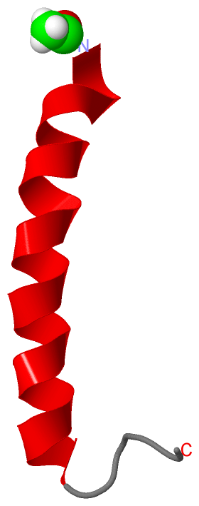
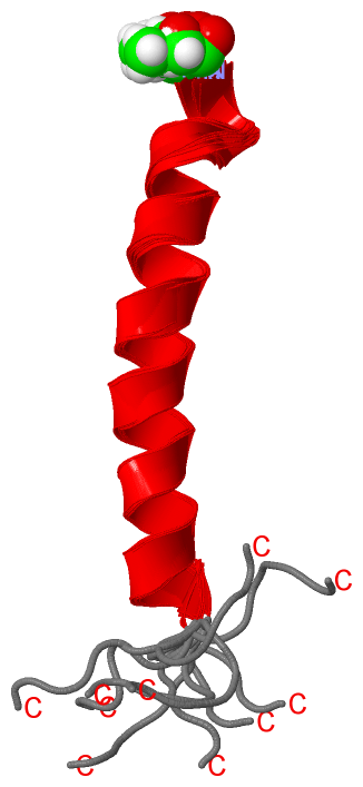
NMR Structure (1, 1)
No.NameCountTypeFull Name
1ACE1Mod. Amino AcidACETYL GROUP
NMR Structure * (1, 1)
No.NameCountTypeFull Name
1ACE1Mod. Amino AcidACETYL GROUP

(-) Sites  (0, 0)

(no "Site" information available for 2YON)

(-) SS Bonds  (0, 0)

(no "SS Bond" information available for 2YON)

(-) Cis Peptide Bonds  (0, 0)

(no "Cis Peptide Bond" information available for 2YON)

 Sequence-Structure Mapping

(-) SAPs(SNPs)/Variants  (0, 0)

(no "SAP(SNP)/Variant" information available for 2YON)

(-) PROSITE Motifs  (0, 0)

(no "PROSITE Motif" information available for 2YON)

(-) Exons   (0, 0)

(no "Exon" information available for 2YON)

(-) Sequences/Alignments

NMR Structure
   Reformat: Number of residues per line =  ('0' or empty: single-line sequence representation)
  Number of residues per labelling interval =   
  UniProt sequence: complete  aligned part    
   Show mapping: SCOP domains CATH domains Pfam domains Secondary structure (by author)
SAPs(SNPs) PROSITE motifs Exons
(details for a mapped element are shown in a popup box when the mouse pointer rests over it)
Chain A from PDB  Type:PROTEIN  Length:30
 aligned with Q88JB0_PSEPK | Q88JB0 from UniProtKB/TrEMBL  Length:151

    Alignment length:30
                                   131       141       151
         Q88JB0_PSEPK   122 VTAQVFAEERVRELEAEVAELRRQQGQAKH 151
               SCOP domains ------------------------------ SCOP domains
               CATH domains ------------------------------ CATH domains
               Pfam domains ------------------------------ Pfam domains
         Sec.struct. author .hhhhhhhhhhhhhhhhhhhhhhhh..... Sec.struct. author
                 SAPs(SNPs) ------------------------------ SAPs(SNPs)
                    PROSITE ------------------------------ PROSITE
                 Transcript ------------------------------ Transcript
                 2yon A 119 xTAQVFAEERVRELEAEVAELRRQQGQAKH 148
                            |      128       138       148
                            |                             
                          119-ACE                         

   Legend:   → Mismatch (orange background)
  - → Gap (green background, '-', border residues have a numbering label)
    → Modified Residue (blue background, lower-case, 'x' indicates undefined single-letter code, labelled with number + name)
  x → Chemical Group (purple background, 'x', labelled with number + name, e.g. ACE or NH2)
  extra numbering lines below/above indicate numbering irregularities and modified residue names etc., number ends below/above '|'

 Classification and Annotation

(-) SCOP Domains  (0, 0)

(no "SCOP Domain" information available for 2YON)

(-) CATH Domains  (0, 0)

(no "CATH Domain" information available for 2YON)

(-) Pfam Domains  (0, 0)

(no "Pfam Domain" information available for 2YON)

(-) Gene Ontology  (4, 4)

NMR Structure(hide GO term definitions)
Chain A   (Q88JB0_PSEPK | Q88JB0)
molecular function
    GO:0000155    phosphorelay sensor kinase activity    Catalysis of the phosphorylation of a histidine residue in response to detection of an extracellular signal such as a chemical ligand or change in environment, to initiate a change in cell state or activity. The two-component sensor is a histidine kinase that autophosphorylates a histidine residue in its active site. The phosphate is then transferred to an aspartate residue in a downstream response regulator, to trigger a response.
biological process
    GO:0000160    phosphorelay signal transduction system    A conserved series of molecular signals found in prokaryotes and eukaryotes; involves autophosphorylation of a histidine kinase and the transfer of the phosphate group to an aspartate that then acts as a phospho-donor to response regulator proteins.
    GO:0023014    signal transduction by protein phosphorylation    A process in which the transfer of one or more phosphate groups to a substrate transmits a signal to the phosphorylated substrate.
cellular component
    GO:0005622    intracellular    The living contents of a cell; the matter contained within (but not including) the plasma membrane, usually taken to exclude large vacuoles and masses of secretory or ingested material. In eukaryotes it includes the nucleus and cytoplasm.

 Visualization

(-) Interactive Views

NMR Structure
  Complete Structure
    Jena3D(integrated viewing of ligand, site, SAP, PROSITE, SCOP information)
    WebMol | AstexViewer[tm]@PDBe
(Java Applets, require no local installation except for Java; loading may be slow)
    STRAP
(Java WebStart application, automatic local installation, requires Java; full application with system access!)
    RasMol
(require local installation)
    Molscript (VRML)
(requires installation of a VRML viewer; select preferred view via VRML and generate a mono or stereo PDF format file)
 
  Ligands, Modified Residues, Ions
    ACE  [ RasMol | Jena3D ]  +environment [ RasMol | Jena3D ]
 
  Sites
(no "Sites" information available for 2yon)
 
  Cis Peptide Bonds
(no "Cis Peptide Bonds" information available for 2yon)
 

(-) Still Images

Jmol
  protein: cartoon or spacefill or dots and stick; nucleic acid: cartoon and stick; ligands: spacefill; active site: stick
Molscript
  protein, nucleic acid: cartoon; ligands: spacefill; active site: ball and stick

 Databases and Analysis Tools

(-) Databases

Access by PDB/NDB ID
  2yon
    Family and Domain InformationProDom | SYSTERS
    General Structural InformationGlycoscienceDB | MMDB | NDB | OCA | PDB | PDBe | PDBj | PDBsum | PDBWiki | PQS | PROTEOPEDIA
    Orientation in MembranesOPM
    Protein SurfaceSURFACE
    Secondary StructureDSSP (structure derived) | HSSP (homology derived)
    Structural GenomicsGeneCensus
    Structural NeighboursCE | VAST
    Structure ClassificationCATH | Dali | SCOP
    Validation and Original DataBMRB Data View | BMRB Restraints Grid | EDS | PROCHECK | RECOORD | WHAT_CHECK
 
Access by UniProt ID/Accession number
  Q88JB0_PSEPK | Q88JB0
    Comparative Protein Structure ModelsModBase
    Genomic InformationEnsembl
    Protein-protein InteractionDIP
    Sequence, Family and Domain InformationInterPro | Pfam | SMART | UniProtKB/TrEMBL
 
Access by Enzyme Classificator   (EC Number)
  (no 'Enzyme Classificator' available)
    General Enzyme InformationBRENDA | EC-PDB | Enzyme | IntEnz
    PathwayKEGG | MetaCyc
 
Access by Disease Identifier   (MIM ID)
  (no 'MIM ID' available)
    Disease InformationOMIM
 
Access by GenAge ID
  (no 'GenAge ID' available)
    Age Related InformationGenAge

(-) Analysis Tools

Access by PDB/NDB ID
    Domain InformationXDom
    Interatomic Contacts of Structural UnitsCSU
    Ligand-protein ContactsLPC
    Protein CavitiescastP
    Sequence and Secondary StructurePDBCartoon
    Structure AlignmentSTRAP(Java WebStart application, automatic local installation, requires Java; full application with system access!)
    Structure and Sequence BrowserSTING
 
Access by UniProt ID/Accession number
  Q88JB0_PSEPK | Q88JB0
    Protein Disorder PredictionDisEMBL | FoldIndex | GLOBPLOT (for more information see DisProt)

 Related Entries

(-) Entries Sharing at Least One Protein Chain (UniProt ID)

(no "Entries Sharing at Least One Protein Chain" available for 2YON)

(-) Related Entries Specified in the PDB File

(no "Related Entries Specified in the PDB File" available for 2YON)