Show PDB file:   
         Plain Text   HTML   (compressed file size)
QuickSearch:   
by PDB,NDB,UniProt,PROSITE Code or Search Term(s)  
(-)Asymmetric Unit
(-)Asym. Unit - sites
(-)Biological Unit 1
(-)Biol. Unit 1 - sites
collapse expand < >
Image Asymmetric Unit
Asymmetric Unit  (Jmol Viewer)
Image Asym. Unit - sites
Asym. Unit - sites  (Jmol Viewer)
Image Biological Unit 1
Biological Unit 1  (Jmol Viewer)
Image Biol. Unit 1 - sites
Biol. Unit 1 - sites  (Jmol Viewer)

(-) Description

Title :  CRYSTAL STRUCTURE OF THE N-TERMINAL IG1-3 MODULE OF HUMAN RECEPTOR PROTEIN TYROSINE PHOSPHATASE SIGMA
 
Authors :  C. H. Coles, Y. Shen, A. P. Tenney, C. Siebold, G. C. Sutton, W. Lu, J. T. Ga E. Y. Jones, J. G. Flanagan, A. R. Aricescu
Date :  18 Mar 11  (Deposition) - 13 Apr 11  (Release) - 04 May 11  (Revision)
Method :  X-RAY DIFFRACTION
Resolution :  2.60
Chains :  Asym. Unit :  A
Biol. Unit 1:  A  (2x)
Keywords :  Hydrolase (Keyword Search: [Gene Ontology, PubMed, Web (Google))
 
Reference :  C. H. Coles, Y. Shen, A. P. Tenney, C. Siebold, G. C. Sutton, W. Lu, J. T. Gallagher, E. Y. Jones, J. G. Flanagan, A. R. Aricescu
Proteoglycan-Specific Molecular Switch For Rptp Sigma Clustering And Neuronal Extension.
Science V. 332 484 2011
PubMed-ID: 21454754  |  Reference-DOI: 10.1126/SCIENCE.1200840

(-) Compounds

Molecule 1 - RECEPTOR-TYPE TYROSINE-PROTEIN PHOSPHATASE S
    ChainsA
    EC Number3.1.3.48
    EngineeredYES
    Expression SystemHOMO SAPIENS
    Expression System Cell LineHEK293T
    Expression System CommonHUMAN
    Expression System Taxid9606
    Expression System VectorPHLSEC
    Expression System Vector TypePLASMID
    FragmentIG1-3, RESIDUES 30-321
    MutationYES
    Organism CommonHUMAN
    Organism ScientificHOMO SAPIENS
    Organism Taxid9606
    SynonymR-PTP-S, RECEPTOR-TYPE TYROSINE-PROTEIN PHOSPHATASE SIGMA R-PTP-SIGMA, RECEPTOR PROTEIN TYROSINE PHOSPHATASE SIGMA

 Structural Features

(-) Chains, Units

  1
Asymmetric Unit A
Biological Unit 1 (2x)A

Summary Information (see also Sequences/Alignments below)

(-) Ligands, Modified Residues, Ions  (4, 11)

Asymmetric Unit (4, 11)
No.NameCountTypeFull Name
1B3P1Ligand/Ion2-[3-(2-HYDROXY-1,1-DIHYDROXYMETHYL-ETHYLAMINO)-PROPYLAMINO]-2-HYDROXYMETHYL-PROPANE-1,3-DIOL
2CL4Ligand/IonCHLORIDE ION
3IOD4Ligand/IonIODIDE ION
4NAG2Ligand/IonN-ACETYL-D-GLUCOSAMINE
Biological Unit 1 (3, 14)
No.NameCountTypeFull Name
1B3P2Ligand/Ion2-[3-(2-HYDROXY-1,1-DIHYDROXYMETHYL-ETHYLAMINO)-PROPYLAMINO]-2-HYDROXYMETHYL-PROPANE-1,3-DIOL
2CL-1Ligand/IonCHLORIDE ION
3IOD8Ligand/IonIODIDE ION
4NAG4Ligand/IonN-ACETYL-D-GLUCOSAMINE

(-) Sites  (9, 9)

Asymmetric Unit (9, 9)
No.NameEvidenceResiduesDescription
1AC1SOFTWAREARG A:233 , PHE A:234BINDING SITE FOR RESIDUE CL A1316
2AC2SOFTWARELYS A:40 , VAL A:53BINDING SITE FOR RESIDUE CL A1317
3AC3SOFTWARESER A:219BINDING SITE FOR RESIDUE CL A1318
4AC4SOFTWARELEU A:155 , SER A:189BINDING SITE FOR RESIDUE IOD A1319
5AC5SOFTWAREGLN A:228 , MET A:260BINDING SITE FOR RESIDUE IOD A1321
6AC6SOFTWAREGLN A:142 , LYS A:144 , LEU A:155 , ARG A:188BINDING SITE FOR RESIDUE IOD A1322
7AC7SOFTWARESER A:293 , ASN A:295BINDING SITE FOR RESIDUE NAG A1323
8AC8SOFTWAREASN A:248 , ASN A:250 , GLU A:286BINDING SITE FOR RESIDUE NAG A1324
9AC9SOFTWAREASP A:138 , CYS A:156 , ALA A:157 , ALA A:158 , ASP A:163 , PRO A:164 , ILE A:166 , ARG A:188 , SER A:189BINDING SITE FOR RESIDUE B3P A1325

(-) SS Bonds  (3, 3)

Asymmetric Unit
No.Residues
1A:54 -A:107
2A:156 -A:207
3A:253 -A:298

(-) Cis Peptide Bonds  (6, 6)

Asymmetric Unit
No.Residues
1Asp A:59 -Pro A:60
2Gln A:94 -Pro A:95
3Thr A:98 -Pro A:99
4Asn A:161 -Pro A:162
5Gly A:246 -Gly A:247
6Ser A:258 -Pro A:259

 Sequence-Structure Mapping

(-) SAPs(SNPs)/Variants  (0, 0)

(no "SAP(SNP)/Variant" information available for 2YD9)

(-) PROSITE Motifs  (0, 0)

(no "PROSITE Motif" information available for 2YD9)

(-) Exons   (0, 0)

(no "Exon" information available for 2YD9)

(-) Sequences/Alignments

Asymmetric Unit
   Reformat: Number of residues per line =  ('0' or empty: single-line sequence representation)
  Number of residues per labelling interval =   
  UniProt sequence: complete  aligned part    
   Show mapping: SCOP domains CATH domains Pfam domains Secondary structure (by author)
SAPs(SNPs) PROSITE motifs Exons
(details for a mapped element are shown in a popup box when the mouse pointer rests over it)
Chain A from PDB  Type:PROTEIN  Length:278
 aligned with PTPRS_HUMAN | Q13332 from UniProtKB/Swiss-Prot  Length:1948

    Alignment length:298
                                    39        49        59        69        79        89        99       109       119       129       139       149       159       169       179       189       199       209       219       229       239       249       259       269       279       289       299       309       319        
          PTPRS_HUMAN    30 EEPPRFIKEPKDQIGVSGGVASFVCQATGDPKPRVTWNKKGKKVNSQRFETIEFDESAGAVLRIQPLRTPRDENVYECVAQNSVGEITVHAKLTVLREDQLPSGFPNIDMGPQLKVVERTRTATMLCAASGNPDPEITWFKDFLPVDPSASNGRIKQLRSETFESTPIRGALQIESSEETDQGKYECVATNSAGVRYSSPANLYVRELREVRRVAPRFSILPMSHEIMPGGNVNITCVAVGSPMPYVKWMQGAEDLTPEDDMPVGRNVLELTDVKDSANYTCVAMSSLGVIEAVAQIT 327
               SCOP domains ---------------------------------------------------------------------------------------------------------------------------------------------------------------------------------------------------------------------------------------------------------------------------------------------------------- SCOP domains
               CATH domains ---------------------------------------------------------------------------------------------------------------------------------------------------------------------------------------------------------------------------------------------------------------------------------------------------------- CATH domains
           Pfam domains (1) -----------------------------------------------------------------------------------------------------------------------------------------------------------------------------------------------------------------------I-set-2yd9A01 A:232-308                                                      ------ Pfam domains (1)
           Pfam domains (2) -----------------------------------------------------------------------------------------------------------------------------------------------------------------------------------------------------------------------I-set-2yd9A02 A:232-308                                                      ------ Pfam domains (2)
           Pfam domains (3) -----------------------------------------------------------------------------------------------------------------------------------------------------------------------------------------------------------------------I-set-2yd9A03 A:232-308                                                      ------ Pfam domains (3)
         Sec.struct. author ....eeee....eeee....eeeeeeee.....eeeee-------...eeeeee....eeeeee..........eeeeeee....ee..eeeeeeehhhhh.....eeee....eeee....eee..eee.....eeeeee..ee.hhhhhh..eee...---------..eee...hhhhheeeeeeeee..eeee...eeeeee----...eeeeeee....ee......eeeeeeeeee...eeeeee..ee..........eeeeee......eeeeeeeee..eeeeeeeeee Sec.struct. author
                 SAPs(SNPs) ---------------------------------------------------------------------------------------------------------------------------------------------------------------------------------------------------------------------------------------------------------------------------------------------------------- SAPs(SNPs)
                    PROSITE ---------------------------------------------------------------------------------------------------------------------------------------------------------------------------------------------------------------------------------------------------------------------------------------------------------- PROSITE
                 Transcript ---------------------------------------------------------------------------------------------------------------------------------------------------------------------------------------------------------------------------------------------------------------------------------------------------------- Transcript
                 2yd9 A  30 EEPPRFIKEPKDQIGVSGGVASFVCQATGDPKPRVTWN-------SQRFETIEFDESAGAVLRIQPLRTPRDENVYECVAQNSVGEITVHAKLTVLREDQLPSGFPNIDMGPQLKVVERTRTATMLCAASGNPDPEITWFKDFLPVDPSASNGRIKQLRS---------GALQIESSEETDQGKYECVATNSAGVRYSSPANLYVR----VQNVAPRFSILPMSHEIMPGGNVNITCVAVGSPMPYVKWMQGAEDLTPEDDMPVGRNVLELTDVKDSANYTCVAMSSLGVIEAVAQIT 314
                                    39        49        59       | -     |  79        89        99       109       119       129       139       149       159       169       179       189       190       200       210       220     |   -|      236       246       256       266       276       286       296       306        
                                                                67      75                                                                                                               189       190                                 226  227                                                                                       

   Legend:   → Mismatch (orange background)
  - → Gap (green background, '-', border residues have a numbering label)
    → Modified Residue (blue background, lower-case, 'x' indicates undefined single-letter code, labelled with number + name)
  x → Chemical Group (purple background, 'x', labelled with number + name, e.g. ACE or NH2)
  extra numbering lines below/above indicate numbering irregularities and modified residue names etc., number ends below/above '|'

 Classification and Annotation

(-) SCOP Domains  (0, 0)

(no "SCOP Domain" information available for 2YD9)

(-) CATH Domains  (0, 0)

(no "CATH Domain" information available for 2YD9)

(-) Pfam Domains  (1, 3)

Asymmetric Unit
(-)
Clan: Ig (577)
(-)
Family: I-set (67)
1aI-set-2yd9A01A:232-308
1bI-set-2yd9A02A:232-308
1cI-set-2yd9A03A:232-308

(-) Gene Ontology  (22, 22)

Asymmetric Unit(hide GO term definitions)
Chain A   (PTPRS_HUMAN | Q13332)
molecular function
    GO:0016787    hydrolase activity    Catalysis of the hydrolysis of various bonds, e.g. C-O, C-N, C-C, phosphoric anhydride bonds, etc. Hydrolase is the systematic name for any enzyme of EC class 3.
    GO:0016791    phosphatase activity    Catalysis of the hydrolysis of phosphoric monoesters, releasing inorganic phosphate.
    GO:0004721    phosphoprotein phosphatase activity    Catalysis of the reaction: a phosphoprotein + H2O = a protein + phosphate. Together with protein kinases, these enzymes control the state of phosphorylation of cell proteins and thereby provide an important mechanism for regulating cellular activity.
    GO:0005515    protein binding    Interacting selectively and non-covalently with any protein or protein complex (a complex of two or more proteins that may include other nonprotein molecules).
    GO:0004725    protein tyrosine phosphatase activity    Catalysis of the reaction: protein tyrosine phosphate + H2O = protein tyrosine + phosphate.
    GO:0005001    transmembrane receptor protein tyrosine phosphatase activity    Combining with a signal and transmitting the signal from one side of the membrane to the other to initiate a change in cell activity by catalysis of the reaction: protein tyrosine phosphate + H2O = protein tyrosine + phosphate.
biological process
    GO:0007155    cell adhesion    The attachment of a cell, either to another cell or to an underlying substrate such as the extracellular matrix, via cell adhesion molecules.
    GO:0021549    cerebellum development    The process whose specific outcome is the progression of the cerebellum over time, from its formation to the mature structure. The cerebellum is the portion of the brain in the back of the head between the cerebrum and the pons. In mice, the cerebellum controls balance for walking and standing, modulates the force and range of movement and is involved in the learning of motor skills.
    GO:0021987    cerebral cortex development    The progression of the cerebral cortex over time from its initial formation until its mature state. The cerebral cortex is the outer layered region of the telencephalon.
    GO:0007268    chemical synaptic transmission    The vesicular release of classical neurotransmitter molecules from a presynapse, across a chemical synapse, the subsequent activation of neurotransmitter receptors at the postsynapse of a target cell (neuron, muscle, or secretory cell) and the effects of this activation on the postsynaptic membrane potential and ionic composition of the postsynaptic cytosol. This process encompasses both spontaneous and evoked release of neurotransmitter and all parts of synaptic vesicle exocytosis. Evoked transmission starts with the arrival of an action potential at the presynapse.
    GO:0022038    corpus callosum development    The process whose specific outcome is the progression of the corpus callosum over time, from its formation to the mature structure. The corpus callosum is a thick bundle of nerve fibers comprising a commissural plate connecting the two cerebral hemispheres. It consists of contralateral axon projections that provide communication between the right and left cerebral hemispheres.
    GO:0016311    dephosphorylation    The process of removing one or more phosphoric (ester or anhydride) residues from a molecule.
    GO:0090557    establishment of endothelial intestinal barrier    The establishment of a barrier between endothelial cell layers of the intestine to exert specific and selective control over the passage of water and solutes, thus allowing formation and maintenance of compartments that differ in fluid and solute composition.
    GO:0021766    hippocampus development    The progression of the hippocampus over time from its initial formation until its mature state.
    GO:0035335    peptidyl-tyrosine dephosphorylation    The removal of phosphoric residues from peptidyl-O-phospho-tyrosine to form peptidyl-tyrosine.
    GO:0006470    protein dephosphorylation    The process of removing one or more phosphoric residues from a protein.
    GO:0021510    spinal cord development    The process whose specific outcome is the progression of the spinal cord over time, from its formation to the mature structure. The spinal cord primarily conducts sensory and motor nerve impulses between the brain and the peripheral nervous tissues.
cellular component
    GO:0070062    extracellular exosome    A vesicle that is released into the extracellular region by fusion of the limiting endosomal membrane of a multivesicular body with the plasma membrane. Extracellular exosomes, also simply called exosomes, have a diameter of about 40-100 nm.
    GO:0016021    integral component of membrane    The component of a membrane consisting of the gene products and protein complexes having at least some part of their peptide sequence embedded in the hydrophobic region of the membrane.
    GO:0005887    integral component of plasma membrane    The component of the plasma membrane consisting of the gene products and protein complexes having at least some part of their peptide sequence embedded in the hydrophobic region of the membrane.
    GO:0016020    membrane    A lipid bilayer along with all the proteins and protein complexes embedded in it an attached to it.
    GO:0005886    plasma membrane    The membrane surrounding a cell that separates the cell from its external environment. It consists of a phospholipid bilayer and associated proteins.
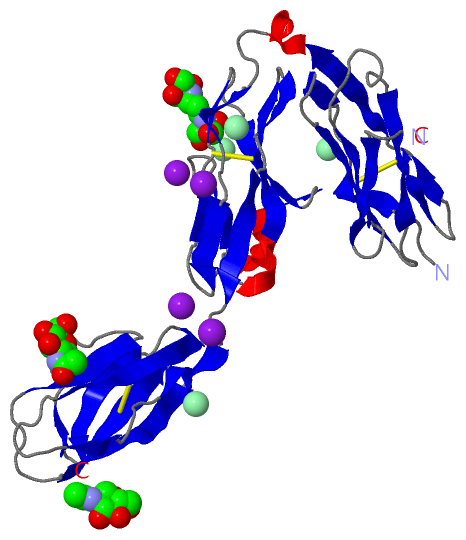
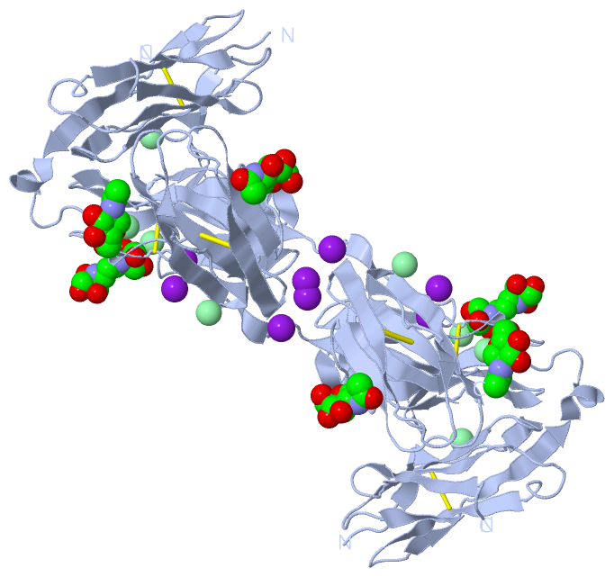
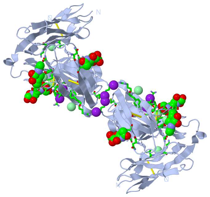
 Visualization

(-) Interactive Views

Asymmetric Unit
  Complete Structure
    Jena3D(integrated viewing of ligand, site, SAP, PROSITE, SCOP information)
    WebMol | AstexViewer[tm]@PDBe
(Java Applets, require no local installation except for Java; loading may be slow)
    STRAP
(Java WebStart application, automatic local installation, requires Java; full application with system access!)
    RasMol
(require local installation)
    Molscript (VRML)
(requires installation of a VRML viewer; select preferred view via VRML and generate a mono or stereo PDF format file)
 
  Ligands, Modified Residues, Ions
    B3P  [ RasMol | Jena3D ]  +environment [ RasMol | Jena3D ]
    CL  [ RasMol | Jena3D ]  +environment [ RasMol | Jena3D ]
    IOD  [ RasMol | Jena3D ]  +environment [ RasMol | Jena3D ]
    NAG  [ RasMol | Jena3D ]  +environment [ RasMol | Jena3D ]
 
  Sites
    AC1  [ RasMol ]  +environment [ RasMol ]
    AC2  [ RasMol ]  +environment [ RasMol ]
    AC3  [ RasMol ]  +environment [ RasMol ]
    AC4  [ RasMol ]  +environment [ RasMol ]
    AC5  [ RasMol ]  +environment [ RasMol ]
    AC6  [ RasMol ]  +environment [ RasMol ]
    AC7  [ RasMol ]  +environment [ RasMol ]
    AC8  [ RasMol ]  +environment [ RasMol ]
    AC9  [ RasMol ]  +environment [ RasMol ]
 
  Cis Peptide Bonds
    Asn A:161 - Pro A:162   [ RasMol ]  
    Asp A:59 - Pro A:60   [ RasMol ]  
    Gln A:94 - Pro A:95   [ RasMol ]  
    Gly A:246 - Gly A:247   [ RasMol ]  
    Ser A:258 - Pro A:259   [ RasMol ]  
    Thr A:98 - Pro A:99   [ RasMol ]  
 
Biological Unit
  Complete Structure
    Biological Unit 1  [ Jena3D ]

(-) Still Images

Jmol
  protein: cartoon or spacefill or dots and stick; nucleic acid: cartoon and stick; ligands: spacefill; active site: stick
Molscript
  protein, nucleic acid: cartoon; ligands: spacefill; active site: ball and stick

 Databases and Analysis Tools

(-) Databases

Access by PDB/NDB ID
  2yd9
    Family and Domain InformationProDom | SYSTERS
    General Structural InformationGlycoscienceDB | MMDB | NDB | OCA | PDB | PDBe | PDBj | PDBsum | PDBWiki | PQS | PROTEOPEDIA
    Orientation in MembranesOPM
    Protein SurfaceSURFACE
    Secondary StructureDSSP (structure derived) | HSSP (homology derived)
    Structural GenomicsGeneCensus
    Structural NeighboursCE | VAST
    Structure ClassificationCATH | Dali | SCOP
    Validation and Original DataBMRB Data View | BMRB Restraints Grid | EDS | PROCHECK | RECOORD | WHAT_CHECK
 
Access by UniProt ID/Accession number
  PTPRS_HUMAN | Q13332
    Comparative Protein Structure ModelsModBase
    Genomic InformationEnsembl
    Protein-protein InteractionDIP
    Sequence, Family and Domain InformationInterPro | Pfam | SMART | UniProtKB/SwissProt
 
Access by Enzyme Classificator   (EC Number)
  3.1.3.48
    General Enzyme InformationBRENDA | EC-PDB | Enzyme | IntEnz
    PathwayKEGG | MetaCyc
 
Access by Disease Identifier   (MIM ID)
  (no 'MIM ID' available)
    Disease InformationOMIM
 
Access by GenAge ID
  (no 'GenAge ID' available)
    Age Related InformationGenAge

(-) Analysis Tools

Access by PDB/NDB ID
    Domain InformationXDom
    Interatomic Contacts of Structural UnitsCSU
    Ligand-protein ContactsLPC
    Protein CavitiescastP
    Sequence and Secondary StructurePDBCartoon
    Structure AlignmentSTRAP(Java WebStart application, automatic local installation, requires Java; full application with system access!)
    Structure and Sequence BrowserSTING
 
Access by UniProt ID/Accession number
  PTPRS_HUMAN | Q13332
    Protein Disorder PredictionDisEMBL | FoldIndex | GLOBPLOT (for more information see DisProt)

 Related Entries

(-) Entries Sharing at Least One Protein Chain (UniProt ID)

UniProtKB/Swiss-Prot
        PTPRS_HUMAN | Q133322fh7 2yd2 2yd3 4bpc 4pbx

(-) Related Entries Specified in the PDB File

2fh7 CRYSTAL STRUCTURE OF THE PHOSPHATASE DOMAINS OF HUMAN PTPSIGMA
2yd1 CRYSTAL STRUCTURE OF THE N-TERMINAL IG1-2 MODULE OF DROSOPHILA RECEPTOR PROTEIN TYROSINE PHOSPHATASE DLAR
2yd2 CRYSTAL STRUCTURE OF THE N-TERMINAL IG1-2 MODULE OF HUMAN RECEPTOR PROTEIN TYROSINE PHOSPHATASE SIGMA
2yd3 CRYSTAL STRUCTURE OF THE N-TERMINAL IG1-2 MODULE OF HUMAN RECEPTOR PROTEIN TYROSINE PHOSPHATASE SIGMA
2yd4 CRYSTAL STRUCTURE OF THE N-TERMINAL IG1-2 MODULE OF CHICKEN RECEPTOR PROTEIN TYROSINE PHOSPHATASE SIGMA
2yd5 CRYSTAL STRUCTURE OF THE N-TERMINAL IG1-2 MODULE OF HUMAN RECEPTOR PROTEIN TYROSINE PHOSPHATASE LAR
2yd6 CRYSTAL STRUCTURE OF THE N-TERMINAL IG1-2 MODULE OF HUMAN RECEPTOR PROTEIN TYROSINE PHOSPHATASE DELTA
2yd7 CRYSTAL STRUCTURE OF THE N-TERMINAL IG1-2 MODULE OF HUMAN RECEPTOR PROTEIN TYROSINE PHOSPHATASE DELTA
2yd8 CRYSTAL STRUCTURE OF THE N-TERMINAL IG1-2 MODULE OF HUMAN RECEPTOR PROTEIN TYROSINE PHOSPHATASE LAR IN COMPLEX WITH SUCROSE OCTASULPHATE