|
|
|
|
 Description
|
|
Compounds
|
||||||||||||||||||||||||||||||||||||
Chains, Units
Summary Information (see also Sequences/Alignments below) |
Ligands, Modified Residues, Ions (1, 4)
Asymmetric Unit (1, 4)
|
Sites (4, 4)
Asymmetric Unit (4, 4)
|
SS Bonds (0, 0)| (no "SS Bond" information available for 2XZR) |
Cis Peptide Bonds (0, 0)| (no "Cis Peptide Bond" information available for 2XZR) |
SAPs(SNPs)/Variants (0, 0)| (no "SAP(SNP)/Variant" information available for 2XZR) |
PROSITE Motifs (0, 0)| (no "PROSITE Motif" information available for 2XZR) |
Exons (0, 0)| (no "Exon" information available for 2XZR) |
Sequences/Alignments
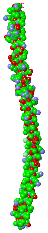
Asymmetric UnitChain A from PDB Type:PROTEIN Length:108 aligned with Q9MCI8_9CAUD | Q9MCI8 from UniProtKB/TrEMBL Length:511 Alignment length:165 342 352 362 372 382 392 402 412 422 432 442 452 462 472 482 492 Q9MCI8_9CAUD 333 KYTDQKTSEVNEKVEARTTVGVDSDGKLTRAEGATKTIAVNDGLVALSGRTDRIDYAVGAIDGRVTRNTQSIEKNSKAIAANTRTLQQHSARLDSQQRQINENHKEMKRAAAQSAALTGLFQPYSVGKFNATAAVGGYSDQQALAVGVGYRFNEQTAAKAGVAFS 497 SCOP domains --------------------------------------------------------------------------------------------------------------------------------------------------------------------- SCOP domains CATH domains --------------------------------------------------------------------------------------------------------------------------------------------------------------------- CATH domains Pfam domains --------------------------------------------------------------------------------------------------------------------------------------------------------------------- Pfam domains SAPs(SNPs) --------------------------------------------------------------------------------------------------------------------------------------------------------------------- SAPs(SNPs) PROSITE --------------------------------------------------------------------------------------------------------------------------------------------------------------------- PROSITE Transcript --------------------------------------------------------------------------------------------------------------------------------------------------------------------- Transcript 2xzr A 363 KQIEDKIEEILSKIYH------------------------------IENEIARIKKLIGAIDGRVTRNTQSIEKNSKAIAANTRTLQQHSARLDSQQRQINENHKEMKQIEDKIEEI--LSKIYHIE-------------------------NEIARIKKLIKLH 470 372 | - - - |382 392 402 412 422 432 442 |450 | - - - | 465 378 379 449 | 457 458 450
|
||||||||||||||||||||
SCOP Domains (0, 0)| (no "SCOP Domain" information available for 2XZR) |
CATH Domains (0, 0)| (no "CATH Domain" information available for 2XZR) |
Pfam Domains (0, 0)| (no "Pfam Domain" information available for 2XZR) |
Gene Ontology (2, 2)|
Asymmetric Unit(hide GO term definitions) Chain A (Q9MCI8_9CAUD | Q9MCI8)
|
||||||||||||||||||||||||
Interactive Views
|
|||||||||||||||||||||||||||||||||||||||||||||||||||||||||||||||||||||||||||||||||||||||||||||||||||||||||||||||||||||||||||||||||||||||||||||||||||||||||||||
Still Images
|
||||||||||||||||
Databases
|
||||||||||||||||||||||||||||||||||||||||||||||||||||||||||||||||||||||||||||||||||||||||||||||||||||||||||||||||||||||||||||||||||||||||||||||||||||||||||||||||
Analysis Tools
|
|||||||||||||||||||||||||||||||||||||||||||||||||||||||||||||
Entries Sharing at Least One Protein Chain (UniProt ID)
Related Entries Specified in the PDB File
|
|