|
|
|
|
 Description
|
|
Compounds
|
||||||||||||||||||||||||
Chains, Units
Summary Information (see also Sequences/Alignments below) |
Ligands, Modified Residues, Ions (3, 4)| Asymmetric/Biological Unit (3, 4) |
Sites (4, 4)
Asymmetric Unit (4, 4)
|
SS Bonds (1, 1)
Asymmetric/Biological Unit
|
||||||||
Cis Peptide Bonds (0, 0)| (no "Cis Peptide Bond" information available for 2XFD) |
SAPs(SNPs)/Variants (0, 0)| (no "SAP(SNP)/Variant" information available for 2XFD) |
PROSITE Motifs (0, 0)| (no "PROSITE Motif" information available for 2XFD) |
Exons (0, 0)| (no "Exon" information available for 2XFD) |
Sequences/Alignments
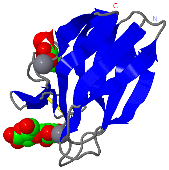
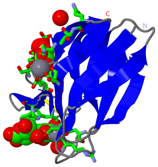
Asymmetric/Biological UnitChain A from PDB Type:PROTEIN Length:109 aligned with D7GNB4_9BACT | D7GNB4 from UniProtKB/TrEMBL Length:111 Alignment length:109 11 21 31 41 51 61 71 81 91 101 D7GNB4_9BACT 2 NSITVRARGVNGQESVSLQVGGTTVQTWTLTTAMQDYTASTSLTGEIRVAFTNDATGRDVQVDYIVVNGQTRQAENQSVNTGVWANNQCGGSGNSEWLHCNGYISFGNV 110 SCOP domains ------------------------------------------------------------------------------------------------------------- SCOP domains CATH domains ------------------------------------------------------------------------------------------------------------- CATH domains Pfam domains ------------------------------------------------------------------------------------------------------------- Pfam domains SAPs(SNPs) ------------------------------------------------------------------------------------------------------------- SAPs(SNPs) PROSITE ------------------------------------------------------------------------------------------------------------- PROSITE Transcript ------------------------------------------------------------------------------------------------------------- Transcript 2xfd A 2 NSITVRARGVNGQESVSLQVGGTTVQTWTLTTAMQDYTASTSLTGEIRVAFTNDATGRDVQVDYIVVNGQTRQAENQSVNTGVWANNQCGGSGNSEWLHCNGYISFGNV 110 11 21 31 41 51 61 71 81 91 101
|
||||||||||||||||||||
SCOP Domains (0, 0)| (no "SCOP Domain" information available for 2XFD) |
CATH Domains (0, 0)| (no "CATH Domain" information available for 2XFD) |
Pfam Domains (0, 0)| (no "Pfam Domain" information available for 2XFD) |
Gene Ontology (1, 1)|
Asymmetric/Biological Unit(hide GO term definitions) Chain A (D7GNB4_9BACT | D7GNB4)
|
||||||||||||
Interactive Views
|
|||||||||||||||||||||||||||||||||||||||||||||||||||||||||||||||||||||||||||||||||||||||||||||||||||||||||||||||||||||||||||||||||||||||||||||||||||||||||
Still Images
|
||||||||||||||||
Databases
|
||||||||||||||||||||||||||||||||||||||||||||||||||||||||||||||||||||||||||||||||||||||||||||||||||||||||||||||||||||||||||||||||||||||||||||||||||||||||||||||||
Analysis Tools
|
|||||||||||||||||||||||||||||||||||||||||||||||||||||||||||||
Entries Sharing at Least One Protein Chain (UniProt ID)
Related Entries Specified in the PDB File
|
|