|
|
|
|
 Description
|
|
Compounds
|
||||||||||||||||||||||||||||||||||||
Chains, Units
Summary Information (see also Sequences/Alignments below) |
Ligands, Modified Residues, Ions (1, 4)
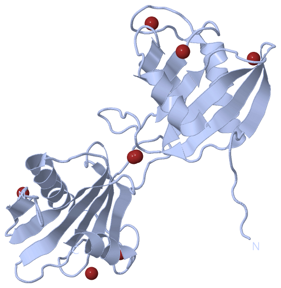
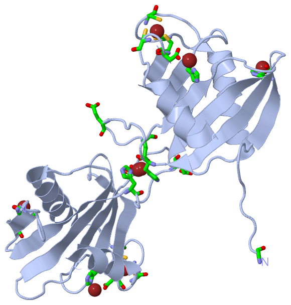
Asymmetric Unit (1, 4)
|
Sites (4, 4)
Asymmetric Unit (4, 4)
|
SS Bonds (0, 0)| (no "SS Bond" information available for 2X5R) |
Cis Peptide Bonds (0, 0)| (no "Cis Peptide Bond" information available for 2X5R) |
SAPs(SNPs)/Variants (0, 0)| (no "SAP(SNP)/Variant" information available for 2X5R) |
PROSITE Motifs (0, 0)| (no "PROSITE Motif" information available for 2X5R) |
Exons (0, 0)| (no "Exon" information available for 2X5R) |
Sequences/Alignments
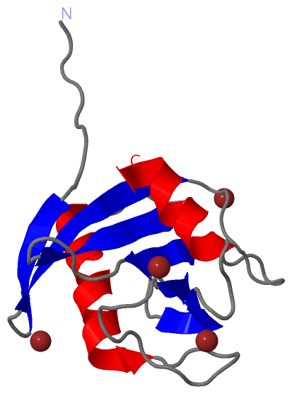
Asymmetric UnitChain A from PDB Type:PROTEIN Length:124 aligned with Q6ZYF6_PSVY | Q6ZYF6 from UniProtKB/TrEMBL Length:125 Alignment length:124 1 | 8 18 28 38 48 58 68 78 88 98 108 118 Q6ZYF6_PSVY - --MARVGPKIEITHGGKKYTVFSKVTHLVPRTENGEEAEYVVFGPEKEGVISVVVLAPKDLNEEALALRVKWFNDTKPRCVKCGAAYNGKNHFRVVAIRNGTYYLDAVCDKCEPRITWLSAIVI 122 SCOP domains ---------------------------------------------------------------------------------------------------------------------------- SCOP domains CATH domains ---------------------------------------------------------------------------------------------------------------------------- CATH domains Pfam domains ---------------------------------------------------------------------------------------------------------------------------- Pfam domains SAPs(SNPs) ---------------------------------------------------------------------------------------------------------------------------- SAPs(SNPs) PROSITE ---------------------------------------------------------------------------------------------------------------------------- PROSITE Transcript ---------------------------------------------------------------------------------------------------------------------------- Transcript 2x5r A 1 GAMARVGPKIEITHGGKKYTVFSKVTHLVPRTENGEEAEYVVFGPEKEGVISVVVLAPKDLNEEALALRVKWFNDTKPRCVKCGAAYNGKNHFRVVAIRNGTYYLDAVCDKCEPRITWLSAIVI 124 10 20 30 40 50 60 70 80 90 100 110 120
|
||||||||||||||||||||
SCOP Domains (0, 0)| (no "SCOP Domain" information available for 2X5R) |
CATH Domains (0, 0)| (no "CATH Domain" information available for 2X5R) |
Pfam Domains (0, 0)| (no "Pfam Domain" information available for 2X5R) |
Gene Ontology (1, 1)|
Asymmetric Unit(hide GO term definitions) Chain A (Q6ZYF6_PSVY | Q6ZYF6)
|
||||||||||||
Interactive Views
|
|||||||||||||||||||||||||||||||||||||||||||||||||||||||||||||||||||||||||||||||||||||||||||||||||||||||||||||||||||||||||||||||||||||||||||||||||||||||||||||
Still Images
|
||||||||||||||||
Databases
|
||||||||||||||||||||||||||||||||||||||||||||||||||||||||||||||||||||||||||||||||||||||||||||||||||||||||||||||||||||||||||||||||||||||||||||||||||||||||||||||||
Analysis Tools
|
|||||||||||||||||||||||||||||||||||||||||||||||||||||||||||||
Entries Sharing at Least One Protein Chain (UniProt ID)
Related Entries Specified in the PDB File
|
|