|
|
|
|
 Description
|
|
Compounds
|
||||||||||||||||||||||||||||||||||||||||||||
Chains, Units
Summary Information (see also Sequences/Alignments below) |
Ligands, Modified Residues, Ions (3, 4)
Asymmetric Unit (3, 4)
|
Sites (4, 4)
Asymmetric Unit (4, 4)
|
SS Bonds (0, 0)| (no "SS Bond" information available for 2WCI) |
Cis Peptide Bonds (2, 2)
Asymmetric Unit
|
||||||||||||
SAPs(SNPs)/Variants (0, 0)| (no "SAP(SNP)/Variant" information available for 2WCI) |
PROSITE Motifs (1, 2)
Asymmetric Unit (1, 2)
|
||||||||||||||||||||||||||||||||||||||||||||||||||||||||||||||||||||||||
Exons (0, 0)| (no "Exon" information available for 2WCI) |
Sequences/Alignments
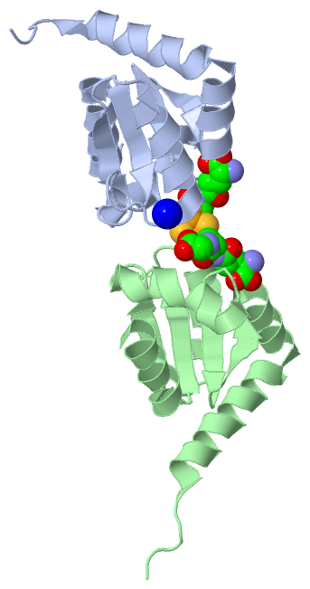
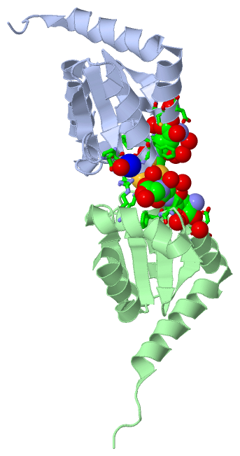
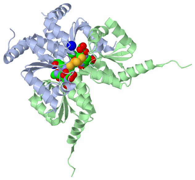
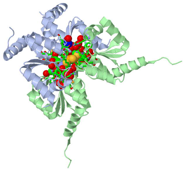
Asymmetric UnitChain A from PDB Type:PROTEIN Length:113 aligned with GLRX4_ECOLI | P0AC69 from UniProtKB/Swiss-Prot Length:115 Alignment length:113 10 20 30 40 50 60 70 80 90 100 110 GLRX4_ECOLI 1 MSTTIEKIQRQIAENPILLYMKGSPKLPSCGFSAQAVQALAACGERFAYVDILQNPDIRAELPKYANWPTFPQLWVDGELVGGCDIVIEMYQRGELQQLIKETAAKYKSEEPD 113 SCOP domains d2wcia_ A: automated matches SCOP domains CATH domains ----------------------------------------------------------------------------------------------------------------- CATH domains Pfam domains ----------------------------------------------------------------------------------------------------------------- Pfam domains SAPs(SNPs) ----------------------------------------------------------------------------------------------------------------- SAPs(SNPs) PROSITE ----GLUTAREDOXIN_2 PDB: A:5-107 UniProt: 5-107 ------ PROSITE Transcript ----------------------------------------------------------------------------------------------------------------- Transcript 2wci A 1 MSTTIEKIQRQIAENPILLYMKGSPKLPSCGFSAQAVQALAACGERFAYVDILQNPDIRAELPKYANWPTFPQLWVDGELVGGCDIVIEMYQRGELQQLIKETAAKYKSEEPD 113 10 20 30 40 50 60 70 80 90 100 110 Chain B from PDB Type:PROTEIN Length:112 aligned with GLRX4_ECOLI | P0AC69 from UniProtKB/Swiss-Prot Length:115 Alignment length:112 12 22 32 42 52 62 72 82 92 102 112 GLRX4_ECOLI 3 TTIEKIQRQIAENPILLYMKGSPKLPSCGFSAQAVQALAACGERFAYVDILQNPDIRAELPKYANWPTFPQLWVDGELVGGCDIVIEMYQRGELQQLIKETAAKYKSEEPDA 114 SCOP domains d2wcib_ B: automated matches SCOP domains CATH domains ---------------------------------------------------------------------------------------------------------------- CATH domains Pfam domains (1) --------------Glutaredoxin-2wciB01 B:17-81 --------------------------------- Pfam domains (1) Pfam domains (2) --------------Glutaredoxin-2wciB02 B:17-81 --------------------------------- Pfam domains (2) SAPs(SNPs) ---------------------------------------------------------------------------------------------------------------- SAPs(SNPs) PROSITE --GLUTAREDOXIN_2 PDB: B:5-107 UniProt: 5-107 ------- PROSITE Transcript ---------------------------------------------------------------------------------------------------------------- Transcript 2wci B 3 TTIEKIQRQIAENPILLYMKGSPKLPSCGFSAQAVQALAACGERFAYVDILQNPDIRAELPKYANWPTFPQLWVDGELVGGCDIVIEMYQRGELQQLIKETAAKYKSEEPDA 114 12 22 32 42 52 62 72 82 92 102 112
|
||||||||||||||||||||
SCOP Domains (1, 2)
Asymmetric Unit
 |
CATH Domains (0, 0)| (no "CATH Domain" information available for 2WCI) |
Pfam Domains (1, 2)
Asymmetric Unit
|
Gene Ontology (10, 10)|
Asymmetric Unit(hide GO term definitions) Chain A,B (GLRX4_ECOLI | P0AC69)
|
||||||||||||||||||||||||||||||||||||||||||||||||||||||||||||||||||||||||||||||
Interactive Views
|
||||||||||||||||||||||||||||||||||||||||||||||||||||||||||||||||||||||||||||||||||||||||||||||||||||||||||||||||||||||||||||||||||||||||||||||||||||||||||||||||||||||||||||||||||||||||
Still Images
|
||||||||||||||||
Databases
|
||||||||||||||||||||||||||||||||||||||||||||||||||||||||||||||||||||||||||||||||||||||||||||||||||||||||||||||||||||||||||||||||||||||||||||||||||||||||||||||||
Analysis Tools
|
|||||||||||||||||||||||||||||||||||||||||||||||||||||||||||||
Entries Sharing at Least One Protein Chain (UniProt ID)
Related Entries Specified in the PDB File
|
|