Show PDB file:   
         Plain Text   HTML   (compressed file size)
QuickSearch:   
by PDB,NDB,UniProt,PROSITE Code or Search Term(s)  
(-)Asymmetric Unit
(-)Asym. Unit - sites
(-)Biological Unit 1
(-)Biol. Unit 1 - sites
(-)Biological Unit 2
collapse expand < >
Image Asymmetric Unit
Asymmetric Unit  (Jmol Viewer)
Image Asym. Unit - sites
Asym. Unit - sites  (Jmol Viewer)
Image Biological Unit 1
Biological Unit 1  (Jmol Viewer)
Image Biol. Unit 1 - sites
Biol. Unit 1 - sites  (Jmol Viewer)
Image Biological Unit 2
Biological Unit 2  (Jmol Viewer)

(-) Description

Title :  PERIPLASMIC DOMAIN OF EPSL FROM VIBRIO PARAHAEMOLYTICUS
 
Authors :  J. Abendroth, A. C. Kreger, H. Abendroth, M. Sandkvist, W. G. J. Hol
Date :  06 Jan 09  (Deposition) - 31 Mar 10  (Release) - 28 Jun 17  (Revision)
Method :  X-RAY DIFFRACTION
Resolution :  2.30
Chains :  Asym. Unit :  A,B
Biol. Unit 1:  A  (2x)
Biol. Unit 2:  B  (2x)
Keywords :  Transport, Type Ii Secretion, Transport Protein (Keyword Search: [Gene Ontology, PubMed, Web (Google))
 
Reference :  J. Abendroth, A. C. Kreger, W. G. J. Hol
The Dimer Formed By The Periplasmic Domain Of Epsl From The Type 2 Secretion System Of Vibrio Parahaemolyticus.
J. Struct. Biol. V. 168 313 2009
PubMed-ID: 19646531  |  Reference-DOI: 10.1016/J.JSB.2009.07.022

(-) Compounds

Molecule 1 - GENERAL SECRETION PATHWAY PROTEIN L
    ChainsA, B
    EngineeredYES
    Expression SystemESCHERICHIA COLI
    Expression System PlasmidPJA-080
    Expression System StrainBL21GOLD(DE3)
    Expression System Taxid469008
    Expression System VectorPET21A
    FragmentPERIPLASMIC DOMAIN, RESIDUES 319-404
    Organism ScientificVIBRIO PARAHAEMOLYTICUS
    Organism Taxid670

 Structural Features

(-) Chains, Units

  12
Asymmetric Unit AB
Biological Unit 1 (2x)A 
Biological Unit 2 (2x) B

Summary Information (see also Sequences/Alignments below)

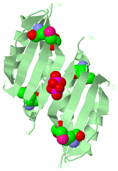
(-) Ligands, Modified Residues, Ions  (3, 8)

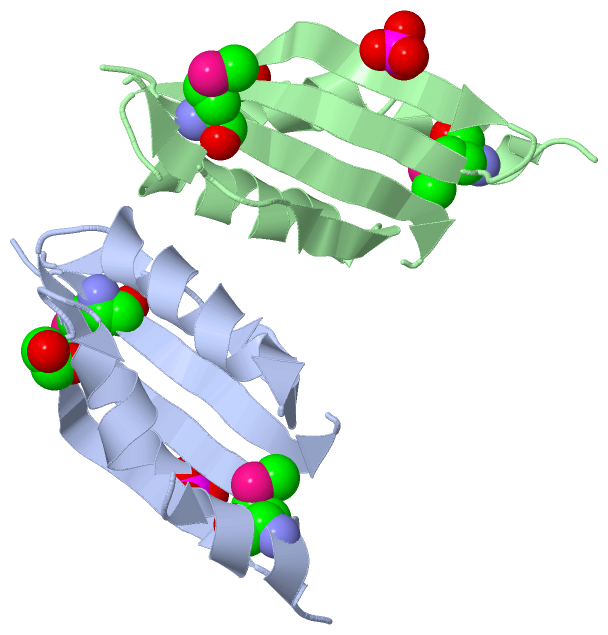
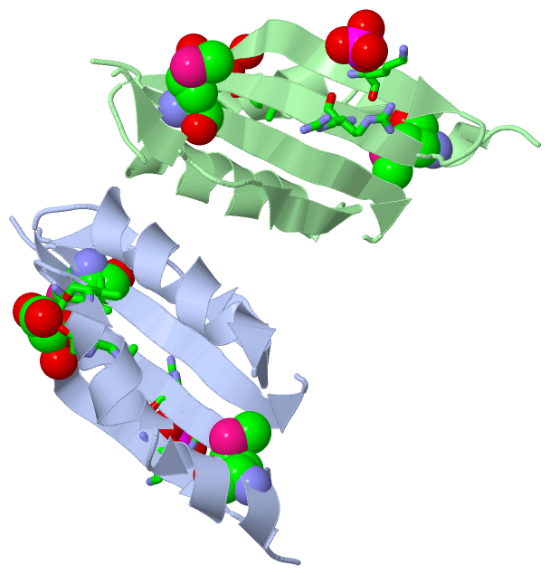
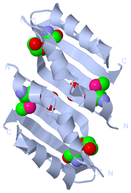
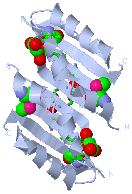
Asymmetric Unit (3, 8)
No.NameCountTypeFull Name
1EDO2Ligand/Ion1,2-ETHANEDIOL
2MSE4Mod. Amino AcidSELENOMETHIONINE
3PO42Ligand/IonPHOSPHATE ION
Biological Unit 1 (3, 8)
No.NameCountTypeFull Name
1EDO2Ligand/Ion1,2-ETHANEDIOL
2MSE4Mod. Amino AcidSELENOMETHIONINE
3PO42Ligand/IonPHOSPHATE ION
Biological Unit 2 (3, 8)
No.NameCountTypeFull Name
1EDO2Ligand/Ion1,2-ETHANEDIOL
2MSE4Mod. Amino AcidSELENOMETHIONINE
3PO42Ligand/IonPHOSPHATE ION

(-) Sites  (4, 4)

Asymmetric Unit (4, 4)
No.NameEvidenceResiduesDescription
1AC1SOFTWARELEU A:338 , GLY A:339 , VAL A:341 , LEU A:344 , HOH A:2005 , HOH A:2024BINDING SITE FOR RESIDUE EDO A 1405
2AC2SOFTWARELEU B:338 , GLY B:339 , VAL B:341 , LEU B:344 , HOH B:2005 , HOH B:2025BINDING SITE FOR RESIDUE EDO B 1405
3AC3SOFTWARELYS A:350 , ARG A:359 , HOH A:2025 , LYS B:350 , ARG B:359 , PO4 B:1406BINDING SITE FOR RESIDUE PO4 A 1406
4AC4SOFTWARELYS A:350 , PO4 A:1406 , LYS B:350BINDING SITE FOR RESIDUE PO4 B 1406

(-) SS Bonds  (0, 0)

(no "SS Bond" information available for 2W7V)

(-) Cis Peptide Bonds  (0, 0)

(no "Cis Peptide Bond" information available for 2W7V)

 Sequence-Structure Mapping

(-) SAPs(SNPs)/Variants  (0, 0)

(no "SAP(SNP)/Variant" information available for 2W7V)

(-) PROSITE Motifs  (0, 0)

(no "PROSITE Motif" information available for 2W7V)

(-) Exons   (0, 0)

(no "Exon" information available for 2W7V)

(-) Sequences/Alignments

Asymmetric Unit
   Reformat: Number of residues per line =  ('0' or empty: single-line sequence representation)
  Number of residues per labelling interval =   
  UniProt sequence: complete  aligned part    
   Show mapping: SCOP domains CATH domains Pfam domains Secondary structure (by author)
SAPs(SNPs) PROSITE motifs Exons
(details for a mapped element are shown in a popup box when the mouse pointer rests over it)
Chain A from PDB  Type:PROTEIN  Length:82
 aligned with Q87TC9_VIBPA | Q87TC9 from UniProtKB/TrEMBL  Length:404

    Alignment length:83
                                   331       341       351       361       371       381       391       401   
         Q87TC9_VIBPA   322 STDVAMLSWLAALPATLGQVKDLEITSFKYDGQRGEVRIHARSSDFQPFEQARVKLAEKFNVEQGQLNRSDNVVMGSFVLKRQ 404
               SCOP domains ----------------------------------------------------------------------------------- SCOP domains
               CATH domains ----------------------------------------------------------------------------------- CATH domains
               Pfam domains ----------------------------------------------------------------------------------- Pfam domains
         Sec.struct. author ..hhhhhhhh.hhhhhhhh....eeeeeeee....eeeeeeee..hhhhhhhhhhhhh.eeeee...ee.-..eeeeeeeee. Sec.struct. author
                 SAPs(SNPs) ----------------------------------------------------------------------------------- SAPs(SNPs)
                    PROSITE ----------------------------------------------------------------------------------- PROSITE
                 Transcript ----------------------------------------------------------------------------------- Transcript
                 2w7v A 322 STDVAmLSWLAALPATLGQVKDLEITSFKYDGQRGEVRIHARSSDFQPFEQARVKLAEKFNVEQGQLNRS-NVVmGSFVLKRQ 404
                                 | 331       341       351       361       371       381       391 |  |  401   
                                 |                                                             391 |  |        
                               327-MSE                                                           393  |        
                                                                                                    396-MSE    

Chain B from PDB  Type:PROTEIN  Length:82
 aligned with Q87TC9_VIBPA | Q87TC9 from UniProtKB/TrEMBL  Length:404

    Alignment length:83
                                   331       341       351       361       371       381       391       401   
         Q87TC9_VIBPA   322 STDVAMLSWLAALPATLGQVKDLEITSFKYDGQRGEVRIHARSSDFQPFEQARVKLAEKFNVEQGQLNRSDNVVMGSFVLKRQ 404
               SCOP domains ----------------------------------------------------------------------------------- SCOP domains
               CATH domains ----------------------------------------------------------------------------------- CATH domains
           Pfam domains (1) GspL_C-2w7vB01 B:322-404                                                            Pfam domains (1)
           Pfam domains (2) GspL_C-2w7vB02 B:322-404                                                            Pfam domains (2)
         Sec.struct. author ..hhhhhhhh.hhhhhhhh....eeeeeeee....eeeeeeee..hhhhhhhhhhhhh.eeeee...ee.-..eeeeeeeee. Sec.struct. author
                 SAPs(SNPs) ----------------------------------------------------------------------------------- SAPs(SNPs)
                    PROSITE ----------------------------------------------------------------------------------- PROSITE
                 Transcript ----------------------------------------------------------------------------------- Transcript
                 2w7v B 322 STDVAmLSWLAALPATLGQVKDLEITSFKYDGQRGEVRIHARSSDFQPFEQARVKLAEKFNVEQGQLNRS-NVVmGSFVLKRQ 404
                                 | 331       341       351       361       371       381       391 |  |  401   
                               327-MSE                                                         391 |  |        
                                                                                                 393  |        
                                                                                                    396-MSE    

   Legend:   → Mismatch (orange background)
  - → Gap (green background, '-', border residues have a numbering label)
    → Modified Residue (blue background, lower-case, 'x' indicates undefined single-letter code, labelled with number + name)
  x → Chemical Group (purple background, 'x', labelled with number + name, e.g. ACE or NH2)
  extra numbering lines below/above indicate numbering irregularities and modified residue names etc., number ends below/above '|'

 Classification and Annotation

(-) SCOP Domains  (0, 0)

(no "SCOP Domain" information available for 2W7V)

(-) CATH Domains  (0, 0)

(no "CATH Domain" information available for 2W7V)

(-) Pfam Domains  (1, 2)

Asymmetric Unit
(-)
Clan: EpsM (2)

(-) Gene Ontology  (5, 5)

Asymmetric Unit(hide GO term definitions)
Chain A,B   (Q87TC9_VIBPA | Q87TC9)
molecular function
    GO:0008565    protein transporter activity    Enables the directed movement of proteins into, out of or within a cell, or between cells.
biological process
    GO:0015031    protein transport    The directed movement of proteins into, out of or within a cell, or between cells, by means of some agent such as a transporter or pore.
    GO:0006810    transport    The directed movement of substances (such as macromolecules, small molecules, ions) or cellular components (such as complexes and organelles) into, out of or within a cell, or between cells, or within a multicellular organism by means of some agent such as a transporter, pore or motor protein.
cellular component
    GO:0009276    Gram-negative-bacterium-type cell wall    The peptidoglycan layer of the Gram-negative cell envelope. In Gram-negative cells the peptidoglycan is relatively thin (1-2nm) and is linked to the outer membrane by lipoproteins. In Gram-negative cells the peptidoglycan is too thin to retain the primary stain in the Gram staining procedure and therefore cells appear red after Gram stain.
    GO:0016021    integral component of membrane    The component of a membrane consisting of the gene products and protein complexes having at least some part of their peptide sequence embedded in the hydrophobic region of the membrane.

 Visualization

(-) Interactive Views

Asymmetric Unit
  Complete Structure
    Jena3D(integrated viewing of ligand, site, SAP, PROSITE, SCOP information)
    WebMol | AstexViewer[tm]@PDBe
(Java Applets, require no local installation except for Java; loading may be slow)
    STRAP
(Java WebStart application, automatic local installation, requires Java; full application with system access!)
    RasMol
(require local installation)
    Molscript (VRML)
(requires installation of a VRML viewer; select preferred view via VRML and generate a mono or stereo PDF format file)
 
  Ligands, Modified Residues, Ions
    EDO  [ RasMol | Jena3D ]  +environment [ RasMol | Jena3D ]
    MSE  [ RasMol | Jena3D ]  +environment [ RasMol | Jena3D ]
    PO4  [ RasMol | Jena3D ]  +environment [ RasMol | Jena3D ]
 
  Sites
    AC1  [ RasMol ]  +environment [ RasMol ]
    AC2  [ RasMol ]  +environment [ RasMol ]
    AC3  [ RasMol ]  +environment [ RasMol ]
    AC4  [ RasMol ]  +environment [ RasMol ]
 
  Cis Peptide Bonds
(no "Cis Peptide Bonds" information available for 2w7v)
 
Biological Units
  Complete Structure
    Biological Unit 1  [ Jena3D ]
    Biological Unit 2  [ Jena3D ]

(-) Still Images

Jmol
  protein: cartoon or spacefill or dots and stick; nucleic acid: cartoon and stick; ligands: spacefill; active site: stick
Molscript
  protein, nucleic acid: cartoon; ligands: spacefill; active site: ball and stick

 Databases and Analysis Tools

(-) Databases

Access by PDB/NDB ID
  2w7v
    Family and Domain InformationProDom | SYSTERS
    General Structural InformationGlycoscienceDB | MMDB | NDB | OCA | PDB | PDBe | PDBj | PDBsum | PDBWiki | PQS | PROTEOPEDIA
    Orientation in MembranesOPM
    Protein SurfaceSURFACE
    Secondary StructureDSSP (structure derived) | HSSP (homology derived)
    Structural GenomicsGeneCensus
    Structural NeighboursCE | VAST
    Structure ClassificationCATH | Dali | SCOP
    Validation and Original DataBMRB Data View | BMRB Restraints Grid | EDS | PROCHECK | RECOORD | WHAT_CHECK
 
Access by UniProt ID/Accession number
  Q87TC9_VIBPA | Q87TC9
    Comparative Protein Structure ModelsModBase
    Genomic InformationEnsembl
    Protein-protein InteractionDIP
    Sequence, Family and Domain InformationInterPro | Pfam | SMART | UniProtKB/TrEMBL
 
Access by Enzyme Classificator   (EC Number)
  (no 'Enzyme Classificator' available)
    General Enzyme InformationBRENDA | EC-PDB | Enzyme | IntEnz
    PathwayKEGG | MetaCyc
 
Access by Disease Identifier   (MIM ID)
  (no 'MIM ID' available)
    Disease InformationOMIM
 
Access by GenAge ID
  (no 'GenAge ID' available)
    Age Related InformationGenAge

(-) Analysis Tools

Access by PDB/NDB ID
    Domain InformationXDom
    Interatomic Contacts of Structural UnitsCSU
    Ligand-protein ContactsLPC
    Protein CavitiescastP
    Sequence and Secondary StructurePDBCartoon
    Structure AlignmentSTRAP(Java WebStart application, automatic local installation, requires Java; full application with system access!)
    Structure and Sequence BrowserSTING
 
Access by UniProt ID/Accession number
  Q87TC9_VIBPA | Q87TC9
    Protein Disorder PredictionDisEMBL | FoldIndex | GLOBPLOT (for more information see DisProt)

 Related Entries

(-) Entries Sharing at Least One Protein Chain (UniProt ID)

(no "Entries Sharing at Least One Protein Chain" available for 2W7V)

(-) Related Entries Specified in the PDB File

(no "Related Entries Specified in the PDB File" available for 2W7V)