|
|
 Description
|
|
Compounds
|
||||||||||||||||||||||||||||||||||||||||||||||||||||||||||||||||||||||||||||||||
Chains, Units
Summary Information (see also Sequences/Alignments below) |
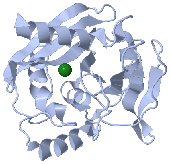
Ligands, Modified Residues, Ions (1, 2)
Asymmetric Unit (1, 2)
|
Sites (2, 2)
Asymmetric Unit (2, 2)
|
SS Bonds (0, 0)| (no "SS Bond" information available for 2W35) |
Cis Peptide Bonds (2, 2)
Asymmetric Unit
|
||||||||||||
SAPs(SNPs)/Variants (0, 0)| (no "SAP(SNP)/Variant" information available for 2W35) |
PROSITE Motifs (0, 0)| (no "PROSITE Motif" information available for 2W35) |
Exons (0, 0)| (no "Exon" information available for 2W35) |
Sequences/Alignments
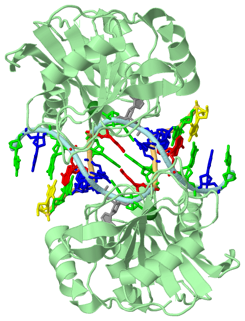
Asymmetric UnitChain A from PDB Type:PROTEIN Length:224 aligned with NFI_THEMA | Q9X2H9 from UniProtKB/Swiss-Prot Length:225 Alignment length:224 10 20 30 40 50 60 70 80 90 100 110 120 130 140 150 160 170 180 190 200 210 220 NFI_THEMA 1 MDYRQLHRWDLPPEEAIKVQNELRKKIKLTPYEGEPEYVAGVDLSFPGKEEGLAVIVVLEYPSFKILEVVSERGEITFPYIPGLLAFREGPLFLKAWEKLRTKPDVVVFDGQGLAHPRKLGIASHMGLFIEIPTIGVAKSRLYGTFKMPEDKRCSWSYLYDGEEIIGCVIRTKEGSAPIFVSPGHLMDVESSKRLIKAFTLPGRRIPEPTRLAHIYTQRLKKGL 224 SCOP domains -------------------------------------------------------------------------------------------------------------------------------------------------------------------------------------------------------------------------------- SCOP domains CATH domains -------------------------------------------------------------------------------------------------------------------------------------------------------------------------------------------------------------------------------- CATH domains Pfam domains -------------------------------------------------------------------------------------------------------------------------------------------------------------------------------------------------------------------------------- Pfam domains SAPs(SNPs) -------------------------------------------------------------------------------------------------------------------------------------------------------------------------------------------------------------------------------- SAPs(SNPs) PROSITE -------------------------------------------------------------------------------------------------------------------------------------------------------------------------------------------------------------------------------- PROSITE Transcript -------------------------------------------------------------------------------------------------------------------------------------------------------------------------------------------------------------------------------- Transcript 2w35 A 1 MDYRQLHRWDLPPEEAIKVQNELRKKIKLTPYEGEPEYVAGVDLSFPGKEEGLAVIVVLEYPSFKILEVVSERGEITFPYIPGLLAFREGPLFLKAWEKLRTKPDVVVFDGQGLAHPRKLGIASHMGLFIEIPTIGVAKSRLYGTFKMPEDKRCSWSYLYDGEEIIGCVIRTKEGSAPIFVSPGHLMDVESSKRLIKAFTLPGRRIPEPTRLAHIYTQRLKKGL 224 10 20 30 40 50 60 70 80 90 100 110 120 130 140 150 160 170 180 190 200 210 220 Chain B from PDB Type:PROTEIN Length:224 aligned with NFI_THEMA | Q9X2H9 from UniProtKB/Swiss-Prot Length:225 Alignment length:224 10 20 30 40 50 60 70 80 90 100 110 120 130 140 150 160 170 180 190 200 210 220 NFI_THEMA 1 MDYRQLHRWDLPPEEAIKVQNELRKKIKLTPYEGEPEYVAGVDLSFPGKEEGLAVIVVLEYPSFKILEVVSERGEITFPYIPGLLAFREGPLFLKAWEKLRTKPDVVVFDGQGLAHPRKLGIASHMGLFIEIPTIGVAKSRLYGTFKMPEDKRCSWSYLYDGEEIIGCVIRTKEGSAPIFVSPGHLMDVESSKRLIKAFTLPGRRIPEPTRLAHIYTQRLKKGL 224 SCOP domains -------------------------------------------------------------------------------------------------------------------------------------------------------------------------------------------------------------------------------- SCOP domains CATH domains -------------------------------------------------------------------------------------------------------------------------------------------------------------------------------------------------------------------------------- CATH domains Pfam domains (1) ------------------Endonuclease_5-2w35B01 B:19-217 ------- Pfam domains (1) Pfam domains (2) ------------------Endonuclease_5-2w35B02 B:19-217 ------- Pfam domains (2) SAPs(SNPs) -------------------------------------------------------------------------------------------------------------------------------------------------------------------------------------------------------------------------------- SAPs(SNPs) PROSITE -------------------------------------------------------------------------------------------------------------------------------------------------------------------------------------------------------------------------------- PROSITE Transcript -------------------------------------------------------------------------------------------------------------------------------------------------------------------------------------------------------------------------------- Transcript 2w35 B 1 MDYRQLHRWDLPPEEAIKVQNELRKKIKLTPYEGEPEYVAGVDLSFPGKEEGLAVIVVLEYPSFKILEVVSERGEITFPYIPGLLAFREGPLFLKAWEKLRTKPDVVVFDGQGLAHPRKLGIASHMGLFIEIPTIGVAKSRLYGTFKMPEDKRCSWSYLYDGEEIIGCVIRTKEGSAPIFVSPGHLMDVESSKRLIKAFTLPGRRIPEPTRLAHIYTQRLKKGL 224 10 20 30 40 50 60 70 80 90 100 110 120 130 140 150 160 170 180 190 200 210 220
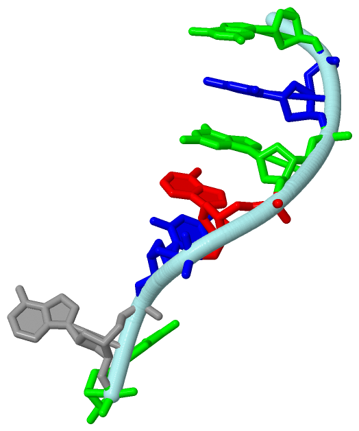
Chain C from PDB  Type:DNA  Length:4

                 2w35 C   6 ACIG   9
Chain D from PDB  Type:DNA  Length:4

                 2w35 D  10 AGCC  13
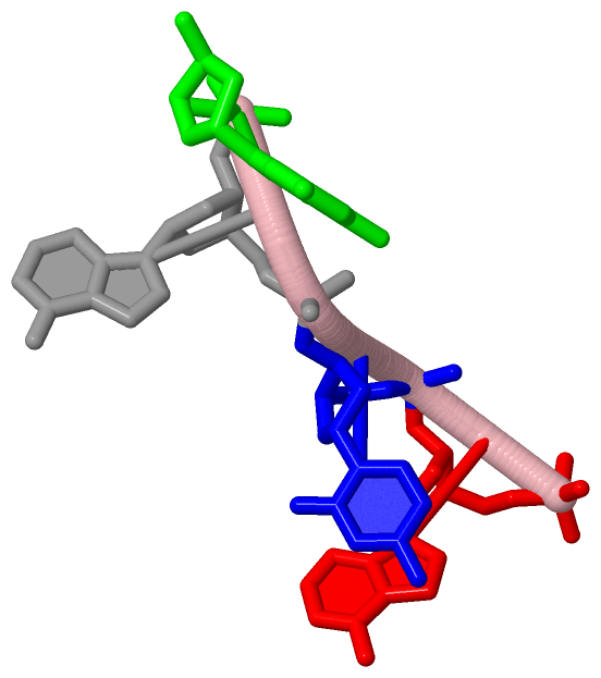
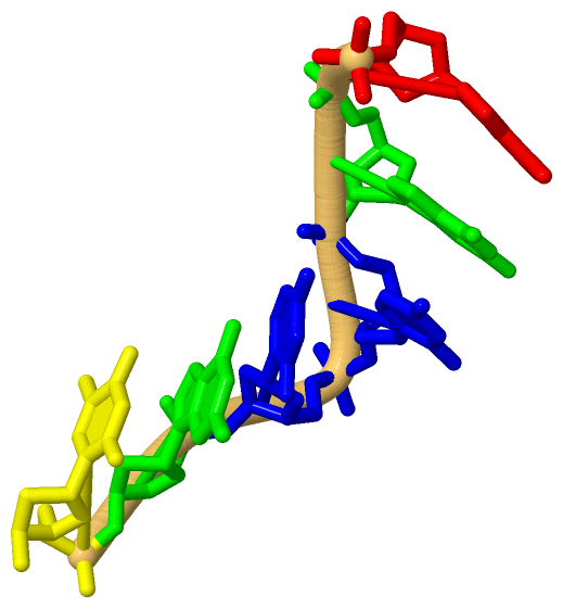
Chain F from PDB  Type:DNA  Length:7

                 2w35 F   3 GCGACIG   9
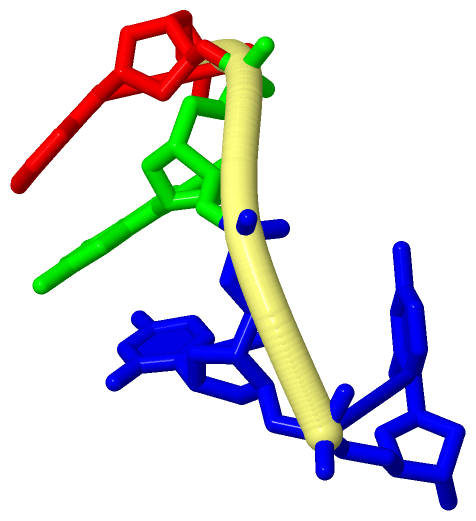
Chain G from PDB  Type:DNA  Length:6

                 2w35 G  10 AGCCGT  15
|
||||||||||||||||||||
SCOP Domains (0, 0)| (no "SCOP Domain" information available for 2W35) |
CATH Domains (0, 0)| (no "CATH Domain" information available for 2W35) |
Pfam Domains (1, 2)
Asymmetric Unit

|
Gene Ontology (10, 10)|
Asymmetric Unit(hide GO term definitions) Chain A,B (NFI_THEMA | Q9X2H9)
|
||||||||||||||||||||||||||||||||||||||||||||||||||||||||||||||||||||||||||||||
Interactive Views
|
||||||||||||||||||||||||||||||||||||||||||||||||||||||||||||||||||||||||||||||||||||||||||||||||||||||||||||||||||||||||||||||||||||||||||||||||||||||||||||||||||||||||||||||||||||||||||
Still Images
|
||||||||||||||||
Databases
|
||||||||||||||||||||||||||||||||||||||||||||||||||||||||||||||||||||||||||||||||||||||||||||||||||||||||||||||||||||||||||||||||||||||||||||||||||||||||||||||||
Analysis Tools
|
|||||||||||||||||||||||||||||||||||||||||||||||||||||||||||||
Entries Sharing at Least One Protein Chain (UniProt ID)
Related Entries Specified in the PDB File
|
|