|
|
|
|
 Description
|
|
Compounds
|
||||||||||||||||||||||||||||||||||||
Chains, Units
Summary Information (see also Sequences/Alignments below) |
Ligands, Modified Residues, Ions (4, 4)| Asymmetric/Biological Unit (4, 4) |
Sites (3, 3)
Asymmetric Unit (3, 3)
|
SS Bonds (3, 3)
Asymmetric/Biological Unit
|
||||||||||||||||
Cis Peptide Bonds (0, 0)| (no "Cis Peptide Bond" information available for 2W12) |
SAPs(SNPs)/Variants (0, 0)| (no "SAP(SNP)/Variant" information available for 2W12) |
PROSITE Motifs (2, 2)
Asymmetric/Biological Unit (2, 2)
|
||||||||||||||||||||||||||||||||
Exons (0, 0)| (no "Exon" information available for 2W12) |
Sequences/Alignments
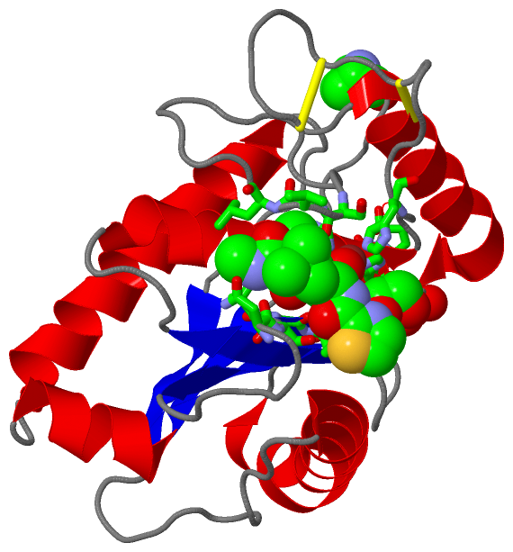
Asymmetric/Biological UnitChain A from PDB Type:PROTEIN Length:202 aligned with VM1B1_BOTAS | P83512 from UniProtKB/Swiss-Prot Length:408 Alignment length:202 202 212 222 232 242 252 262 272 282 292 302 312 322 332 342 352 362 372 382 392 VM1B1_BOTAS 193 QRFSPRYIELAVVADHGIFTKYNSNLNTIRTRVHEMLNTVNGFYRSVDVHAPLANLEVWSKQDLIKVQKDSSKTLKSFGEWRERDLLPRISHDHAQLLTAVVFDGNTIGRAYTGGMCDPRHSVGVVRDHSKNNLWVAVTMAHELGHNLGIHHDTGSCSCGAKSCIMASVLSKVLSYEFSDCSQNQYETYLTNHNPQCILNKP 394 SCOP domains d2w12a_ A: Snake venom metalloprotease SCOP domains CATH domains -2w12A00 A:2-202 Collagenase (Catalytic Domain) CATH domains Pfam domains ---------------------------------------------------------------------------------------------------------------------------------------------------------------------------------------------------------- Pfam domains SAPs(SNPs) ---------------------------------------------------------------------------------------------------------------------------------------------------------------------------------------------------------- SAPs(SNPs) PROSITE (1) -----ADAM_MEPRO PDB: A:6-202 UniProt: 198-394 PROSITE (1) PROSITE (2) ------------------------------------------------------------------------------------------------------------------------------------------ZINC_PROTE------------------------------------------------------ PROSITE (2) Transcript ---------------------------------------------------------------------------------------------------------------------------------------------------------------------------------------------------------- Transcript 2w12 A 1 xRFSPRYIELAVVADHGIFTKYNSNLNTIRTRVHEMLNTVNGFYRSVDVHAPLANLEVWSKQDLIKVQKDSSKTLKSFGEWRERDLLPRISHDHAQLLTAVVFDGNTIGRAYTGGMCDPRHSVGVVRDHSKNNLWVAVTMAHELGHNLGIHHDTGSCSCGAKSCIMASVLSKVLSYEFSDCSQNQYETYLTNHNPQCILNKP 202 | 10 20 30 40 50 60 70 80 90 100 110 120 130 140 150 160 170 180 190 200 | 1-PCA
|
||||||||||||||||||||
SCOP Domains (1, 1)
Asymmetric/Biological Unit
 |
CATH Domains (1, 1)
Asymmetric/Biological Unit
|
Pfam Domains (0, 0)| (no "Pfam Domain" information available for 2W12) |
Gene Ontology (9, 9)|
Asymmetric/Biological Unit(hide GO term definitions) Chain A (VM1B1_BOTAS | P83512)
|
||||||||||||||||||||||||||||||||||||||||||||||||||||||||||||||||||||||||
Interactive Views
|
|||||||||||||||||||||||||||||||||||||||||||||||||||||||||||||||||||||||||||||||||||||||||||||||||||||||||||||||||||||||||||||||||||||||||||||||||||||||||
Still Images
|
||||||||||||||||
Databases
|
||||||||||||||||||||||||||||||||||||||||||||||||||||||||||||||||||||||||||||||||||||||||||||||||||||||||||||||||||||||||||||||||||||||||||||||||||||||||||||||||
Analysis Tools
|
|||||||||||||||||||||||||||||||||||||||||||||||||||||||||||||
Entries Sharing at Least One Protein Chain (UniProt ID)
Related Entries Specified in the PDB File
|
|