|
|
|
|
 Description
|
|
Compounds
|
||||||||||||||||||||||||||||||||||||||||
Chains, Units
Summary Information (see also Sequences/Alignments below) |
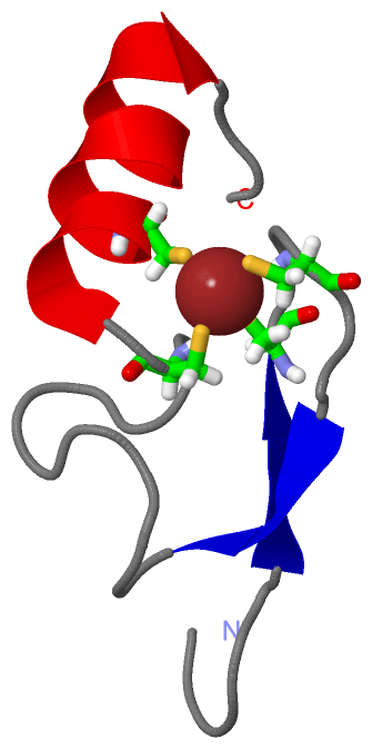
Ligands, Modified Residues, Ions (1, 1)
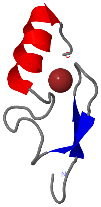
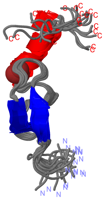
NMR Structure (1, 1)
|
Sites (1, 1)
NMR Structure (1, 1)
|
SS Bonds (0, 0)| (no "SS Bond" information available for 2W0T) |
Cis Peptide Bonds (0, 0)| (no "Cis Peptide Bond" information available for 2W0T) |
SAPs(SNPs)/Variants (0, 0)| (no "SAP(SNP)/Variant" information available for 2W0T) |
PROSITE Motifs (0, 0)| (no "PROSITE Motif" information available for 2W0T) |
Exons (0, 0)| (no "Exon" information available for 2W0T) |
Sequences/Alignments
NMR StructureChain A from PDB Type:PROTEIN Length:43 aligned with LMBL2_HUMAN | Q969R5 from UniProtKB/Swiss-Prot Length:705 Alignment length:43 91 101 111 121 LMBL2_HUMAN 82 GSGSEPAVCEMCGIVGTREAFFSKTKRFCSVSCSRSYSSNSKK 124 SCOP domains ------------------------------------------- SCOP domains CATH domains ------------------------------------------- CATH domains Pfam domains ------------------------------------------- Pfam domains SAPs(SNPs) ------------------------------------------- SAPs(SNPs) PROSITE ------------------------------------------- PROSITE Transcript ------------------------------------------- Transcript 2w0t A 82 GSGSEPAVCEMCGIVGTREAFFSKTKRFCSVSCSRSYSSNSKK 124 91 101 111 121
|
||||||||||||||||||||
SCOP Domains (0, 0)| (no "SCOP Domain" information available for 2W0T) |
CATH Domains (0, 0)| (no "CATH Domain" information available for 2W0T) |
Pfam Domains (0, 0)| (no "Pfam Domain" information available for 2W0T) |
Gene Ontology (10, 10)|
NMR Structure(hide GO term definitions) Chain A (LMBL2_HUMAN | Q969R5)
|
||||||||||||||||||||||||||||||||||||||||||||||||||||||||||||||||||||||||||||||
Interactive Views
|
||||||||||||||||||||||||||||||||||||||||||||||||||||||||||||||||||||||||||||||||||||||||||||||||||||||||||||||||||||||
Still Images
|
||||||||||||||||
Databases
|
||||||||||||||||||||||||||||||||||||||||||||||||||||||||||||||||||||||||||||||||||||||||||||||||||||||||||||||||||||||||||||||||||||||||||||||||||||||||||||||||
Analysis Tools
|
|||||||||||||||||||||||||||||||||||||||||||||||||||||||||||||
Entries Sharing at Least One Protein Chain (UniProt ID)
Related Entries Specified in the PDB File
|
|