|
|
|
|
 Description
|
|
Compounds
|
||||||||||||||||||||||||||||||||||||||||||||||||||||
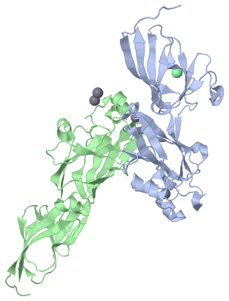
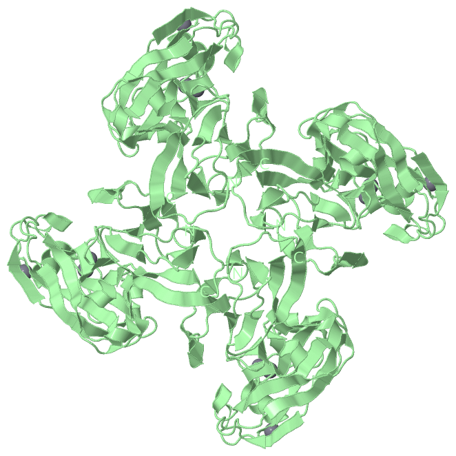
Chains, Units
Summary Information (see also Sequences/Alignments below) |
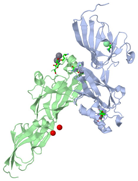
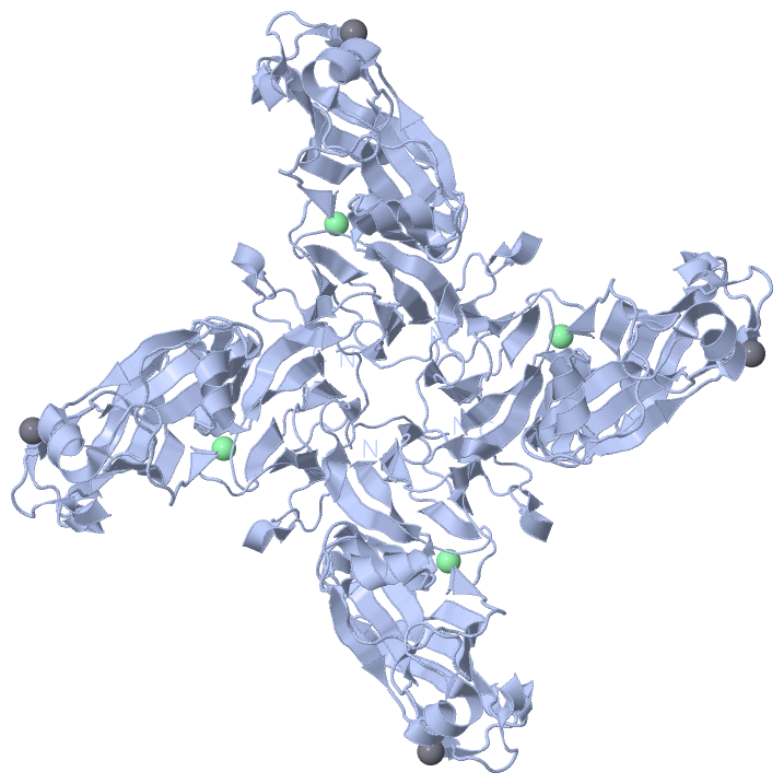
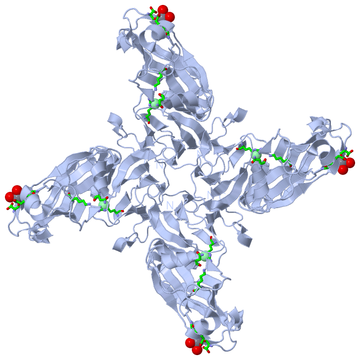
Ligands, Modified Residues, Ions (2, 5)| Asymmetric Unit (2, 5) Biological Unit 1 (0, 0) Biological Unit 2 (0, 0) |
Sites (5, 5)
Asymmetric Unit (5, 5)
|
SS Bonds (0, 0)| (no "SS Bond" information available for 2VVE) |
Cis Peptide Bonds (0, 0)| (no "Cis Peptide Bond" information available for 2VVE) |
SAPs(SNPs)/Variants (0, 0)| (no "SAP(SNP)/Variant" information available for 2VVE) |
PROSITE Motifs (0, 0)| (no "PROSITE Motif" information available for 2VVE) |
Exons (0, 0)| (no "Exon" information available for 2VVE) |
Sequences/Alignments
Asymmetric UnitChain A from PDB Type:PROTEIN Length:252 aligned with SPIKE_BPPM2 | Q9XJR3 from UniProtKB/Swiss-Prot Length:335 Alignment length:252 93 103 113 123 133 143 153 163 173 183 193 203 213 223 233 243 253 263 273 283 293 303 313 323 333 SPIKE_BPPM2 84 QEQTTKSRDVNSFQIPLRDGVRELLPEDASRNRASIKSPVDIWIGGENMTALNGIVDGGRKFEAGQEFQINTFGSVNYWVSDEEIRVFKEYSARAKYAQNEGRTALEANNVPFFDIDVPPELDGVPFSLKARVRHKSKGVDGLGDYTSISVKPAFYITEGDETTDTLIKYTSYGSTGSHSGYDFDDNTLDVMVTLSAGVHRVFPVETELDYDAVQEVQHDWYDESFTTFIEVYSDDPLLTVKGYAQILMERT 335 SCOP domains ------------------------------------------------------------------------------------------------------------------------------------------------------------------------------------------------------------------------------------------------------------ SCOP domains CATH domains ------------------------------------------------------------------------------------------------------------------------------------------------------------------------------------------------------------------------------------------------------------ CATH domains Pfam domains ------------------------------------------------------------------------------------------------------------------------------------------------------------------------------------------------------------------------------------------------------------ Pfam domains SAPs(SNPs) ------------------------------------------------------------------------------------------------------------------------------------------------------------------------------------------------------------------------------------------------------------ SAPs(SNPs) PROSITE ------------------------------------------------------------------------------------------------------------------------------------------------------------------------------------------------------------------------------------------------------------ PROSITE Transcript ------------------------------------------------------------------------------------------------------------------------------------------------------------------------------------------------------------------------------------------------------------ Transcript 2vve A 84 QEQTTKSRDVNSFQIPLRDGVRELLPEDASRNRASIKSPVDIWIGGENMTALNGIVDGGRKFEAGQEFQINTFGSVNYWVSDEEIRVFKEYSARAKYAQNEGRTALEANNVPFFDIDVPPELDGVPFSLKARVRHKSKGVDGLGDYTSISVKPAFYITEGDETTDTLIKYTSYGSTGSHSGYDFDDNTLDVMVTLSAGVHRVFPVETELDYDAVQEVQHDWYDESFTTFIEVYSDDPLLTVKGYAQILMERT 335 93 103 113 123 133 143 153 163 173 183 193 203 213 223 233 243 253 263 273 283 293 303 313 323 333 Chain B from PDB Type:PROTEIN Length:251 aligned with SPIKE_BPPM2 | Q9XJR3 from UniProtKB/Swiss-Prot Length:335 Alignment length:251 94 104 114 124 134 144 154 164 174 184 194 204 214 224 234 244 254 264 274 284 294 304 314 324 334 SPIKE_BPPM2 85 EQTTKSRDVNSFQIPLRDGVRELLPEDASRNRASIKSPVDIWIGGENMTALNGIVDGGRKFEAGQEFQINTFGSVNYWVSDEEIRVFKEYSARAKYAQNEGRTALEANNVPFFDIDVPPELDGVPFSLKARVRHKSKGVDGLGDYTSISVKPAFYITEGDETTDTLIKYTSYGSTGSHSGYDFDDNTLDVMVTLSAGVHRVFPVETELDYDAVQEVQHDWYDESFTTFIEVYSDDPLLTVKGYAQILMERT 335 SCOP domains ----------------------------------------------------------------------------------------------------------------------------------------------------------------------------------------------------------------------------------------------------------- SCOP domains CATH domains ----------------------------------------------------------------------------------------------------------------------------------------------------------------------------------------------------------------------------------------------------------- CATH domains Pfam domains ----------------------------------------------------------------------------------------------------------------------------------------------------------------------------------------------------------------------------------------------------------- Pfam domains SAPs(SNPs) ----------------------------------------------------------------------------------------------------------------------------------------------------------------------------------------------------------------------------------------------------------- SAPs(SNPs) PROSITE ----------------------------------------------------------------------------------------------------------------------------------------------------------------------------------------------------------------------------------------------------------- PROSITE Transcript ----------------------------------------------------------------------------------------------------------------------------------------------------------------------------------------------------------------------------------------------------------- Transcript 2vve B 85 EQTTKSRDVNSFQIPLRDGVRELLPEDASRNRASIKSPVDIWIGGENMTALNGIVDGGRKFEAGQEFQINTFGSVNYWVSDEEIRVFKEYSARAKYAQNEGRTALEANNVPFFDIDVPPELDGVPFSLKARVRHKSKGVDGLGDYTSISVKPAFYITEGDETTDTLIKYTSYGSTGSHSGYDFDDNTLDVMVTLSAGVHRVFPVETELDYDAVQEVQHDWYDESFTTFIEVYSDDPLLTVKGYAQILMERT 335 94 104 114 124 134 144 154 164 174 184 194 204 214 224 234 244 254 264 274 284 294 304 314 324 334
|
||||||||||||||||||||
SCOP Domains (0, 0)| (no "SCOP Domain" information available for 2VVE) |
CATH Domains (0, 0)| (no "CATH Domain" information available for 2VVE) |
Pfam Domains (0, 0)| (no "Pfam Domain" information available for 2VVE) |
Gene Ontology (1, 1)|
Asymmetric Unit(hide GO term definitions) Chain A,B (SPIKE_BPPM2 | Q9XJR3)
|
||||||||||||
Interactive Views
|
||||||||||||||||||||||||||||||||||||||||||||||||||||||||||||||||||||||||||||||||||||||||||||||||||||||||||||||||||||||||||||||||||||||||||||||||||||||||||||||||||||||||||||||||
Still Images
|
||||||||||||||||
Databases
|
||||||||||||||||||||||||||||||||||||||||||||||||||||||||||||||||||||||||||||||||||||||||||||||||||||||||||||||||||||||||||||||||||||||||||||||||||||||||||||||||
Analysis Tools
|
|||||||||||||||||||||||||||||||||||||||||||||||||||||||||||||
Entries Sharing at Least One Protein Chain (UniProt ID)
Related Entries Specified in the PDB File
|
|