Show PDB file:   
         Plain Text   HTML   (compressed file size)
QuickSearch:   
by PDB,NDB,UniProt,PROSITE Code or Search Term(s)  
(-)Asymmetric Unit
(-)Asym. Unit - sites
(-)Biological Unit 1
(-)Biol. Unit 1 - sites
(-)Biological Unit 2
(-)Biological Unit 3
collapse expand < >
Image Asymmetric Unit
Asymmetric Unit  (Jmol Viewer)
Image Asym. Unit - sites
Asym. Unit - sites  (Jmol Viewer)
Image Biological Unit 1
Biological Unit 1  (Jmol Viewer)
Image Biol. Unit 1 - sites
Biol. Unit 1 - sites  (Jmol Viewer)
Image Biological Unit 2
Biological Unit 2  (Jmol Viewer)
Image Biological Unit 3
Biological Unit 3  (Jmol Viewer)

(-) Description

Title :  APO FUTA2 FROM SYNECHOCYSTIS PCC6803
 
Authors :  A. Badarau, S. J. Firbank, M. J. Banfield, C. Dennison
Date :  25 Feb 08  (Deposition) - 25 Mar 08  (Release) - 24 Feb 09  (Revision)
Method :  X-RAY DIFFRACTION
Resolution :  1.70
Chains :  Asym. Unit :  A,B
Biol. Unit 1:  A,B  (2x)
Biol. Unit 2:  A  (1x)
Biol. Unit 3:  B  (1x)
Keywords :  Ferric Binding Protein, Metal-Binding Protein, Tat, Iron, Futa2, Synechocystis (Keyword Search: [Gene Ontology, PubMed, Web (Google))
 
Reference :  A. Badarau, S. J. Firbank, K. J. Waldron, S. Yanagisawa, N. J. Robinson, M. J. Banfield, C. Dennison
Futa2 Is A Ferric Binding Protein From Synechocystis Pcc 6803.
J. Biol. Chem. V. 283 12520 2008
PubMed-ID: 18252722  |  Reference-DOI: 10.1074/JBC.M709907200

(-) Compounds

Molecule 1 - PERIPLASMIC IRON-BINDING PROTEIN
    ChainsA, B
    Organism ScientificSYNECHOCYSTIS SP.
    Organism Taxid1148
    StrainPCC 6803
    SynonymFUTA2

 Structural Features

(-) Chains, Units

  12
Asymmetric Unit AB
Biological Unit 1 (2x)AB
Biological Unit 2 (1x)A 
Biological Unit 3 (1x) B

Summary Information (see also Sequences/Alignments below)

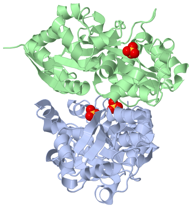
(-) Ligands, Modified Residues, Ions  (1, 3)

Asymmetric Unit (1, 3)
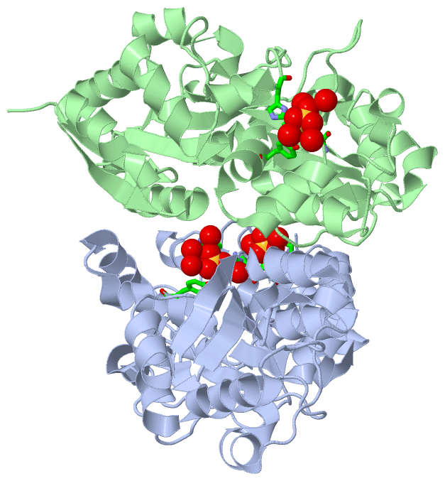
No.NameCountTypeFull Name
1SO43Ligand/IonSULFATE ION
Biological Unit 1 (1, 6)
No.NameCountTypeFull Name
1SO46Ligand/IonSULFATE ION
Biological Unit 2 (1, 2)
No.NameCountTypeFull Name
1SO42Ligand/IonSULFATE ION
Biological Unit 3 (1, 1)
No.NameCountTypeFull Name
1SO41Ligand/IonSULFATE ION

(-) Sites  (3, 3)

Asymmetric Unit (3, 3)
No.NameEvidenceResiduesDescription
1AC1SOFTWAREARG A:163 , ASN A:204 , ASP A:205 , TYR A:226 , HOH A:2196 , HOH A:2238 , HOH A:2381 , HOH A:2382 , HOH A:2383BINDING SITE FOR RESIDUE SO4 A1347
2AC2SOFTWARESER A:166 , ASN A:167 , HOH A:2384 , HOH A:2386 , HOH B:2218BINDING SITE FOR RESIDUE SO4 A1348
3AC3SOFTWAREARG A:157 , TYR B:227 , SER B:246 , LEU B:247 , HIS B:316 , HOH B:2275 , HOH B:2411 , HOH B:2412 , HOH B:2413BINDING SITE FOR RESIDUE SO4 B1347

(-) SS Bonds  (0, 0)

(no "SS Bond" information available for 2VOZ)

(-) Cis Peptide Bonds  (0, 0)

(no "Cis Peptide Bond" information available for 2VOZ)

 Sequence-Structure Mapping

(-) SAPs(SNPs)/Variants  (0, 0)

(no "SAP(SNP)/Variant" information available for 2VOZ)

(-) PROSITE Motifs  (0, 0)

(no "PROSITE Motif" information available for 2VOZ)

(-) Exons   (0, 0)

(no "Exon" information available for 2VOZ)

(-) Sequences/Alignments

Asymmetric Unit
   Reformat: Number of residues per line =  ('0' or empty: single-line sequence representation)
  Number of residues per labelling interval =   
  UniProt sequence: complete  aligned part    
   Show mapping: SCOP domains CATH domains Pfam domains Secondary structure (by author)
SAPs(SNPs) PROSITE motifs Exons
(details for a mapped element are shown in a popup box when the mouse pointer rests over it)
Chain A from PDB  Type:PROTEIN  Length:313
 aligned with FUTA2_SYNY3 | Q55835 from UniProtKB/Swiss-Prot  Length:346

    Alignment length:313
                                    43        53        63        73        83        93       103       113       123       133       143       153       163       173       183       193       203       213       223       233       243       253       263       273       283       293       303       313       323       333       343   
          FUTA2_SYNY3    34 RTINLYSSRHYNTDDALYDAFGEVNLIEASAEELIERIQSEGANSPGDILFTVDAGMLWRAEQAGLFQPVRSGKLNERIPENLRHPDGLWYGFTQRARVLYYSRDRVNPADLSTYEALADPQWRGKILVRPSSNVYNLSLTASRIAIHGEPETRRWLQGLVGNFARQPEGNDTAQIRAIAAGIGDVAIANSYYYIRLQKSTDPADQEVVEKVSLFFPNTGSGERGTHVNVSGAGVLKNAPNRDAAIAFLEYLASDDAQRYFAEGNNEYPVIPGVPIDPVLAAHGQLKGDPLNVSNLGRYQPDSARLMNEVGWQ 346
               SCOP domains d2voza_ A: automated matches                                                                                                                                                                                                                                                                                              SCOP domains
               CATH domains 2vozA01 A:34-129,A:263-309 Periplasmic binding protein-like II                                  -------------------------------------------------------------------------------------------------------------------------------------2vozA01 A:34-129,A:263-309                     ------------------------------------- CATH domains
               Pfam domains ------------------------------------------------------------------------------------------------------------------------------------------------------------------------------------------------------------------------------------------------------------------------------------------------------------------------- Pfam domains
         Sec.struct. author .eeeeee.....hhhhhhhhhheeeeee.hhhhhhhhhhhhhhhh...eeeeehhhhhhhhhhh.......hhhhhhhhhhhh.......eeeeeeeeeeeee....hhhhh..hhhhhhhhhh..ee.....hhhhhhhhhhhhhhhhhhhhhhhhhhhhhh.......hhhhhhhhhhh....eeeeehhhhhhhhhh.hhhhhhhhhh................eeeeeeee.....hhhhhhhhhhhhhhhhhhhhhhhh...ee.......hhhhhh..........hhhhhhhhhhhhhhhhhh... Sec.struct. author
                 SAPs(SNPs) ------------------------------------------------------------------------------------------------------------------------------------------------------------------------------------------------------------------------------------------------------------------------------------------------------------------------- SAPs(SNPs)
                    PROSITE ------------------------------------------------------------------------------------------------------------------------------------------------------------------------------------------------------------------------------------------------------------------------------------------------------------------------- PROSITE
                 Transcript ------------------------------------------------------------------------------------------------------------------------------------------------------------------------------------------------------------------------------------------------------------------------------------------------------------------------- Transcript
                 2voz A  34 RTINLYSSRHYNTDDALYDAFGEVNLIEASAEELIERIQSEGANSPGDILFTVDAGMLWRAEQAGLFQPVRSGKLNERIPENLRHPDGLWYGFTQRARVLYYSRDRVNPADLSTYEALADPQWRGKILVRPSSNVYNLSLTASRIAIHGEPETRRWLQGLVGNFARQPEGNDTAQIRAIAAGIGDVAIANSYYYIRLQKSTDPADQEVVEKVSLFFPNTGSGERGTHVNVSGAGVLKNAPNRDAAIAFLEYLASDDAQRYFAEGNNEYPVIPGVPIDPVLAAHGQLKGDPLNVSNLGRYQPDSARLMNEVGWQ 346
                                    43        53        63        73        83        93       103       113       123       133       143       153       163       173       183       193       203       213       223       233       243       253       263       273       283       293       303       313       323       333       343   

Chain B from PDB  Type:PROTEIN  Length:315
 aligned with FUTA2_SYNY3 | Q55835 from UniProtKB/Swiss-Prot  Length:346

    Alignment length:315
                                    41        51        61        71        81        91       101       111       121       131       141       151       161       171       181       191       201       211       221       231       241       251       261       271       281       291       301       311       321       331       341     
          FUTA2_SYNY3    32 QSRTINLYSSRHYNTDDALYDAFGEVNLIEASAEELIERIQSEGANSPGDILFTVDAGMLWRAEQAGLFQPVRSGKLNERIPENLRHPDGLWYGFTQRARVLYYSRDRVNPADLSTYEALADPQWRGKILVRPSSNVYNLSLTASRIAIHGEPETRRWLQGLVGNFARQPEGNDTAQIRAIAAGIGDVAIANSYYYIRLQKSTDPADQEVVEKVSLFFPNTGSGERGTHVNVSGAGVLKNAPNRDAAIAFLEYLASDDAQRYFAEGNNEYPVIPGVPIDPVLAAHGQLKGDPLNVSNLGRYQPDSARLMNEVGWQ 346
               SCOP domains d2vozb_ B: automated matches                                                                                                                                                                                                                                                                                                SCOP domains
               CATH domains 2vozB01 B:32-129,B:263-309 Periplasmic binding protein-like II                                    -------------------------------------------------------------------------------------------------------------------------------------2vozB01 B:32-129,B:263-309                     ------------------------------------- CATH domains
           Pfam domains (1) ---------------------------------------------SBP_bac_6-2vozB01 B:77-326                                                                                                                                                                                                                                -------------------- Pfam domains (1)
           Pfam domains (2) ---------------------------------------------SBP_bac_6-2vozB02 B:77-326                                                                                                                                                                                                                                -------------------- Pfam domains (2)
         Sec.struct. author ...eeeeee...hhhhhhhhhhhheeeeee.hhhhhhhhhhhhhhhh...eeeeehhhhhhhhhhh.......hhhhhhhhhhhh.......eeeeeeeeeeeee....hhhhh..hhhhhhhhhh..ee.....hhhhhhhhhhhhhhhhhhhhhhhhhhhhhh.......hhhhhhhhhhh....eeeeehhhhhhhhhh.hhhhhhhhhh................eeeeeeee.....hhhhhhhhhhhhhhhhhhhhhhhhh..ee.......hhhhhh..........hhhhhhhhhhhhhhhhhh... Sec.struct. author
                 SAPs(SNPs) --------------------------------------------------------------------------------------------------------------------------------------------------------------------------------------------------------------------------------------------------------------------------------------------------------------------------- SAPs(SNPs)
                    PROSITE --------------------------------------------------------------------------------------------------------------------------------------------------------------------------------------------------------------------------------------------------------------------------------------------------------------------------- PROSITE
                 Transcript --------------------------------------------------------------------------------------------------------------------------------------------------------------------------------------------------------------------------------------------------------------------------------------------------------------------------- Transcript
                 2voz B  32 QSRTINLYSSRHYNTDDALYDAFGEVNLIEASAEELIERIQSEGANSPGDILFTVDAGMLWRAEQAGLFQPVRSGKLNERIPENLRHPDGLWYGFTQRARVLYYSRDRVNPADLSTYEALADPQWRGKILVRPSSNVYNLSLTASRIAIHGEPETRRWLQGLVGNFARQPEGNDTAQIRAIAAGIGDVAIANSYYYIRLQKSTDPADQEVVEKVSLFFPNTGSGERGTHVNVSGAGVLKNAPNRDAAIAFLEYLASDDAQRYFAEGNNEYPVIPGVPIDPVLAAHGQLKGDPLNVSNLGRYQPDSARLMNEVGWQ 346
                                    41        51        61        71        81        91       101       111       121       131       141       151       161       171       181       191       201       211       221       231       241       251       261       271       281       291       301       311       321       331       341     

   Legend:   → Mismatch (orange background)
  - → Gap (green background, '-', border residues have a numbering label)
    → Modified Residue (blue background, lower-case, 'x' indicates undefined single-letter code, labelled with number + name)
  x → Chemical Group (purple background, 'x', labelled with number + name, e.g. ACE or NH2)
  extra numbering lines below/above indicate numbering irregularities and modified residue names etc., number ends below/above '|'

 Classification and Annotation

(-) SCOP Domains  (1, 2)

Asymmetric Unit

(-) CATH Domains  (1, 2)

Asymmetric Unit
(-)
Class: Alpha Beta (26913)

(-) Pfam Domains  (1, 2)

Asymmetric Unit
(-)
Clan: PBP (391)

(-) Gene Ontology  (9, 9)

Asymmetric Unit(hide GO term definitions)
Chain A,B   (FUTA2_SYNY3 | Q55835)
molecular function
    GO:0046872    metal ion binding    Interacting selectively and non-covalently with any metal ion.
biological process
    GO:0006811    ion transport    The directed movement of charged atoms or small charged molecules into, out of or within a cell, or between cells, by means of some agent such as a transporter or pore.
    GO:0055072    iron ion homeostasis    Any process involved in the maintenance of an internal steady state of iron ions within an organism or cell.
    GO:0006810    transport    The directed movement of substances (such as macromolecules, small molecules, ions) or cellular components (such as complexes and organelles) into, out of or within a cell, or between cells, or within a multicellular organism by means of some agent such as a transporter, pore or motor protein.
cellular component
    GO:0016020    membrane    A lipid bilayer along with all the proteins and protein complexes embedded in it an attached to it.
    GO:0030288    outer membrane-bounded periplasmic space    The region between the inner (cytoplasmic or plasma) membrane and outer membrane of organisms with two membranes such as Gram negative bacteria. These periplasmic spaces are relatively thick and contain a thin peptidoglycan layer (PGL), also referred to as a thin cell wall.
    GO:0042597    periplasmic space    The region between the inner (cytoplasmic) and outer membrane (Gram-negative Bacteria) or cytoplasmic membrane and cell wall (Fungi and Gram-positive Bacteria).
    GO:0009579    thylakoid    A membranous cellular structure that bears the photosynthetic pigments in plants, algae, and cyanobacteria. In cyanobacteria thylakoids are of various shapes and are attached to, or continuous with, the plasma membrane. In eukaryotes they are flattened, membrane-bounded disk-like structures located in the chloroplasts; in the chloroplasts of higher plants the thylakoids form dense stacks called grana. Isolated thylakoid preparations can carry out photosynthetic electron transport and the associated phosphorylation.
    GO:0042651    thylakoid membrane    The pigmented membrane of any thylakoid.

 Visualization

(-) Interactive Views

Asymmetric Unit
  Complete Structure
    Jena3D(integrated viewing of ligand, site, SAP, PROSITE, SCOP information)
    WebMol | AstexViewer[tm]@PDBe
(Java Applets, require no local installation except for Java; loading may be slow)
    STRAP
(Java WebStart application, automatic local installation, requires Java; full application with system access!)
    RasMol
(require local installation)
    Molscript (VRML)
(requires installation of a VRML viewer; select preferred view via VRML and generate a mono or stereo PDF format file)
 
  Ligands, Modified Residues, Ions
    SO4  [ RasMol | Jena3D ]  +environment [ RasMol | Jena3D ]
 
  Sites
    AC1  [ RasMol ]  +environment [ RasMol ]
    AC2  [ RasMol ]  +environment [ RasMol ]
    AC3  [ RasMol ]  +environment [ RasMol ]
 
  Cis Peptide Bonds
(no "Cis Peptide Bonds" information available for 2voz)
 
Biological Units
  Complete Structure
    Biological Unit 1  [ Jena3D ]
    Biological Unit 2  [ Jena3D ]
    Biological Unit 3  [ Jena3D ]

(-) Still Images

Jmol
  protein: cartoon or spacefill or dots and stick; nucleic acid: cartoon and stick; ligands: spacefill; active site: stick
Molscript
  protein, nucleic acid: cartoon; ligands: spacefill; active site: ball and stick

 Databases and Analysis Tools

(-) Databases

Access by PDB/NDB ID
  2voz
    Family and Domain InformationProDom | SYSTERS
    General Structural InformationGlycoscienceDB | MMDB | NDB | OCA | PDB | PDBe | PDBj | PDBsum | PDBWiki | PQS | PROTEOPEDIA
    Orientation in MembranesOPM
    Protein SurfaceSURFACE
    Secondary StructureDSSP (structure derived) | HSSP (homology derived)
    Structural GenomicsGeneCensus
    Structural NeighboursCE | VAST
    Structure ClassificationCATH | Dali | SCOP
    Validation and Original DataBMRB Data View | BMRB Restraints Grid | EDS | PROCHECK | RECOORD | WHAT_CHECK
 
Access by UniProt ID/Accession number
  FUTA2_SYNY3 | Q55835
    Comparative Protein Structure ModelsModBase
    Genomic InformationEnsembl
    Protein-protein InteractionDIP
    Sequence, Family and Domain InformationInterPro | Pfam | SMART | UniProtKB/SwissProt
 
Access by Enzyme Classificator   (EC Number)
  (no 'Enzyme Classificator' available)
    General Enzyme InformationBRENDA | EC-PDB | Enzyme | IntEnz
    PathwayKEGG | MetaCyc
 
Access by Disease Identifier   (MIM ID)
  (no 'MIM ID' available)
    Disease InformationOMIM
 
Access by GenAge ID
  (no 'GenAge ID' available)
    Age Related InformationGenAge

(-) Analysis Tools

Access by PDB/NDB ID
    Domain InformationXDom
    Interatomic Contacts of Structural UnitsCSU
    Ligand-protein ContactsLPC
    Protein CavitiescastP
    Sequence and Secondary StructurePDBCartoon
    Structure AlignmentSTRAP(Java WebStart application, automatic local installation, requires Java; full application with system access!)
    Structure and Sequence BrowserSTING
 
Access by UniProt ID/Accession number
  FUTA2_SYNY3 | Q55835
    Protein Disorder PredictionDisEMBL | FoldIndex | GLOBPLOT (for more information see DisProt)

 Related Entries

(-) Entries Sharing at Least One Protein Chain (UniProt ID)

UniProtKB/Swiss-Prot
        FUTA2_SYNY3 | Q558352vp1

(-) Related Entries Specified in the PDB File

2vp1 FE-FUTA2 FROM SYNECHOCYSTIS PCC6803