|
|
|
|
 Description
|
|
Compounds
|
||||||||||||||||||||||||||||||||||||||||||||||||||||||||
Chains, Units
Summary Information (see also Sequences/Alignments below) |
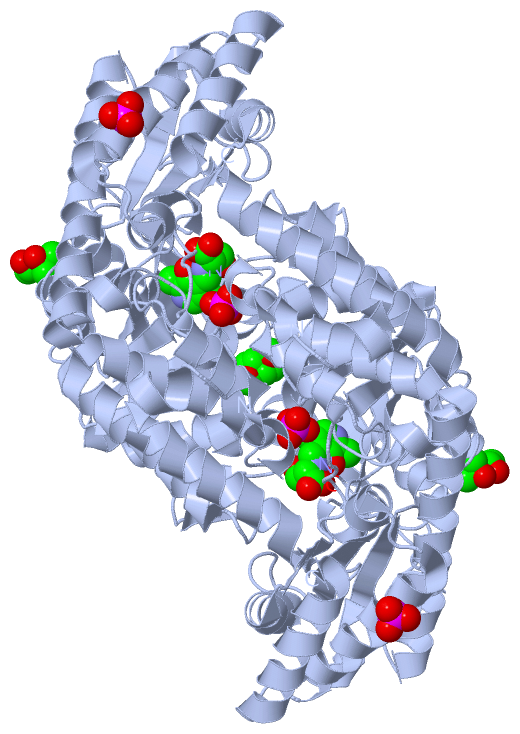
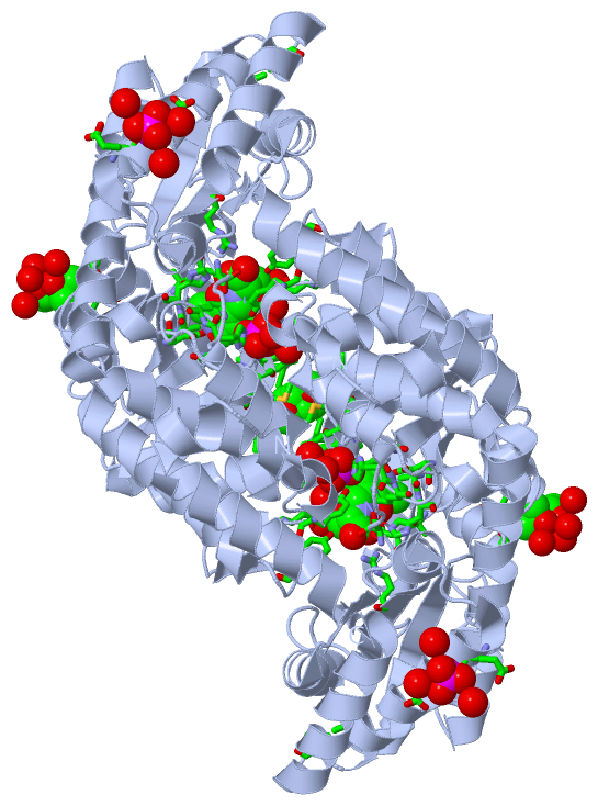
Ligands, Modified Residues, Ions (4, 5)| Asymmetric Unit (4, 5) Biological Unit 1 (4, 10) |
Sites (5, 5)
Asymmetric Unit (5, 5)
|
SS Bonds (0, 0)| (no "SS Bond" information available for 2VMW) |
Cis Peptide Bonds (1, 1)
Asymmetric Unit
|
||||||||
SAPs(SNPs)/Variants (0, 0)| (no "SAP(SNP)/Variant" information available for 2VMW) |
PROSITE Motifs (0, 0)| (no "PROSITE Motif" information available for 2VMW) |
Exons (0, 0)| (no "Exon" information available for 2VMW) |
Sequences/Alignments
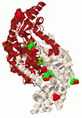
Asymmetric UnitChain A from PDB Type:PROTEIN Length:406 aligned with Q7SIB6_GEOSE | Q7SIB6 from UniProtKB/TrEMBL Length:419 Alignment length:407 10 20 30 40 50 60 70 80 90 100 110 120 130 140 150 160 170 180 190 200 210 220 230 240 250 260 270 280 290 300 310 320 330 340 350 360 370 380 390 400 Q7SIB6_GEOSE 1 MKYLPQQDPQVFAAIEQERKRQHAKIELIASENFVSRAVMEAQGSVLTNKYAEGYPGRRYYGGCEYVDIVEELARERAKQLFGAEHANVQPHSGAQANMAVYFTVLEHGDTVLGMNLSHGGHLTHGSPVNFSGVQYNFVAYGVDPETHVIDYDDVREKARLHRPKLIVAAASAYPRIIDFAKFREIADEVGAYLMVDMAHIAGLVAAGLHPNPVPYAHFVTTTTHKTLRGPRGGMILCQEQFAKQIDKAIFPGIQGGPLMHVIAAKAVAFGEALQDDFKAYAKRVVDNAKRLASALQNEGFTLVSGGTDNHLLLVDLRPQQLTGKTAEKVLDEVGITVNKNTIPYDPESPFVTSGIRIGTAAVTTRGFGLEEMDEIAAIIGLVLKNVGSEQALEEARQRVAALTDPT 407 SCOP domains d2vmwa_ A: Serine hydroxymethyltransferase SCOP domains CATH domains 2vmwA01 A:1-32,A:284-405 2vmwA02 A:33-283 Type I PLP-dependent aspartate aminotransferase-like (Major domain) 2vmwA01 A:1-32,A:284-405 Aspartate Aminotransferase, domain 1 -- CATH domains Pfam domains ---SHMT-2vmwA01 A:4-380 --------------------------- Pfam domains SAPs(SNPs) ----------------------------------------------------------------------------------------------------------------------------------------------------------------------------------------------------------------------------------------------------------------------------------------------------------------------------------------------------------------------------------------------------------------------- SAPs(SNPs) PROSITE ----------------------------------------------------------------------------------------------------------------------------------------------------------------------------------------------------------------------------------------------------------------------------------------------------------------------------------------------------------------------------------------------------------------------- PROSITE Transcript ----------------------------------------------------------------------------------------------------------------------------------------------------------------------------------------------------------------------------------------------------------------------------------------------------------------------------------------------------------------------------------------------------------------------- Transcript 2vmw A 1 MKYLPQQDPQVFAAIEQERKRQHAKIELIASENFVSRAVMEAQGSVLTNKYAEGYPGRRYYGGCEYVDIVEELARERAKQLFGAEHANVQPHSGAQANMAVYFTVLEHGDTVLGMNLSHGGHLTHGSPVNFSGVQYNFVAYGVDPETHVIDYDDVREKARLHRPKLIVAAASAYPRIIDFAKFREIADEVGAYLMVDMAHIAGLVAAGLHPNPVPYAHFVTTTTHKTLRGPRGGMILCQEQFAKQIDKAIFPGIQGGPLMHVIAAKAVAFGEALQDDFKAYAKRVVDNAKRLASALQNEGFTLVSGGTDNHLLLVDLRPQQLTGKTAEKVLDEVGITVNKNTIPYDPESPGVTSGIRIGTAAVTTRGFGLEEMDEIAAIIGLVLKNVGSEQALEEARQRVAALTD-S 502 10 20 30 40 50 60 70 80 90 100 110 120 130 140 150 160 170 180 190 200 210 220 230 240 250 260 270 280 290 300 310 320 330 340 350 360 370 380 390 400 | | 405 | 502
|
||||||||||||||||||||
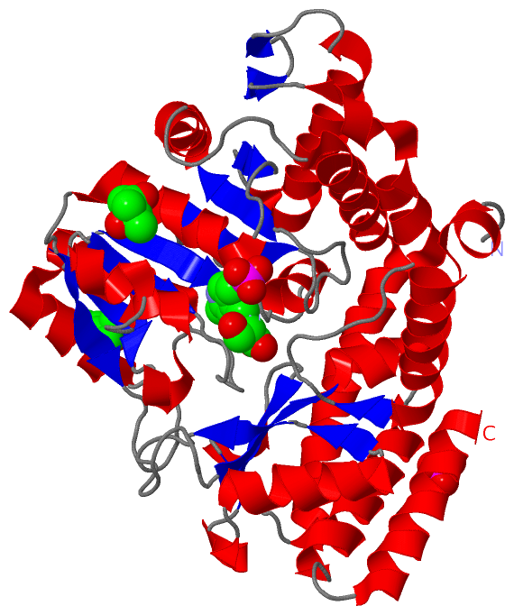
SCOP Domains (1, 1)
Asymmetric Unit
 |
CATH Domains (2, 2)
Asymmetric Unit
|
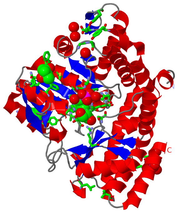
Pfam Domains (1, 1)| Asymmetric Unit |
Gene Ontology (12, 12)|
Asymmetric Unit(hide GO term definitions) Chain A (Q7SIB6_GEOSE | Q7SIB6)
|
||||||||||||||||||||||||||||||||||||||||||||||||||||||||||||||||||||||||||||||||||||||||||
Interactive Views
|
||||||||||||||||||||||||||||||||||||||||||||||||||||||||||||||||||||||||||||||||||||||||||||||||||||||||||||||||||||||||||||||||||||||||||||||||||||||||||||||||||||||||||||||||||||||||||
Still Images
|
||||||||||||||||
Databases
|
||||||||||||||||||||||||||||||||||||||||||||||||||||||||||||||||||||||||||||||||||||||||||||||||||||||||||||||||||||||||||||||||||||||||||||||||||||||||||||||||
Analysis Tools
|
|||||||||||||||||||||||||||||||||||||||||||||||||||||||||||||
Entries Sharing at Least One Protein Chain (UniProt ID)
Related Entries Specified in the PDB File
|
|