Show PDB file:   
         Plain Text   HTML   (compressed file size)
QuickSearch:   
by PDB,NDB,UniProt,PROSITE Code or Search Term(s)  
(-)Asymmetric Unit
(-)Asym. Unit - sites
(-)Biological Unit 1
(-)Biol. Unit 1 - sites
collapse expand < >
Image Asymmetric Unit
Asymmetric Unit  (Jmol Viewer)
Image Asym. Unit - sites
Asym. Unit - sites  (Jmol Viewer)
Image Biological Unit 1
Biological Unit 1  (Jmol Viewer)
Image Biol. Unit 1 - sites
Biol. Unit 1 - sites  (Jmol Viewer)

(-) Description

Title :  STRUCTURE OF THE COMPLEX OF DISCOIDIN II FROM DICTYOSTELIUM DISCOIDEUM WITH BETA-METHYL-GALACTOSE
 
Authors :  K. S. Aragao, M. Satre, A. Imberty, A. Varrot
Date :  25 Jan 08  (Deposition) - 01 Jul 08  (Release) - 08 Jun 11  (Revision)
Method :  X-RAY DIFFRACTION
Resolution :  1.90
Chains :  Asym. Unit :  A
Biol. Unit 1:  A  (3x)
Keywords :  Ddr, Lectin, Aggregation, Cell Adhesion (Keyword Search: [Gene Ontology, PubMed, Web (Google))
 
Reference :  K. S. Aragao, M. Satre, A. Imberty, A. Varrot
Structure Determination Of Discoidin Ii From Dictyostelium Discoideum And Carbohydrate Binding Properties Of The Lectin Domain.
Proteins V. 73 43 2008
PubMed-ID: 18384150  |  Reference-DOI: 10.1002/PROT.22038

(-) Compounds

Molecule 1 - DISCOIDIN-2
    ChainsA
    EngineeredYES
    Expression SystemESCHERICHIA COLI
    Expression System PlasmidPPROEXHTB
    Expression System StrainBL21(DE3)
    Expression System Taxid469008
    Expression System VariantROSETTA2 PLYSS
    Expression System Vector TypePLASMID
    Organism CommonSLIME MOLD
    Organism ScientificDICTYOSTELIUM DISCOIDEUM
    Organism Taxid366501
    StrainAX2
    SynonymDISCOIDIN II

 Structural Features

(-) Chains, Units

  1
Asymmetric Unit A
Biological Unit 1 (3x)A

Summary Information (see also Sequences/Alignments below)

(-) Ligands, Modified Residues, Ions  (5, 6)

Asymmetric Unit (5, 6)
No.NameCountTypeFull Name
11PG2Ligand/Ion2-(2-{2-[2-(2-METHOXY-ETHOXY)-ETHOXY]-ETHOXY}-ETHOXY)-ETHANOL
2CA1Ligand/IonCALCIUM ION
3CL1Ligand/IonCHLORIDE ION
4EDO1Ligand/Ion1,2-ETHANEDIOL
5MBG1Ligand/IonMETHYL-BETA-GALACTOSE
Biological Unit 1 (3, 12)
No.NameCountTypeFull Name
11PG6Ligand/Ion2-(2-{2-[2-(2-METHOXY-ETHOXY)-ETHOXY]-ETHOXY}-ETHOXY)-ETHANOL
2CA-1Ligand/IonCALCIUM ION
3CL-1Ligand/IonCHLORIDE ION
4EDO3Ligand/Ion1,2-ETHANEDIOL
5MBG3Ligand/IonMETHYL-BETA-GALACTOSE

(-) Sites  (6, 6)

Asymmetric Unit (6, 6)
No.NameEvidenceResiduesDescription
1AC1SOFTWARETRP A:219BINDING SITE FOR RESIDUE CL A 499
2AC2SOFTWAREASN A:39 , SER A:40 , ASP A:47 , HOH A:2051 , HOH A:2056 , HOH A:2064 , HOH A:2065BINDING SITE FOR RESIDUE CA A 500
3AC3SOFTWAREASP A:209 , GLN A:216 , ARG A:218 , TRP A:238 , TYR A:244 , HOH A:2226 , HOH A:2227BINDING SITE FOR RESIDUE MBG A 501
4AC4SOFTWAREALA A:12 , ARG A:94 , TRP A:102 , ALA A:135 , HIS A:137BINDING SITE FOR RESIDUE 1PG A 502
5AC5SOFTWARELYS A:59 , LYS A:92 , GLN A:139 , HOH A:2228BINDING SITE FOR RESIDUE 1PG A 503
6AC6SOFTWAREARG A:81 , GLN A:86 , HOH A:2040BINDING SITE FOR RESIDUE EDO A 504

(-) SS Bonds  (0, 0)

(no "SS Bond" information available for 2VMD)

(-) Cis Peptide Bonds  (1, 1)

Asymmetric Unit
No.Residues
1Asn A:126 -Pro A:127

 Sequence-Structure Mapping

(-) SAPs(SNPs)/Variants  (0, 0)

(no "SAP(SNP)/Variant" information available for 2VMD)

(-) PROSITE Motifs  (2, 2)

Asymmetric Unit (2, 2)
 PROSITEUniProtKBPDB
No.IDACDescriptionIDLocationCountLocation
1FA58C_3PS50022 Coagulation factors 5/8 type C domain (FA58C) profile.DIS2_DICDI10-154  1A:10-154
2FA58C_1PS01285 Coagulation factors 5/8 type C domain (FA58C) signature 1.DIS2_DICDI51-80  1A:51-80
Biological Unit 1 (2, 6)
 PROSITEUniProtKBPDB
No.IDACDescriptionIDLocationCountLocation
1FA58C_3PS50022 Coagulation factors 5/8 type C domain (FA58C) profile.DIS2_DICDI10-154  3A:10-154
2FA58C_1PS01285 Coagulation factors 5/8 type C domain (FA58C) signature 1.DIS2_DICDI51-80  3A:51-80

(-) Exons   (0, 0)

(no "Exon" information available for 2VMD)

(-) Sequences/Alignments

Asymmetric Unit
   Reformat: Number of residues per line =  ('0' or empty: single-line sequence representation)
  Number of residues per labelling interval =   
  UniProt sequence: complete  aligned part    
   Show mapping: SCOP domains CATH domains Pfam domains Secondary structure (by author)
SAPs(SNPs) PROSITE motifs Exons
(details for a mapped element are shown in a popup box when the mouse pointer rests over it)
Chain A from PDB  Type:PROTEIN  Length:252
 aligned with DIS2_DICDI | P42530 from UniProtKB/Swiss-Prot  Length:257

    Alignment length:252
                                    15        25        35        45        55        65        75        85        95       105       115       125       135       145       155       165       175       185       195       205       215       225       235       245       255  
           DIS2_DICDI     6 GSVSCLANALLNLRSSTDYNADHGVKNSILNFSNSKDASRFDGSESWSSSVLDKNQFIVAGSDSVKHFVAISTQGRGDHDQWVTSYKLRYTLDNVNWVEYNNGEIINANKDRNSIVTINFNPPIKARSIAIHPQTYNNHISLRWELYALPVKSYSNPSVQVGEVSIGDRSLNSGTGSRTIVRHVKFPVEFLSVPIVSIGCKKVDAHTDNGQMRWEGKSENITTKGFDLTFITWGNNAVYDLTFDYVAVEFNN 257
               SCOP domains ------------------------------------------------------------------------------------------------------------------------------------------------------------------------------------------------------------------------------------------------------------ SCOP domains
               CATH domains ------------------------------------------------------------------------------------------------------------------------------------------------------------------------------------------------------------------------------------------------------------ CATH domains
               Pfam domains -----------F5_F8_type_C-2vmdA01 A:17-151                                                                                                          -------------------------------H_lectin-2vmdA02 A:183-255                                               -- Pfam domains
         Sec.struct. author ....hhhhh..eeee..........................................eeee....eeeeeeeee.......eeeeeeeeee.....eee......eeee......eeeee...eeeeeeeeeeeeee...eeeeeeee.........eeeeeeee............eeeeeeee.........eeeeeeeeeee.........eeeee.....eeeeeeee.....eeeeeeeeeeee... Sec.struct. author
                 SAPs(SNPs) ------------------------------------------------------------------------------------------------------------------------------------------------------------------------------------------------------------------------------------------------------------ SAPs(SNPs)
                PROSITE (1) ----FA58C_3  PDB: A:10-154 UniProt: 10-154                                                                                                           ------------------------------------------------------------------------------------------------------- PROSITE (1)
                PROSITE (2) ---------------------------------------------FA58C_1  PDB: A:51-80         --------------------------------------------------------------------------------------------------------------------------------------------------------------------------------- PROSITE (2)
                 Transcript ------------------------------------------------------------------------------------------------------------------------------------------------------------------------------------------------------------------------------------------------------------ Transcript
                 2vmd A   6 GSVSCLANALLNLRSSTDYNADHGVKNSILNFSNSKDASRFDGSESWSSSVLDKNQFIVAGSDSVKHFVAISTQGRGDHDQWVTSYKLRYTLDNVNWVEYNNGEIINANKDRNSIVTINFNPPIKARSIAIHPQTYNNHISLRWELYALPVKSYSNPSVQVGEVSIGDRSLNSGTGSRTIVRHVKFPVEFLSVPIVSIGCKKVDAHTDNGQMRWEGKSENITTKGFDLTFITWGNNAVYDLTFDYVAVEFNN 257
                                    15        25        35        45        55        65        75        85        95       105       115       125       135       145       155       165       175       185       195       205       215       225       235       245       255  

   Legend:   → Mismatch (orange background)
  - → Gap (green background, '-', border residues have a numbering label)
    → Modified Residue (blue background, lower-case, 'x' indicates undefined single-letter code, labelled with number + name)
  x → Chemical Group (purple background, 'x', labelled with number + name, e.g. ACE or NH2)
  extra numbering lines below/above indicate numbering irregularities and modified residue names etc., number ends below/above '|'

 Classification and Annotation

(-) SCOP Domains  (0, 0)

(no "SCOP Domain" information available for 2VMD)

(-) CATH Domains  (0, 0)

(no "CATH Domain" information available for 2VMD)

(-) Pfam Domains  (2, 2)

Asymmetric Unit
(-)
Clan: GBD (153)

(-) Gene Ontology  (14, 14)

Asymmetric Unit(hide GO term definitions)
Chain A   (DIS2_DICDI | P42530)
molecular function
    GO:0046871    N-acetylgalactosamine binding    Interacting selectively and non-covalently with N-acetylgalactosamine, 2-acetamido-2-deoxygalactopyranose, the n-acetyl derivative of galactosamine.
    GO:0030246    carbohydrate binding    Interacting selectively and non-covalently with any carbohydrate, which includes monosaccharides, oligosaccharides and polysaccharides as well as substances derived from monosaccharides by reduction of the carbonyl group (alditols), by oxidation of one or more hydroxy groups to afford the corresponding aldehydes, ketones, or carboxylic acids, or by replacement of one or more hydroxy group(s) by a hydrogen atom. Cyclitols are generally not regarded as carbohydrates.
    GO:0016936    galactoside binding    Interacting selectively and non-covalently with any glycoside in which the sugar group is galactose.
    GO:0046872    metal ion binding    Interacting selectively and non-covalently with any metal ion.
    GO:0070492    oligosaccharide binding    Interacting selectively and non-covalently with any oligosaccharide, a molecule with between two and (about) 20 monosaccharide residues connected by glycosidic linkages.
    GO:0030247    polysaccharide binding    Interacting selectively and non-covalently with any polysaccharide, a polymer of many (typically more than 10) monosaccharide residues linked glycosidically.
biological process
    GO:0007155    cell adhesion    The attachment of a cell, either to another cell or to an underlying substrate such as the extracellular matrix, via cell adhesion molecules.
    GO:0098609    cell-cell adhesion    The attachment of one cell to another cell via adhesion molecules.
    GO:0044351    macropinocytosis    An endocytosis process that results in the uptake of liquid material by cells from their external environment by the 'ruffling' of the cell membrane to form heterogeneously sized intracellular vesicles called macropinosomes, which can be up to 5 micrometers in size.
    GO:0009617    response to bacterium    Any process that results in a change in state or activity of a cell or an organism (in terms of movement, secretion, enzyme production, gene expression, etc.) as a result of a stimulus from a bacterium.
    GO:0046689    response to mercury ion    Any process that results in a change in state or activity of a cell or an organism (in terms of movement, secretion, enzyme production, gene expression, etc.) as a result of a mercury ion stimulus.
cellular component
    GO:0005615    extracellular space    That part of a multicellular organism outside the cells proper, usually taken to be outside the plasma membranes, and occupied by fluid.
    GO:0045335    phagocytic vesicle    A membrane-bounded intracellular vesicle that arises from the ingestion of particulate material by phagocytosis.
    GO:0031982    vesicle    Any small, fluid-filled, spherical organelle enclosed by membrane.
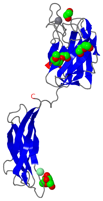
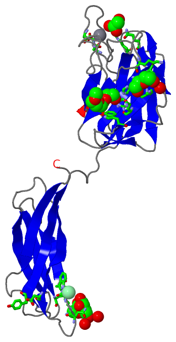
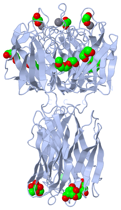
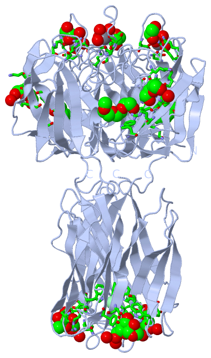
 Visualization

(-) Interactive Views

Asymmetric Unit
  Complete Structure
    Jena3D(integrated viewing of ligand, site, SAP, PROSITE, SCOP information)
    WebMol | AstexViewer[tm]@PDBe
(Java Applets, require no local installation except for Java; loading may be slow)
    STRAP
(Java WebStart application, automatic local installation, requires Java; full application with system access!)
    RasMol
(require local installation)
    Molscript (VRML)
(requires installation of a VRML viewer; select preferred view via VRML and generate a mono or stereo PDF format file)
 
  Ligands, Modified Residues, Ions
    1PG  [ RasMol | Jena3D ]  +environment [ RasMol | Jena3D ]
    CA  [ RasMol | Jena3D ]  +environment [ RasMol | Jena3D ]
    CL  [ RasMol | Jena3D ]  +environment [ RasMol | Jena3D ]
    EDO  [ RasMol | Jena3D ]  +environment [ RasMol | Jena3D ]
    MBG  [ RasMol | Jena3D ]  +environment [ RasMol | Jena3D ]
 
  Sites
    AC1  [ RasMol ]  +environment [ RasMol ]
    AC2  [ RasMol ]  +environment [ RasMol ]
    AC3  [ RasMol ]  +environment [ RasMol ]
    AC4  [ RasMol ]  +environment [ RasMol ]
    AC5  [ RasMol ]  +environment [ RasMol ]
    AC6  [ RasMol ]  +environment [ RasMol ]
 
  Cis Peptide Bonds
    Asn A:126 - Pro A:127   [ RasMol ]  
 
Biological Unit
  Complete Structure
    Biological Unit 1  [ Jena3D ]

(-) Still Images

Jmol
  protein: cartoon or spacefill or dots and stick; nucleic acid: cartoon and stick; ligands: spacefill; active site: stick
Molscript
  protein, nucleic acid: cartoon; ligands: spacefill; active site: ball and stick

 Databases and Analysis Tools

(-) Databases

Access by PDB/NDB ID
  2vmd
    Family and Domain InformationProDom | SYSTERS
    General Structural InformationGlycoscienceDB | MMDB | NDB | OCA | PDB | PDBe | PDBj | PDBsum | PDBWiki | PQS | PROTEOPEDIA
    Orientation in MembranesOPM
    Protein SurfaceSURFACE
    Secondary StructureDSSP (structure derived) | HSSP (homology derived)
    Structural GenomicsGeneCensus
    Structural NeighboursCE | VAST
    Structure ClassificationCATH | Dali | SCOP
    Validation and Original DataBMRB Data View | BMRB Restraints Grid | EDS | PROCHECK | RECOORD | WHAT_CHECK
 
Access by UniProt ID/Accession number
  DIS2_DICDI | P42530
    Comparative Protein Structure ModelsModBase
    Genomic InformationEnsembl
    Protein-protein InteractionDIP
    Sequence, Family and Domain InformationInterPro | Pfam | SMART | UniProtKB/SwissProt
 
Access by Enzyme Classificator   (EC Number)
  (no 'Enzyme Classificator' available)
    General Enzyme InformationBRENDA | EC-PDB | Enzyme | IntEnz
    PathwayKEGG | MetaCyc
 
Access by Disease Identifier   (MIM ID)
  (no 'MIM ID' available)
    Disease InformationOMIM
 
Access by GenAge ID
  (no 'GenAge ID' available)
    Age Related InformationGenAge

(-) Analysis Tools

Access by PDB/NDB ID
    Domain InformationXDom
    Interatomic Contacts of Structural UnitsCSU
    Ligand-protein ContactsLPC
    Protein CavitiescastP
    Sequence and Secondary StructurePDBCartoon
    Structure AlignmentSTRAP(Java WebStart application, automatic local installation, requires Java; full application with system access!)
    Structure and Sequence BrowserSTING
 
Access by UniProt ID/Accession number
  DIS2_DICDI | P42530
    Protein Disorder PredictionDisEMBL | FoldIndex | GLOBPLOT (for more information see DisProt)

 Related Entries

(-) Entries Sharing at Least One Protein Chain (UniProt ID)

UniProtKB/Swiss-Prot
        DIS2_DICDI | P425302vm9 2vmc 2vme

(-) Related Entries Specified in the PDB File

2vm9 NATIVE STRUCTURE OF THE RECOMBINANT DISCOIDIN II OF DICTYOSTELIUM DISCOIDEUM AT 1.75 ANGSTROM
2vmc STRUCTURE OF THE COMPLEX OF DISCOIDIN II FROM DICTYOSTELIUM DISCOIDEUM WITH N-ACETYL-GALACTOSAMINE
2vme STRUCTURE OF THE WILD-TYPE DISCOIDIN II FROM DICTYOSTELIUM DISCOIDEUM