|
|
|
|
 Description
|
|
Compounds
|
||||||||||||||||||||||||||||||||||||
Chains, Units
Summary Information (see also Sequences/Alignments below) |
Ligands, Modified Residues, Ions (3, 5)| Asymmetric/Biological Unit (3, 5) |
Sites (5, 5)
Asymmetric Unit (5, 5)
|
SS Bonds (0, 0)| (no "SS Bond" information available for 2VBU) |
Cis Peptide Bonds (0, 0)| (no "Cis Peptide Bond" information available for 2VBU) |
SAPs(SNPs)/Variants (0, 0)| (no "SAP(SNP)/Variant" information available for 2VBU) |
PROSITE Motifs (0, 0)| (no "PROSITE Motif" information available for 2VBU) |
Exons (0, 0)| (no "Exon" information available for 2VBU) |
Sequences/Alignments
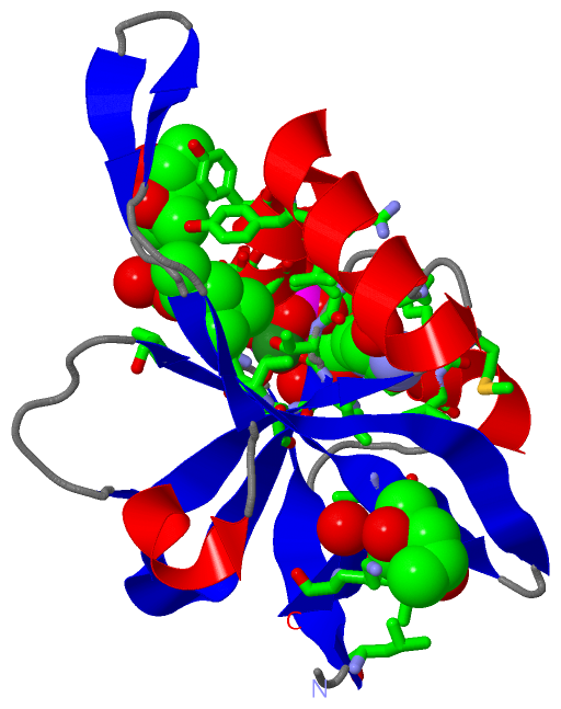
Asymmetric/Biological UnitChain A from PDB Type:PROTEIN Length:131 aligned with RIFK_METJA | Q60365 from UniProtKB/Swiss-Prot Length:132 Alignment length:131 1 | 7 17 27 37 47 57 67 77 87 97 107 117 127 RIFK_METJA - ---MIIEGEVVSGLGEGRYFLSLPPYKEIFKKILGFEPYEGTLNLKLDREFDINKFKYIETEDFEFNGKRFFGVKVLPIKILIGNKKIDGAIVVPKKTYHSSEIIEIIAPMKLREQFNLKDGDVIKILIKG 128 SCOP domains d2vbua_ A: CTP-dependent riboflavin kinase, Rfk SCOP domains CATH domains -2vbuA01 A:3-132 Riboflavin kinase-like CATH domains Pfam domains ---------CTP-dep_RFKase-2vbuA01 A:11-130 -- Pfam domains SAPs(SNPs) ----------------------------------------------------------------------------------------------------------------------------------- SAPs(SNPs) PROSITE ----------------------------------------------------------------------------------------------------------------------------------- PROSITE Transcript ----------------------------------------------------------------------------------------------------------------------------------- Transcript 2vbu A 2 VKLMIIEGEVVSGLGEGRYFLSLPPYKEIFKKILGFEPYEGTLNLKLDREFDINKFKYIETEDFEFNGKRFFGVKVLPIKILIGNKKIDGAIVVPKKTYHSSEIIEIIAPMKLREQFNLKDGDVIKILIKG 132 11 21 31 41 51 61 71 81 91 101 111 121 131
|
||||||||||||||||||||
SCOP Domains (1, 1)
Asymmetric/Biological Unit
 |
CATH Domains (1, 1)
Asymmetric/Biological Unit
|
Pfam Domains (1, 1)
Asymmetric/Biological Unit
|
Gene Ontology (10, 10)|
Asymmetric/Biological Unit(hide GO term definitions) Chain A (RIFK_METJA | Q60365)
|
||||||||||||||||||||||||||||||||||||||||||||||||||||||||||||||||||||||||
Interactive Views
|
||||||||||||||||||||||||||||||||||||||||||||||||||||||||||||||||||||||||||||||||||||||||||||||||||||||||||||||||||||||||||||||||||||||||||||||||||||||||||||||||
Still Images
|
||||||||||||||||
Databases
|
||||||||||||||||||||||||||||||||||||||||||||||||||||||||||||||||||||||||||||||||||||||||||||||||||||||||||||||||||||||||||||||||||||||||||||||||||||||||||||||||
Analysis Tools
|
|||||||||||||||||||||||||||||||||||||||||||||||||||||||||||||
Entries Sharing at Least One Protein Chain (UniProt ID)
Related Entries Specified in the PDB File
|
|